#labcam
Video
Essas pequenas gotas verdes à deriva são as partes fotossintéticas de uma planta chamados cloroplastos. Eles são verdes devido à presença de um pigmento chamado clorofila. Esses cloroplastos são visualizados dentro das células vegetais individuais de uma planta aquática chamada Elodea. Durante a fotossíntese, essas pequenas organelas convertem dióxido de carbono e água em glicose e oxigênio. O movimento dos cloroplastos é chamado ciclose ou fluxo citoplasmático. . Texto completo em inglês @my.microscopic.world . #Repost @my.microscopic.world • • • • • • These small green drifting blobs are the photosynthetic parts of a plant called chloroplasts. They are green due to the presence of a pigment called chlorophyll. These chloroplasts are viewed inside the individual plant cells of a water plant called Elodea, commonly known as waterweed. During photosynthesis these small organelles convert carbon dioxide and water into glucose and oxygen. The movement of the chloroplasts is called cyclosis or cytoplasmic streaming. The chloroplasts move along actin filaments using a molecule called myosin and these two molecules are actually also what makes you and your muscles able to move. So, you might have more in common with plants than you thought! . This was as always filmed with my iPhone mounted on my microscope with a @ilabcam and the beautiful music is made by @animamusic1 . . When I make these photos and videos, I use a lot of time and materials, like petri dishes, microscope slides, sample tubes, pippets and so on. If you want to help me make more of this content you can find me on patreon.com/mymicroscopicworld (link in bio) and support me with as little or as much as you like, which would help me a lot in making these photos and videos and getting new materials! . #science #biology #microscopy #plant #plantcells #cell #labcam #shotoniphone #underthemicroscope #nature #biologo #bio #biologosnarede #biologia #Biology https://www.instagram.com/p/B2yqH80nBK1/?igshid=4wjopfff3me9
#repost#science#biology#microscopy#plant#plantcells#cell#labcam#shotoniphone#underthemicroscope#nature#biologo#bio#biologosnarede#biologia
5 notes
·
View notes
Text
LabCam Pro: Is this the ultimate handphone microscope adapter?
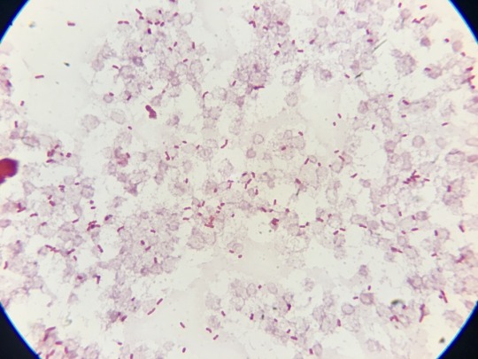
We have previously blogged about using the Olympus Air for taking images of Gram stains. This gave the image quality of a dedicated microscope camera but the added convenience of quickly sharing the image because it was transferred to a handphone. But why not just use a handphone to capture the image directly?

Anyone who has tried this will realise it is not as easy as it sounds. If you just hold the handphone lens flush against the microscope eyepiece, it is very difficult to get a good image of the slide because the wide angle of the camera lens tends to include the sides of the eyepiece barrel. You can obtain a better field of view of the slide by holding the handphone a small distance away from the eyepiece but then you need steady hands...
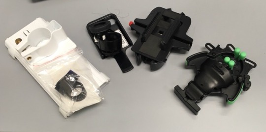
We have experimented with a number of commercial microscope-handphone adapters without much joy. They all suffer from 2 problems. Firstly, they all hold the handphone flush with the eyepiece so you don’t solve the field of view problem mentioned above. Secondly, the attachment method does not result in a stable platform.
We were so frustrated with the results that we considered designing and 3D-printing our own adapter. It would have set the handphone lens a bit further back from the eyepiece and would involve a redesign of the attachment method that would leverage somehow on the binocular eyepiece of the microscope to keep the whole platform steady.
Someone has now come up with a better design based on a similar thought process.
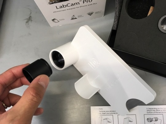

The LabCam addressed the field of view issue optically by including a lens which replaces the microscope eyepiece (2 are provided which should fit most microscopes). Both the replacement eyepiece and the design of the adapter itself ensure a stable platform. It is easy to set up and use.
The LabCam was actually designed by an undergraduate student and converted to viable product by a Bench to Bedside Initiative.
There remain a number of issues.
Because it includes lenses, it is more expensive than the other adapters (but the ease of use and the results obtained will trump price in our view).
The lens could be of higher quality. Sharpness is good centrally but falls away at the edges. There also appears to be chromatic aberration at the edges (blue fringing around the image).
It is only available for iPhone at the moment, though the modular design does suggest the potential to apply the same concept to other handphones.
Nevertheless, we feel this is the best solution on the market at the moment. You can see more images captured with this system on our Instagram.
https://www.instagram.com/10minus6cosm/
0 notes
Photo

Fruit fly focus stack - Shot with my iPhone mounted on my microscope with a LabCam (150 times total magnification)
1 note
·
View note
Photo

LabCam for iPhone (Microscope and Telescope) $239 USD
0 notes
Text


Wow
Thursday 17 June 2021
Wokelate
Took Manjot to labcame h
Mepar
Packed and cleaned
Drove to Colorado with anish and Pranav
Then used hot tub
Got ramen
0 notes
Text
Ata da reunião do Centro Acadêmico de Biologia 08 de maio de 2017
Local: Sala 43 Hora de início: 18:05h
Hora de término: 19:00h
Presentes: Matheus Ganiko, Bruno Sandri, Cahique, Letícia Vasconcelos, Geovane Gaia, Kevin Elias, Alessandra Custodio, Fernanda Borges, Bruno Francisco, Guilherme Bevilacqua, Raissa Rotelli, Nathalia Marinho, Juliana Xavier, Leonardo Dota, Gabriela.
Informes:
1) Semana do Meio Ambiente: Sandri foi na reunião do SIMAB, disse que a reunião foi longa e disse que passaram todos os dias os eventos que ia ter, no dia 6 vamos fazer uma palestra junto com os alunos da USP na USP, e ele disse que terá uma feira de trocas e que você pode entregar algo que não queira como um livro ou CD, por exemplo, e você poderá fazer a troca. Na quinta, dia 8, terá o passeio pelos laboratórios pelos alunos do 9º ano, Sandri propôs que apenas 4 laboratórios participassem: LABCAM, LAPALMA, GEBA E ORNITOLOGIA. Guilherme B. irá verificar 7 monitores para participar da ronda.
2) Semana da Bio: Sandri questionou se alguém sabia como era decidido o presidente da Semana da Bio. Ele disse andaram reclamando e algumas pessoas que entraram para a organização sem participar do processo seletivo e uma delas ainda indicada para ser a presidente da comissão de apoio. Pediram para o CA levar esse problema para o Conselho de Curso. Guilherme B. comentou que o Sheldon já havia apontado esse problema anteriormente.
3) Identidade Visual Bio na Rua: Cahique disse que o OB avisou que identidade visual está pronta e que logo deve nos enviar a arte.
4) Fábio: Fernanda disse que as pessoas que faltaram na aula de genética para ir ao bio no bosque receberam uma bronca do Fábio inclusive ela que estava contribuindo para o evento.
5) Camiseta do CA: Cahique propôs que pensássemos na possibilidade de ter uma camiseta do CA para participar das reuniões.
6) Moldes das Camisetas: Marina, Kevin e Letícia irão verificar esses detalhes de moldes.
Pautas e discussões:
1) Semana da Bio: Letícia informou que o pessoal da comissão da SEMBIO gostaria de ter uma sala no departamento de bio e que não sabia com quem conversar. Cahique disse que essa informação não compete ao CA e que eles precisarão verificar com a Anne.
2) Visita Estação de Tratamento: Kevin disse que a Estação de Tratamento vai estar aberta durante a semana da bio e que as pessoas do locar comentou dos alunos da biologia se interessarem em ir lá.
3) Patrocínio Ecológico: Kevin disse para ir atrás de empresas sustentáveis para patrocinar o bio na rua.
4) Sala 43: Koji disse que sempre chega o professor Guilherme para usar essa sala para dar aula e se não seria bom reservar e começar a fazer reunião em outra sala. Foi resolvido continuar as reuniões na mesma sala pois assim teria um limite para a reunião terminar.
5) Identificação CABio: Letícia disse que precisamos dar um jeito de reforçar a identificação do CABio porque todo evento chegam as mesmas pessoas perguntando o que é.
6) Membro do Mês: Os membros do conselho fizeram uma votação para escolher o membro do mês e o Koji foi o mais votado sendo eleito o membro do mês e receberá um doce da Marina.
7) Sujinho 2.0: Koji disse que o Sheldon propôs que cada um doasse um real para comprar um jaleco para deixar disponível para quem esquecer, usar. Sandri e Cahique vão ver de doar os deles.
8) Bancadas Bio na Rua: Koji disse que ele, Kevin e Letícia conversaram com os professores e eles pediram para enviar a lista com os participantes do Bio na Rua. Koji informou a quantidade de pessoas, de mesas e materiais por bancada. Sushi disse que pedirá mesas para o museu também. O prazo para inscrição para as bancadas será até a próxima sexta, dia 12.
PRÓXIMAS AÇÕES:
Bruno Sandri:
- Marcar reunião com a Lótus na quarta-feira na hora do almoço para conversar sobre a apresentação de projetos sustentáveis na USP
Coordenadoria de Políticas (Bruno Sandri e Letícia Vasconcelos):
- Conversar com a Anne sobre o passeio nos laboratórios (Laboratório de Ornitologia, LABCAM, GEBA e LAPALMA)
- Após aprovação da Anne, conversar com os laboratórios para elaborar apresentação
- Reservar outra sala para reuniões
Coordenadoria de Comunicação (Cahique Daneluz e Guilherme Martinez):
- Divulgar Mesa redonda do dia 07/06
Guilherme Bevilacqua:
- Encontrar monitores para passeio nos laboratórios
Bruno Sandri e Cahique Daneluz:
- Dar jaleco alternativo ao sujinho
PRÓXIMAS DATAS:
12/05 – Prazo final para inscrições no Bio na Rua
SEMANA DO MEIO AMBIENTE:
(VAMOS MARCAR PRESENÇA PELA UNESP!!!)
06/06 – Encontro Educativo “Biodiversidade e as questões do meio ambiente na UNESP e na USP” e Feira de Trocas
07/06 – Mesa redonda sobre a interdisciplinaridade nos estudos de biodiversidade
08/06 – Circuito da Biodiversidade UNESP - A ciência leva ao conhecimento
09/06 – Bio na Rua na Feira Ambiental
0 notes
Video
youtube
Bear swims the Suwannee river GoPro style.
0 notes
Video
#Repost @my.microscopic.world @download.ins --- The term ‘water flea’ is used a bit different depending on who you ask. Some places a water flea is described as any member of the order cladocera. Other places only members of the suborder anomopoda is considered water fleas. And some will even refer to almost all tiny crustaceans as water fleas. However, despite all this confusion, all animals in this clip are water fleas, and they are some of my favorite animals to observe under the microscope! . This was as always filmed with my iPhone mounted on my microscope with a @ilabcam and the music is made by the talented @derekmarquez1 . When I make these videos, I use a lot of time and materials, like petri dishes, microscope slides, sample tubes, pippets and so on. If you want to help me make these videos you can find me on patreon.com and support me with as little or as much as you like, which would help me a lot in making these videos and getting new materials. . #science #microscopy #biology #underthemicroscope #labcam #shotoniphone #waterflea #cladocera #animals #beautiful #nature #microworld #pondlife #microscope #darkfieldmicroscopy #bugs #ecology #freshwater #microorganisms #microcosmos #pond (at NeuralNet) https://www.instagram.com/p/B7cYhnlJmlj/?igshid=hy9v3ybomk
#repost#science#microscopy#biology#underthemicroscope#labcam#shotoniphone#waterflea#cladocera#animals#beautiful#nature#microworld#pondlife#microscope#darkfieldmicroscopy#bugs#ecology#freshwater#microorganisms#microcosmos#pond
0 notes
Video
#Repost @my.microscopic.world @download.ins --- A couple of days ago I took a moss sample from a tree and it was filled with tardigrades of different species, I counted at least six. This clip is a compilation of some of the different ones I found, can you tell them apart? There are around 1200 known species of tardigrade in the world divided on three classes, eutardigrada, heterotardigrada and mesotardigrada. The last class, mesotardigrada, only has one species which was found in Japan in 1937 but got destroyed by an earthquake so some say there are only two classes! In this clip you can see examples of both of these two classes. . This was as always filmed with my iPhone mounted on my microscope with a @ilabcam and the beautiful music was made by @derekmarquez1 . . If you would like to help me make more and better videos like this one, you can support me on patreon.com which would help me a lot! There is a link in my bio. . #mymicroscopictardigrade #tardigrade #waterbear #science #biology #microbiology #ecology #zoology #nature #animals #species #discovery #microscopic #microworld #labcam #shotoniphone #underthemicroscope #microscope #microscopy #darkfieldmicroscopy (at Urb Los Ficus - Santa Anita) https://www.instagram.com/p/B7cVaOBJLBF/?igshid=18szlkb0rg284
#repost#mymicroscopictardigrade#tardigrade#waterbear#science#biology#microbiology#ecology#zoology#nature#animals#species#discovery#microscopic#microworld#labcam#shotoniphone#underthemicroscope#microscope#microscopy#darkfieldmicroscopy
0 notes
Video
#Repost @my.microscopic.world @download.ins --- These tiny organisms are fairly common inhabitants in ponds. All of them are just barely visible to the naked eye, and only when the light is right. If you look straight down in a pond you will have a very hard time finding them. But when the water is in a glass jar or so, you are able to see a lot of the tiny life. I took a micropipette and started to extract some organisms from my sample and put a few different ones on a slide. All the creatures you can see are swimming around inside a single drop of water. So, you can easily imagine how much life there is able to fit in a large body of water! . This was as always filmed with my iPhone mounted on my microscope with a @ilabcam and the music is once again made by @derekmarquez1 . . If you want to help me make more of these videos you can find me on patreon.com and support me with anything from a single dollar to whatever you like (link in bio)! . #science #biology #microscopy #underthemicroscope #darkfieldmicroscopy #microscope #labcam #pondlife #microworld #microcosmos #nature #animals #microscopic #life #ecology #microbiology #entomology #microorganisms (at Urb Los Ficus - Santa Anita) https://www.instagram.com/p/B7cUBFGJSiq/?igshid=r37ji71n5zmj
#repost#science#biology#microscopy#underthemicroscope#darkfieldmicroscopy#microscope#labcam#pondlife#microworld#microcosmos#nature#animals#microscopic#life#ecology#microbiology#entomology#microorganisms
0 notes
Video
#Repost @my.microscopic.world @download.ins --- This is an amoeba moving around. The way an amoeba moves is by changing its shape and forming projections called pseudopodia. These projections are also used when the amoeba feeds by surrounding an object and ingesting it, essentially hugging its food into its “belly”. If you look closely you are able to see the cytoplasm inside the cell moving around along with ingested material, the nucleus and other cellular components. This footage is sped 5x! . This was as always filmed with my iPhone mounted on my microscope with a @ilabcam and the beautiful music is made by @derekmarquez1 . . #underthemicroscope #science #amoeba #labcam #biology #microscopy #shotoniphone #microbiology #microscope #microworld #microscopic #micro #ecology #facts #unicellular #pondlife #protozoa (at Urb Los Ficus - Santa Anita) https://www.instagram.com/p/B7cTfj-JG6I/?igshid=1015o020iaw79
#repost#underthemicroscope#science#amoeba#labcam#biology#microscopy#shotoniphone#microbiology#microscope#microworld#microscopic#micro#ecology#facts#unicellular#pondlife#protozoa
0 notes
Video
#Repost @my.microscopic.world @download.ins --- This pretty little thing is a marine bristle worm or a polychaete if you are fancy. There are over 10,000 known species, and most of them are marine, only about 150 species are found in freshwater. This one is from a water sample from a beach and is only about 2 mm long! Polychaetes can be found in all oceans at all depths, one species has even been found at the deepest point on the planet over 10 km beneath the ocean surface! Even though this one is very small the smallest ones are about 1 mm and the largest ones can be as long as 3 meters! These types of worms have been around for over 500 million years, and with such a diversity in habitats, from the deepest ocean to the highest bodies of freshwater, they will probably be around for a long, long time! This was as always filmed with my iPhone mounted on my microscope with a @ilabcam and the music is made by @derekmarquez1 #underthemicroscope #shotoniphone #labcam #polychaete #worm #biology #science #facts #microbiology #ecology #microworld #microscopic #micro #microscopy #microscope #darkfieldmicroscopy #microorganisms #bug #nature #bristleworm (at Urb Los Ficus - Santa Anita) https://www.instagram.com/p/B7cMdoyJUBi/?igshid=1xp3c6hr0u739
#repost#underthemicroscope#shotoniphone#labcam#polychaete#worm#biology#science#facts#microbiology#ecology#microworld#microscopic#micro#microscopy#microscope#darkfieldmicroscopy#microorganisms#bug#nature#bristleworm
0 notes
Video
#Repost @my.microscopic.world @download.ins --- This pretty little thing is a marine bristle worm or a polychaete if you are fancy. There are over 10,000 known species, and most of them are marine, only about 150 species are found in freshwater. This one is from a water sample from a beach and is only about 2 mm long! Polychaetes can be found in all oceans at all depths, one species has even been found at the deepest point on the planet over 10 km beneath the ocean surface! Even though this one is very small the smallest ones are about 1 mm and the largest ones can be as long as 3 meters! These types of worms have been around for over 500 million years, and with such a diversity in habitats, from the deepest ocean to the highest bodies of freshwater, they will probably be around for a long, long time! This was as always filmed with my iPhone mounted on my microscope with a @ilabcam and the music is made by @derekmarquez1 #underthemicroscope #shotoniphone #labcam #polychaete #worm #biology #science #facts #microbiology #ecology #microworld #microscopic #micro #microscopy #microscope #darkfieldmicroscopy #microorganisms #bug #nature #bristleworm (at Urb Los Ficus - Santa Anita) https://www.instagram.com/p/B7cLnS5pYK7/?igshid=1mjh9j7drwji5
#repost#underthemicroscope#shotoniphone#labcam#polychaete#worm#biology#science#facts#microbiology#ecology#microworld#microscopic#micro#microscopy#microscope#darkfieldmicroscopy#microorganisms#bug#nature#bristleworm
0 notes
Video
Reposted from @my.microscopic.world Sound on!🔈Look at this monster, swinging its head from side to side like an alligator of the microworld. This beast is a huge tardigrade, and this species is a predator! Many tardigrades eat plant material, but some prefer animal cells. They eat anything from single celled organisms to tiny animals like rotifers. This tardigrade is one of the biggest and fastest I have ever seen. It looked like a tank powering through the debris on the slide as it speeded around looking for a meal. This was as always filmed with my iPhone mounted on my microscope with a @ilabcam #underthemicroscope #tardigrade #waterbear #shotoniphone #biology #science #microscopy #microworld #predator #animal #nature #microscope #darkfieldmicroscopy #labcam #microscopic #micro #hunt #microorganisms #microbiology #ecology #mymicroscopictardigrade - #regrann (at Urb Los Ficus - Santa Anita) https://www.instagram.com/p/B7cJtO2JCw4/?igshid=19fi4szvd5ph6
#underthemicroscope#tardigrade#waterbear#shotoniphone#biology#science#microscopy#microworld#predator#animal#nature#microscope#darkfieldmicroscopy#labcam#microscopic#micro#hunt#microorganisms#microbiology#ecology#mymicroscopictardigrade#regrann
0 notes
Video
Reposted from @my.microscopic.world (@get_regrann) - These beautiful and amazing blobs are actually multicellular algae called volvox. Volvox is a type of green freshwater algae and each individual sphere can contain several thousand individual cells. The outer layer is made of somatic cells capable of moving the entire colony towards light. This is achieved with “eye-spots” sensitive to light in each cell. The bigger structures inside the volvox are daughter colonies which gets released when the volvox breaks. If you look closely you are maybe able to spot a rotifer or two, taking a spin on the algae. This was as always filmed with my iPhone mounted on my microscope with a @ilabcam and the music is made by @derekmarquez1 #shotoniphone #volvox #algae #microorganisms #microscopy #microscope #microworld #microbiology #biology #science #labcam #beautiful #multicellular #micro #magnification #ecology #pondlife #nature #darkfieldmicroscopy - #regrann (en Urb Los Ficus - Santa Anita) https://www.instagram.com/p/B5ZanVgFc-I/?igshid=6gsvq9mbzqiw
#shotoniphone#volvox#algae#microorganisms#microscopy#microscope#microworld#microbiology#biology#science#labcam#beautiful#multicellular#micro#magnification#ecology#pondlife#nature#darkfieldmicroscopy#regrann
0 notes
Last Seen Blogs
aphrodites-reanimation
“The birds will sing that you are part of everything.”

garudyn3
moved to prismatoxic
paper0
paper hearts

ibblescribbles
Ibble's Scribbles

enegen
Enegen