#osteosarcoma
Text
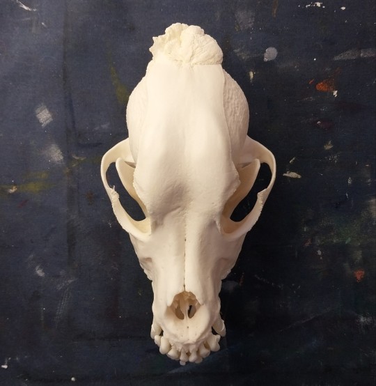
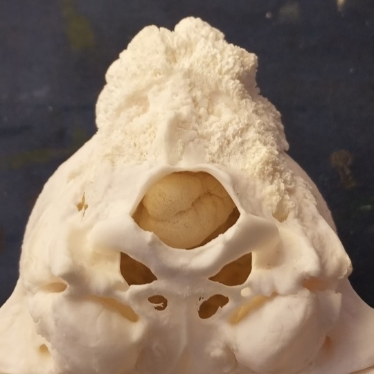
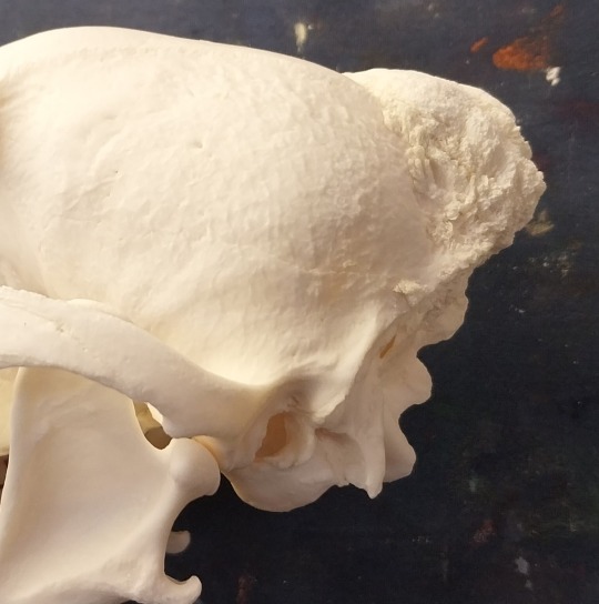
Dog (cocker spaniel mix i believe) skull with cancer. Looks like osteosarcoma. It grew into the braincase 😭
107 notes
·
View notes
Text
Hello and welcome to my desperate groveling for the sake of my sweet girl.
I don’t like to ask for help but I truly would do anything for my dog. We found out today she has bone cancer in her left shoulder and with our current funds we aren’t able to help her.
Without treatment we may get a few more months, at best. With treatment we can get a few years that are well deserved! She’s an absolute angel who spent most of her life on a racetrack rather than in a nice bed and we’ve caught the diagnosis early. With luck and some help we can give her many more deserved years!
We’ve been really fortunate to have a few donations already from some generous friends but literally anything helps!
1k will be going toward surgery fee’d and the rest toward her chemotherapy treatment after to prevent it from spreading.
So far we’ve raised $1,060/$3000!
Any little bit helps and I’m eternally grateful for anything, please help my girl if you can.
46 notes
·
View notes
Text

The cutest reindeer of all 🥹
Blessed to still have my boy with me today.
#catahoula#catahoula leopard dog#reindeer#cutest reindeer of all#dog mom#fuck cancer#osteosarcoma#canine osteosarcoma
6 notes
·
View notes
Link
En este Pódcast estamos solicitando ayuda para un joven de 23 años, colombiano, que requiere una prótesis por desarticulación de extremidad derecha.
Donaciones:
Daviplata y Nequi
3173772751
Bancolombia Ahorros
29983991881
Informes: [email protected]
Responsable:
Giovanni Agudelo Mancera
Periodista
Móvil: +573173772751
0 notes
Text
phases i go through bc little bro has cancer
The initial shock

2. I laugh hysterically when I can get my mind off it

3. Trying to pull it together when it hits again

4. Hopeful that he will be okay

5. It hitting that I might actually lose my brother

0 notes
Text
Exploring Bone Sarcoma: Variants, Therapeutic Approaches, and Future Prospects
Bone sarcoma, a rare type of cancer, affects the bones and surrounding tissues. The condition comprises various types, including chondrosarcoma, Ewing sarcoma, and osteosarcoma, each presenting unique challenges in treatment. However, recent advancements in therapies offer hope for improved outcomes in managing bone sarcoma.
Challenges Associated with Bone Sarcoma Treatment:
Bone sarcoma…
View On WordPress
#Bone Sarcoma#Bone Sarcoma Companies#Bone Sarcoma Types#Chondrosarcoma#Chondrosarcoma Drugs#Chondrosarcoma Market#Chondrosarcoma Treatment#Ewing Sarcoma#Ewing Sarcoma Drugs#Ewing Sarcoma Market#Ewing Sarcoma Treatment#Osteosarcoma#Osteosarcoma Companies#Osteosarcoma Drugs#Osteosarcoma Emerging Therapies#Osteosarcoma Market#Osteosarcoma Treatment#Osteosarcoma Treatment Market
0 notes

Text
Abstract
Osteosarcoma is a malignant bone tumor that is common in children and adolescents. The tumor microenvironment is highly effective in the development and progression of osteosarcoma. Transforming growth factor-β (TGF-β) is one of the most abundant cytokines in the tumor microenvironment, and can regulate tumor initiation, progression, and metastasis promoting extracellular matrix (ECM) remodeling and epithelial-mesenchymal transition (EMT). ADAMTS (ADAM Metallopeptidase with Thrombospondin Motifs) proteases have critical functions in normal and tumor microenvironments by processing individual proteins in the ECM. ADAMTSs contribute to tissue remodeling, inflammation, cell migration and, angiogenesis. Among the family members, ADAMTS-2 is a well-known example for ECM remodeling which cleaves the N-terminal propeptide of procollagen and promotes correct collagen fibrillogenesis. Cytokines can regulate normal and tumor microenvironments by affecting ECM proteins. In this study, the effect of TGF-β1, on the transcriptional regulation of the ADAMTS-2, which is an essential enzyme for ECM remodeling was investigated in Saos-2 cells. TGF-β1 upregulated ADAMTS-2 expression both at mRNA and protein levels. Transient transfection assays revealed that TGF-β1 was also induced ADAMTS-2 promoter activity. According to the pathway inhibition studies, both canonical and non-canonical signaling pathways and post-translational mechanisms were responsible for the induction. These studies will contribute to future research on ADAMTS-2 mediated ECM remodeling in osteosarcoma.
0 notes
Text
Understanding the Stages of Osteosarcoma in Dogs
Osteosarcoma is a word no dog owner wants to hear. This aggressive form of bone cancer is the most common type seen in dogs, often affecting larger breeds. Understanding the stages of osteosarcoma can be vital in helping your dog navigate this challenging journey, equipping you to provide the best possible care and support. In this blog post, we’ll walk through the various stages of osteosarcoma…

View On WordPress
#cancer in dogs#canine cancer#dog cancer#dog cancer treatment#osa in dogs#Osteosarcoma#osteosarcoma dogs#treating cancer in dogs
0 notes
Text
Primary breast osteosarcoma with pulmonary metastases by Guillaume Fahrni in Journal of Clinical and Medical Images, Case Reports

Abstract
Primary breast osteosarcoma is a rare malignant tumor, with only small series reported. We report the case of an 89-year-old woman presenting with a large calcified tumor of the left breast, associated with calcified lung metastases. After non-conclusive biopsy, osteosarcoma was proven at tumorectomy.
Keywords: Osteosarcoma; breast tumor; calcified cancer; lung metastases; breast surgery.
Background
Breast sarcomas are uncommon malignant tumors. Among them, primary breast osteosarcoma is rare with less than 200 cases reported in the literature, representing 12.5% of all breast sarcomas and 1% of all breast tumors [1]. It is a very aggressive tumor that typically produces bone and osteoid matrix. It usually affects elderly patients, with a median age at diagnosis of 64.5 years [3]. The clinical presentation is a palpable mass sometimes associated with a cutaneous rash. Complete resection is the treatment of choice in case of limited disease extension [4]. Survival rate is poor, under 40% at 5 years [5].
Case Presentation
An 89-year-old woman without relevant past medical history was hospitalized for cardiac failure. While performing routine physical examination, a palpable firm and irregular mass of the left breast was evidenced, seemingly unnoticed by the patient. This finding motivated the realization of a mammography showing a regional area of coarse heterogeneous calcifications in the internal quadrants of the left breast. Correlation with ultrasound demonstrated a subcutaneous mass with posterior acoustic shadowing (Figure 1). A fine needle biopsy was performed, however it was not contributive due to the large calcifications within the sample. Due to the high suspicion of primary breast neoplasm, a thoracoabdominal CT was performed that revealed a large heterogeneous and calcified mass of the left breast associated with diffuse and partially calcified lung nodules (Figure 2).
The patient’s case was discussed at the tumor board and a breast tumorectomy was decided and performed without any immediate complication following surgery. On gross observation, the tumorectomy piece (16.5 x 13.5 x 9.5cm) contained a well-defined mass measuring 10. x 8.5 x 7.5 cm, heterogeneous, partially calcified and containing areas of central necrosis (Figure 3). Microscopic examination showed a peripheral zone containing a dense proliferation of spindle-shaped cells. There was a central zone composed of ossification foci, osteoclastic cells and cartilage, as well as a transition zone with osteoid formations and sclerous tissue (Figure 3). Immunohistochemistry was performed, with spindle-shaped cells showing a strong expression of MYC, a moderate expression of MDM2, a heterogenous expression of p63 and no expression of epithelial markers CKAE1/AE3, CK5/6, EMA, MNF-116, CK7, CK19. An intra-mammary lymph node (2.2 cm) was also identified, containing metastatic tissue. The aforementioned findings were consistent with the diagnosis of high-grade malignant mesenchymal neoplasm compatible with an extraskeletal osteosarcoma of the breast with lung metastasis. Patient outcome was unfavorable with rapid decline and death due to advanced metastatic disease progression.
Figure 1: Mammography with medio-lateral oblique (A) and cranio-caudal (B) incidencesviews of the left breast evidencing a regional area of coarse heterogeneous calcifications at the union of the internal quadrants. Breast ultrasound (C) showing a subcutaneous mass with large posterior acoustic shadowing limiting further analysis.
Figure 2: Thoracoabdominal contrast-enhanced CT revealing a large calcified mass involving the internal quadrants of the left breast (A, arrowhead) and diffuse partially calcified pulmonary nodules consistent with lung metastasis (B-C). Note the calcified aspect of the lung metastasis (C, arrow).
Figure 3: Macroscopic examination (A) evidencing a voluminous calcified mass with central necrosis. Microscopic examination (B-C) showed a peripheral zone containing a dense proliferation of spindle-shaped cells (B), and a central zone composed of ossification foci, osteoclastic cells and cartilage (C).
Discussion
Primary breast osteosarcoma is a type of breast tumor with bone producing osteoid matrix [6]. It is not only a very rare breast malignancy but also an unusual location for an extraskeletal sarcoma [7]. Although rare, its diagnosis is important as the imaging features, treatment and prognosis differ from other breast cancers. As with all sarcomas, it is defined by the underlying type of tissue with the most common subtype including fibrohistiosarcoma, myxofibrosarcoma and angiosarcoma [2].
Primary breast osteosarcoma has to be differentiated from metaplastic carcinoma and cystosarcoma phyllodes, that can both present with similar imaging features [8]. The clinical presentation is a breast mass, with rapid growth but rarely associated with pain [9]. Nodal involvement is uncommon, but metastases are frequent. Lung is the most commonly organ involved, followed by bone. The survival is poor, with an overall 5-year survival rate of 38% [10]. On mammography, the primary lesion is usually a large, dense, calcified mass [11]. However, in some cases, calcifications can be absent [12]. Ultrasound is helpful for lesion characterization, staging and biopsy guidance and usually shows important acoustic shadowing limiting analysis [2]. MRI is useful to assess the extent of the mass within the breast, to search out for other masses, assess for chest wall invasion usually showing a high T2 signal mass with enhancement [1]. Whole-body contrast-enhanced CT scan or 18FDG PET-CT are of interest to detect metastatic extension [6]. The final diagnosis of primary breast osteosarcoma is made on pathology, demonstrating osteosarcomatous matrix and showing that the lesion does not arise from adjacent bone (sternum or ribs) [13]. Immunohistochemistry is helpful to establish the diagnosis, with the use of epithelial markers such as CKAE1/AE3, CK5/6, EMA, SMA, CK7, vimentin, MAC-387 and others [14]. Excisional biopsy (complete tumorectomy), as this was done in our patient, is preferred to fine needle biopsy, as the mass can be very firm due to the presence of calcifications that can lead to inconclusive results as in our case [6]. As with any subtype of sarcoma, the treatment is the complete surgical excision with large margins [11]. Local recurrence rates were reported as high as 67% after tumorectomy and 11% after mastectomy [5, 15]. If no local node involvement is seen, axillary node dissection is not recommended, since this type of sarcoma does not spread via lymphatic route [16]. In this regard, our case was unusual as a pathologic intra-mammary lymph node was found. There is no consensus about the administration of adjuvant chemotherapy. However, it is generally recommended as it evidenced to improve survival rates, but no standard dose has been established yet [10]. For more advanced cases with metastasis, chemotherapy is the main treatment. Drugs used are the same as with other sarcomas and include cisplatin, doxorubicin, ifosfamide and methotrexate [17].
Acknowledgments: The authors declare no conflict of interest.
For more details : https://jcmimagescasereports.org/author-guidelines/
#Osteosarcoma#breast tumor;#calcified cancer#lung metastases#breast surgery#Immunohistochemistry#cystosarcoma#Guillaume Fahrni#JCMICR
0 notes
Photo

🧵 Well fuck. My foster-twin has terminal #cancer, and we’re waiting on test results to see if this last round of chemo is working or not before she transitions to hospice. I need to visit before the end, and adults don’t get #MakeAWish. So I need to sell a bunch of diamond paintings to raise ~$1000. I’ve not seen my sister in a decade because we live on opposite coasts and a #disabled artist doesn’t make much. Last minute plane tickets from Virginia to Oregon aren’t easy to afford! If you can, please retweet and look at my shop! www.PuzzlePawsBlog.com/shop If you can’t afford a painting, please retweet! Every little bit helps. Venmo @PuzzlePaws Cashapp $PuzzlePaws Or pm me for my PayPal. #FuckCancer #FoundFamily #Sister #SisterFromAnotherMister #FosterTwin #Osteosarcoma #LastWishes https://www.instagram.com/p/CpoI8YMuroy/?igshid=NGJjMDIxMWI=
#cancer#makeawish#disabled#fuckcancer#foundfamily#sister#sisterfromanothermister#fostertwin#osteosarcoma#lastwishes
0 notes
Text
Activation of microvesicle peripheral blood mononuclear cells by mesenchymal stem cells secretome co-cultivated with osteosarcoma stem cell
Article published in J. Pharm. Pharmacogn. Res., vol. 10, no. 5, pp. 782-790, September-October 2022.
DOI: https://doi.org/10.56499/jppres22.1414_10.5.782
Fachrizal Arfani Prawiragara1,2, Ferdiansyah2*, Mouli Edward2, Dwikora Novembri Utomo2, Mohammad Hardian Basuki2, Alexander Patera Nugraha3, Fedik Abdul Rantam4
1Magister Clinical Medicine Program, Orthopedic and Traumatology Department,…

View On WordPress
0 notes
Link
Cancro ossa, italiani svelano proteina chiave instabilità genomica Studio Igb-Cnr apre a nuove prospettive di cura per l'osteosarcoma
0 notes
Photo

A vida vale apena, não desista nunca. #osteosarcoma #cancer #familia #amigosirmãos #vivaavida (em Piracicaba, Sao Paulo) https://www.instagram.com/p/Cmew0TBJaNS/?igshid=NGJjMDIxMWI=
0 notes
Link
En este Pódcast estamos solicitando ayuda para un joven de 23 años, colombiano, que requiere una prótesis por desarticulación de extremidad derecha.
Donaciones:
Daviplata y Nequi
3173772751
Bancolombia Ahorros
29983991881
Informes: [email protected]
Responsable:
Giovanni Agudelo Mancera
Periodista
Móvil: +573173772751
0 notes
Text
First experiance with Cancer Support Community really fucking sucks.
My brother has cancer. Osteosarcoma. On Tumblr it's one of the most lethal cancers for dogs, but for humans it's survivable.
I feel can't say any more than that in cancer support groups. I have to act like my brother is dying, and these are his final moments.
People: This disease sucks, but he can live.
Me: Yeah, that's what I figure. Google says he has a 60-77% survival rate
People: OH MY GOD NEVER GOOGLE ANYTHING YOU DUMB BITCH, THOSE NUMBERS MEAN SHIT AND YOU NEED TO LISTEN TO YOUR BROTHER HE IS DYING!!!!
like..... wtf! How am I supposed to know ANYTHING about this cancer??? SHUT THE FUCK UP. GOD! I KNOW googling health things is dumb, but I'd like to know if my brother has a chance at life, or if this is "Once they find it it's too late" cancer.
It's very upsetting because, from what I've seen this cancer CAN and often IS treated, and people SURVIVE ALL THE TIME!!!! LITERALLY, I WAS IN A BETTER PLACE BEFORE I REACHED OUT TO THE COMMUNITY. I know there is a chance my brother won't make it, that we are too late, BUT LET ME HAVE HOPE GOD DAMNIT!
FUCK THOSE PEOPLE. I literally don't even want to try again. Just tell me my brother is dying and I am stupid. This isn't support.
0 notes
Last Seen Blogs

kingbasiesimsreblogs
KingBasie Sims 4 Reblogs

2018thelookbookza-blog
#THELOOKBOOKZA

herosofrock
Heros Of Rock

pave-uw
PAVE UW-Madison

mysticsituations
MYSTIC SITUATIONS