#Juniper Publishers Indexing
Text
Palaeoenvironmental Background for the Early Paleolithic Occupation of the Volcanic Upland in the South Caucasus
Abstract
Nowadays, it has been reliably established that the Early Paleolithic people producing both Oldowan and Early Acheulian industries initially occupied the volcanic upland of the South Caucasus not later than 1.9.1.85Myr (Olduvai subchron). This period was characterized by a much lower relief than at present and subtropic climate that favored a dispersal of the early humans. The volcanic activity created there fertile paleosols with grass covers feeding hunted herbivores as well as rocks suitable for manufacturing lithic artifacts. In the subsequent periods of the Early and the Middle Pleistocene the upland underwent uplifting that led to a gradual deterioration of climate and appearance of glacial-interglacial rhythms with an increasingly pronounced difference between warm and cold periods. However, the chronological ranges of the studied Acheulian sites suggest that the humans continued to occupy the region under both more and less favorable conditions. The early humans are believed to have spread most widely in the region in the Late Acheulian, supposedly during in the Holstein interglacial.
Read More about this Article: https://juniperpublishers.com/ijesnr/IJESNR.MS.ID.556189.php
Read More Juniper Publishers Google Scholar: https://scholar.google.com/citations?view_op=view_citation&hl=en&user=4WXzQFMAAAAJ&citation_for_view=4WXzQFMAAAAJ:HoB7MX3m0LUC
#Juniper Publishers in USA#Juniper Publishers group#environmental science#sustainable development#Juniper Publishers PubMed Indexed Journals
0 notes
Text
Let’s Talk and Grow Together: A Bidirectional Communication between Granulosa- and Oocyte Derived Factors in the Ovary

Abstract
Reproduction, one of the most active and appealing area of research for endocrinologists and reproductive biologists since many a years, has several faces that remains to be unmasked in terms of its regulatory aspects. Available information on the regulation of oocyte development and maturational competence are gaping and needs elucidation to achieve utmost quality of eggs, a major area of concern. The notion of the somatic follicular cells providing an appropriate microenvironment for the development of oocyte throughout its journey has been replaced with the current perception of a complex yet regulated cross-talk between the granulosa-and oocyte-derived factors to orchestrate follicle development. Interestingly, actions of FSH and LH are mediated or modulated by these locally produced non-steroidal peptide factors from the follicular layer and the oocyte itself (insulin-like growth factors (IGFs), epidermal growth factor (EGF) family members, TGFβ super family members etc.), forming an intimate regulatory network within the ovarian follicles. Present article will provide a deeper insight into the need and underlying mechanisms of action of these growth factors in the intraovarian network to sustain a healthy oocyte.
Read More About This Article: https://juniperpublishers.com/gjorm/GJORM.MS.ID.555569.php
Read More Juniper Publishers Google Scholar Articles: https://scholar.google.com/citations?view_op=view_citation&hl=en&user=xBu_HGEAAAAJ&authuser=1&citation_for_view=xBu_HGEAAAAJ:Zph67rFs4hoC
#gjorm#overy#reproductive medicine#Juniper Publishers Indexing Sites List#Juniper Publishers PubMed Indexed Articles
0 notes
Text
Hyperparathyroidism in Pregnant Women: Clinical Issues, Laboratory Findings and Relevant Therapeutic Approachesr - Juniper Publishers

Juniper Publishers- Open Access Journal of Annals of Reviews & Research
Hyperparathyroidism in Pregnant Women: Clinical Issues, Laboratory Findings and Relevant Therapeutic Approachesr - Juniper Publishers
Authored by Rosita Fontes
Abstract
Primary hyperparathyroidism (PHPT) is the most common cause of hypercalcemia in outpatient settings, with an incidence in women of reproductive age of 4.7-6.2 cases per 100,000 persons. When untreated in pregnant women, PHPT can lead to maternal and fetal complications. The authors present a case of a patient with a pre-pregnancy hyperparathyroidism diagnosis: she had worsening symptoms during pregnancy, so was referred for surgery due to failure of clinical treatment to keep the disease under control. Clinical issues, laboratory findings, and relevant therapeutic approaches are discussed.
Keywords: Hyperparathyroidism; Hypercalcemia; Multiple endocrine neoplasia syndrome; mTc-sestamibi scintigraphy; Beta chorionic gonadotropin; Parathyroidectomy; nephrolithiasis; calciuria
Abbreviations: PHPT: Primary Hyperparathyroidism; MEN: Multiple Endocrine Neoplasia Syndrome; BP: Blood Pressure; US: Ultrasound
Introduction
Primary hyperparathyroidism (PHPT) is a disorder that results from hypersecretion of parathyroid hormone (PTH), which is the most common cause of hypercalcemia in the outpatient setting. An analysis published in 2013 showed that the incidence in women of reproductive age, within a racially mixed population, is 4.7-6.2 cases per 100,000 persons [1]. In pregnant women, the disease occurs in 0.5-1.4%, and in 80% it is due to adenoma of the parathyroid glands [2]. When untreated, PHPT can lead to maternal complications such as nephrolithiasis, pancreatitis, and eclampsia, and fetal complications such as low birth weight and neonatal seizure [3-6]. Adequate control of patients who become pregnant with hyperparathyroidism can be a challenge, depending on their clinical symptoms and the laboratory and imaging evaluation. According to the trimester of pregnancy and severity of the disease, the treatment may be either clinical or surgical. The authors present a case of a patient with a pre-pregnancy diagnosis, and a worsening of symptoms during pregnancy; she was referred for surgery due to failure of clinical treatment to keep the disease under control.
Case Presentation
A pregnant 45-year-old woman, Afro-descendant, gravida 3, para 1, presented with a history of recurrent bilateral nephrolithiasis since she was 40 years old. She had a left wrist fracture after falling from height. Her past medical history was notable for low calcium intake, asthma and diabetes mellitus type 2, which was under control with metformin. In her surgical history, she underwent ureteral stent double J, and ectopic pregnancy surgery. She also suffered a lithotripsy for treatment of kidney stones. She presented for consultation for investigation of repetitive renal stones, denying any symptoms compatible with hyperparathyroidism. In her family history, there were no cases of hyperparathyroidism, no multiple endocrine neoplasia syndrome (MEN), or other syndromes associated with hypercalcemia. On physical examination, the patient was overweight (BMI 28.72 kg/m2), blood pressure (BP) was 110/75 mm Hg, bone deformities or fractures were not observed, and renal fist-percussion was negative. There were no other abnormalities found.
At admission, she brought examinations showing parathyroid hormone (PTH) of 390 pg/mL (12-65), total calcium corrected for albumin of 9.5 mg/dL (8.4-10.5), phosphorus of 2.3 mg/dL (2.5-4.5), as well as urinary calciuria in 24hours of 359 mg/day (100-250) and 4.78 mg/kg/day (< 4.0). Thyroid function tests were normal, as well as hemogram and biochemistry assessments. In several tests requested to confirm these results, her serum calcium was high (Table 1), along with maintained high PTH, low phosphate, and high calciuria. The parathyroid glands were not visualized on cervical ultrasound (US), but parathyroid 99mTcsestamibi scintigraphy (MIBI) showed hypercaptation of the tracer after 2 hours in the left lower parathyroid projection area, suggestive of a parathyroid adenoma (Figure 1). Renal US showed bilateral cortical stones, without lithiasis in the pyelocalyceal system. Adequate hydration and furosemide 20 mg/day were prescribed.
The 25-hydroxy vitamin D (VD) was low, 7.1 ng/mL (>30 ng/ mL), and the 1.25-dihydroxy vitamin D (1.25(OH)2D) was 193.9 pg/mL (18-78). She had recently presented with visual turbidity, nausea, headache, asthenia and dizziness, and recurrent renal colic, with an episode of pyelonephritis that was treated with antibiotic therapy. As the patient had a 2-week menstrual delay, a beta chorionic gonadotropin (beta-HCG) test was ordered, and the result was compatible with pregnancy. The gestational age calculated by the date of the last menstrual period was 5 weeks and 2 days. PTH and calcium remained elevated during the first trimester of pregnancy (Table 1). The option at that time, after discussion with the obstetrician, was increased furosemide at 40 mg/day and intensification of hydration. During the second trimester, the calcemia increased even more (Table 1), with the symptoms persisting. At that point, VD replacement was prescribed, and surgery was indicated. Parathyroidectomy was performed at 25 weeks of pregnancy, which was carried out without incident. On the first postoperative day, the patient had no symptoms, and the corrected calcium and PTH were normal (Table 1), although anatomopathological exam confirmed parathyroid adenoma. The pregnancy progressed without intercurrences. At 38 weeks and 2 days, she had an uneventful cesarean delivery. The newborn was healthy, with APGAR 9 at 1 minute and 10 at 5 minutes, 3.990 kg, and 49 cm, with no symptoms or laboratory evidence of hypocalcemia.
Discussion
PHPT is a rare condition in pregnancy [2,7].We present a PHPT case who became pregnant even with a diagnosis of the disease previously confirmed, and then underwent surgery for adequate hypercalcemia control. For the general population, hypercalcemia is defined as total serum calcium above 10.5 mg/ dL (>2.6 mmol/L). In a pregnant patient, the serum albumin falls due to hemodilution and remains low until delivery. Calcium is transferred through the placenta to mineralize the skeleton, and the glomerular filtration rate is increased, culminating with lower total calcium levels. Due to these factors, the upper limit of the reference range for corrected calcium in pregnancy is about 9.5 mg/ dL (2.3 mmol/L) [8,9]. This patient became pregnant at 45-yearsold. In addition to increased calcium and PTH, she presented with low phosphate and elevated 1.25(OH)2D. Most patients with PHPT are older than 50, but the disease is diagnosed during childbearing years in 25% of females [10]. Inappropriately high serum PTH concentration leads to increased renal reabsorption of calcium, phosphaturia, and 1.25(OH)2D synthesis, resulting in hypercalcemia and hypophosphatemia, loss of cortical bone, hypercalciuria, and various sequelae of chronic hypercalcemia [11]. During the course of her pregnancy, the patient presented symptoms of hypercalcemia as the serum calcium increased; such symptoms do not depend on gestational age, as they are the same for non-pregnant women [6,12].
A pregnancy in a patient with hyperparathyroidism may have maternal complications of 67%, and in neonates it would be about 80% [2]. The most frequent maternal complications are hyperemesis and nephrolithiasis; less frequently, preeclampsia and pancreatitis occur [2,6]. Fetal complications may include miscarriage, neonatal seizures, low birth weight for gestational age, and hypocalcemia [3,13]. This patient and her newborn did not present complications, probably because gestation was monitored from the beginning and surgery was performed in the second trimester to control calcemia. In a normal pregnancy, PTH levels are low during the first trimester compared to nonpregnant women and remain normal through the rest of the pregnancy. The relatively low PTH may be due to the suppressive effect of raised 1.25(OH)2D [14]. In this patient with primary hyperparathyroidism, high PTH levels were due to the autonomic secretion of the adenoma, preventing its fall as calcium levels raised. As such, continuous reabsorption of calcium of the bones occurs, as well as an increase of the tubular reabsorption of calcium, including intestinal absorption secondary to 1.25(OH)2D levels [15]. This modifies what physiologically occurs in gestation, when maternal calcium falls by about 10%; the current patient presented calcium within reference values at the beginning of the investigation, which increased progressively. The therapeutic measurements in the first trimester were sufficient to maintain serum calcium at the maximum level of 11.6 mg/dL (2.89 mmol/L); however, in the second trimester, calcium increased to 12.76 mg/dL (3.18 mmol/L). The incidence of pregnancy loss and its relationship to calcium elevation is not entirely known. Fetal loss is seen at all levels of elevated maternal calcium, but it is suggested that calcium levels higher than 11.4 mg/dL (2.85 mmol/L) may increase the risk of adverse events for both mother and fetus [16].
The patient urinary calcium was already high before pregnancy, with previous nephrolitiasis. Characteristic of the gestational period is an increase of calciuria [17]. Despite increased fluid intake, the risk of new kidney stones could be high, especially because she was put on furosemide in order to control hypercalcemia, which was an additional concern in deciding on surgery. This patient had low levels of VD. It has been demonstrated that even in tropical countries, its prevalence in pregnant women may exceed 80% [18,19]. In a normal pregnancy, 1.25(OH)2D may be twice as high compared to non-pregnant women [17]. However, this patient’s 1.25(OH)2D was very high: levels of the transporter 1.25(OH)2D protein, secondary to hyperestrogenism proper to gestation, and an increase in free 1.25(OH)2D production (due to increased activity of 1 alpha hydroxylase stimulated by parathyroid hormone-related protein (PTHrp), estrogen, prolactin, and placental lactogen hormone) are relevant factors that contribute to this issue. In addition, the synthesis of 1.25 (OH) D is increased by the activity of placentary 1 alpha VD hydroxylation [20,21]. In this case, elevated PTH was an additional factor that increased the 1.25(OH)2Dto such high levels. We prescribed VD based on the low levels presented, since the patient would need surgical correction of the hyperparathyroidism, with 1.25 (OH) D returning to normal pregnancy levels (and thereby revealing VD deficiency). There are no specific guidelines for treatment of hyperparathyroidism during pregnancy, as the approach is individualized according to the severity of hypercalcemia, the symptoms presented, a worsening of the clinical condition, and laboratory parameters.
During pregnancy hydration, enhancement and forced diuresis are measures that can avoid severe increases in serum calcium. The medications used for this purpose are class C and D for a pregnant woman, including calcitonin, cinacalcet, and furosemide. Bisphosphonates belong to category D [5,22]. We decided to prescribe a category C drug, with furosemide being the most accessible medication for the patient. However, a 40 mg/ day dose was not sufficient to control hypercalcemia, as she had continued clinical symptoms; thus, we decided not to introduce a new medication, but to refer the patient to surgery in the second trimester. Surgery is avoided in the first trimester due to increased risk of miscarriage, and in the third trimester, due to risk of preterm birth [23]. Although uneventful surgery has been done in the third trimester [24], the second trimester is the ideal time, as it is the period with potentially lower risks for fetal complications [12,25]. The patient underwent surgery, with excision of the left inferior parathyroid at the end of the second trimester: both she and the newborn were free of complications.
Conclusion
Patients with hyperparathyroidism who become pregnant need special attention for the development of clinical symptoms, the monitoring of laboratory parameters, and appropriate therapeutic interventions, given the occurrence of certain events. Since the management of pregnant women with hyperparathyroidism is relatively limited, we present a successful case here, which is aimed at discussing its clinical aspects, expected laboratory changes, and therapeutic possibilities.
To know more about Juniper Publishers please click on: https://juniperpublishers.com/aboutus.php
For more articles in Open Access Journal of Reviews & Research please click on: https://juniperpublishers.com/arr/index.php
To know more about Open Access Journals please click on: https://juniperpublishers.com/journals.php
#reviews research paper#Juniper Publisher Reviews#JuniperPublishers#Hyperparathyroidism#Multiple endocrine neoplasia syndrome#juniper publishers indexing juniper publishers journals
0 notes
Text


𝐓𝐇𝐄 𝐒𝐔𝐍 𝐀𝐍𝐃 𝐓𝐇𝐄 𝐌𝐎𝐎𝐍 .
( Synopsis ) And you witnessed the macrocosm in all its dazzling contours.
( Authors Note ) This work is released before Sumeru is added to Genshin Impact, so there is very little lore regarding Al Haitham when this is published. I hope you enjoy this at the end, and constructive criticism is greatly appreciated :)
( Pairing ) Al-Haitham x GN! Reader
( Content Warnings ) Established relationship. Written to satisfy my brain rot. Mentions of the word gorgeous.
( Word Count ) 1.0k words // EDITED!
— THE SUN WAS A MASTERPIECE AND GLIMMERED IN BRILLIANCE,
the sun: a hidden flirt and a massive tease (brightness)
Al Haitham conversed with a hidden charm that seemed to sway the lush leaves outside the library's windows. The sunlight perfectly radiated on the outstanding library shelves, cascading onto dusted and aged books neatly lined up against one another. Grandeur scrolls of knowledge were proudly displayed on the wood walls, making the murmurs of wisdom traverse around the serene room with a flip of a page.
He never bothered worrying about who would interrupt him in his time of research, because only you and he knew of this place. Hidden in plain sight under the watch of a lesser god.
Pale juniper green irises twinkled in the gratification that a few hours were in his hand, unconsciously humming in satisfaction when he spotted a book to his liking. Perhaps he could read this with you?
The thought of the two of you relishing in each other's company put his mind at ease.
He took a step forward, ornaments from his outfit creating a rhythmic sound. He placed his index finger and thumb on the spine of the book to slide it out the shelf—
—nothing.
Push. Pull. Push. Pull. It was almost as if the work was glued to the frame of the furniture. Perhaps if he made an effort to look for another book. There is no point fretting over this.
That one. Al Haitham thought, reaching out to grab a book two rows below the seemingly glued book.
Nothing.
He added a little more force this time, tenderly making sure he won't damage the piece of work. It didn't move. Why is he unable to get it?
"Ugh, why can't I grab this book..." A melody his ears sought reached his ears.
"Gorgeous?"
"Huh? Gorgeous? Wait don't tell me..."
Out of a pinch of amusement, he tipped the third book he laid his hands on— which now successfully rested on his hands— and came into contact with your gaze. He rested his arm on whatever space was left from the book he got and tipped his lips up to a minor smirk.
"You were getting the book I was getting a while ago, weren't you." You're baffled expression occupied his thoughts. Although, it was probably useless to confirm it with him since he most likely connected the puzzle before you.
"You're correct."
"I was going to get it and give it to you since you probably wanted to read it together!"
"Alas, here we are, separated by a wall filled with words spilled from hours of studying. We really were fated to be, gorgeous."
"You're the gorgeous one here and we both know it, shut up."
He adores your flustered self.
— MISTIFIED AND ENTRANCED WITH THE ILLUSIONS OF THE AWAKENING MOON,
the moon: a mysterious gentleman with nothing but respect (calmness)
Bountiful were the imperial potted padisarahs cultivating on the rich soils of his private riad. Other unscrupulous classes would believe that after you have gained control of knowledge— respect, admiration, power, and money is nothing but child's play. All that was left to obtain was ambition and the vessel of power from the gods.
A vision.
But what is wisdom and power when ambition and morale fail to play the role?
Conceivably, this was what separated him from the rest.
He knows, that beneath the raw memorabilia of godly divinity, rested the dead and the falsehoods that happened before miraculous spectacles flourished. A mortal carelessly strolling around the break of dawn with a fraction— or a percentage— of immortals, was a laughable notion. Humans had mortality while divinity had immortality.
Someday it will be revealed that it is nothing but a mere tool.
How foolish. Al Haitham spoke to himself.
( Al Haitham was foolish to underestimate the eye of the gods when the hoaxes end. )
He was far from that fantasy stereotype. He was an enigma. A secret buried under the thick unforgiving hail of Dragonspine. A mystery buried at the bottom of Liyue's mountains after dragons roared amid battle. A paradox thrown to the depths of Inazuma's oceans with the remains of divinity beheaded by the Almighty Shogun's spear. A dilemma sunken in Sumeru's volcanoes that glimpsed battles between warriors and cowards thousands of years ago.
The catastrophes didn't cease you from achieving your destination, though.
You hiked the mountain peaks of Dragonspine under layers of courageousness despite unknowing the secrets of the deceased princess who wished nothing but for someone to wear her crown. You've seen fossils, bones, and remains— the aftermath of Liyue's devastating war. You surfed above the thunderous oceans and danced around the thunderstorm once upon a time. You quelled the bubbling turmoil and made amends co-existing with the strong and the weak.
Then the moon.
Al Haitham was the moon. Galaxies away from arm's reach, it adorned the dark tempest taking hold of Teyvat's twilight, giving mercy and sprouting a second chance. It gave seeds of fake hope of wonder, the moonlight nothing but a mere marvel illusion. It would take light years for mechanics to evolve and when a Teyvatian finally conquered the moon— what then?
Defeat. The moon was just a mere dull stone blessed with the glory of the sun. Receding spears and swords would pierce the shielded heart because their ambition was worthless. A setback— no other forms of life thriving in the abundance of a hidden empire, no sanctuary and grasslands of fading stars unearthing equilibrium.
Al Haitham never minded the negative impact. For he was a young eagle luring his prey who made him loathe their nefarious spirit.
Your sentiment is stated contrarily.
He was the anchor for those lost in a dulcet of eunoia, ridding their sonder of the anchor's chain that lent a second chance.
He was your anchor, your moon, and your whole world. Al Haitham knew this too.
You've seen him stress out, get furious, and all his flawed emotions.
And all he can say is, thank you. I love you.
As Al Haitham prepared his surprise feast for you and him, he only hopes you enjoy it as his thanksgiving. To you, and all his blessings.
The sun and the moon juggling a chance in a circus was a dream to transpire.
in a way, we lived in the same atmosphere shining in our own beautiful ways (an ephemeral dream)
astolary 2023 — do not edit, repost, or translate.
#( ❁ଓ.° )#( ❁ଓ.° ) astro! genshin impact#genshin impact x reader#genshin headcanons#genshin scenarios#genshin x reader#genshin al haitham x reader#al haitham x reader#genshin al-haitham x reader#al-haitham x reader
270 notes
·
View notes
Text
Optical Interconnects Market Growing Popularity and Emerging Trends in the Industry
The Latest Released market study on Global Optical Interconnects market provides information and useful stats on market structure, size and trends. The report is intended to provide cutting-edge market intelligence and strategic insights to help decision makers take sound investment decisions and identify potential gaps and growth opportunities. Besides, the report also identifies and analyses changing dynamics, emerging trends along with essential drivers, challenges, opportunities and restraints in Optical Interconnects market.
What’s keeping II-VI Incorporated (United States), Lumentum Operations LLC (United States), Molex, LLC (United States), InnoLight Technology (Suzhou) Ltd. (China), NVIDIA Corporation (United States), Fujitsu Ltd. (Japan), TE Connectivity (Switzerland), Amphenol Corporation (United States), Accelink Technology Co. Ltd. (China), Juniper Networks, Inc. (United States), Infinera Corporation (United States) Keep Growing in the Market? Benchmark yourself with the strategic moves and latest Market Share and Sizing of Global Optical Interconnects market recently published by AMA
Optical interconnects are a form of communication that uses optical cables instead of conventional cables. Optical cables have a far higher bandwidth than traditional cables. For both telecoms and datacoms, optical technology is a promising, energy-efficient solution for meeting increased bandwidth requirements. Since light can pass through dielectrics and even vacuum, optical interconnect is an interesting alternative to metallic wires. One of the main factors driving the optical interconnect market is the adoption of cloud databases. Storage-as-a-service (SaaS) and platform-as-a-service (PaaS) cloud storage systems have greater scalability by expertly managing complex data and storing it for later use. Optical interconnect is more cost-effective than networking infrastructure because it uses less electricity.
The Optical Interconnects Market segments and Market Data Break Down by Type (Cable Assemblies (Indoor Cable Assemblies, Outdoor Cable Assemblies, Active Optical Cables), Connectors (LC Connectors, SC Connectors, ST Connectors, MPO/MTP Connectors), Optical Transceivers, Fibre and Waveguides, Silicon Photonics, Others), Application (Telecommunication, Data communication), Level Type (Chip- & Board-Level, Board-To-Board and Rack-Level, Metro & long Haul), Fiber Mode (Single Mode Fiber, Multi-Mode Fiber (Step Index Multi-Mode Fiber, Graded Index Multi-Mode Fiber)), End-Use Industry (Energy & Utility, Consumer Electronics, Automotive, Chemicals, IT&Telecommunication, Others)
On the geographical front, the market has been segregated into North America (the United States and Canada), Europe (Germany, France, the United Kingdom, Italy, Spain, Russia and others), Asia Pacific (China, Japan, India, South Korea, Australia, Indonesia and others), Latin America (Brazil, Mexico and others), and Middle East and Africa.
What’s Trending in Market: Continuous Development in the Digital Transformation across the World
Market Opportunities: Increase in Use of Optical Interconnects in High-Performance Computing Applications
Mounting Demand from Emerging Economics such as China, Brazil, India, among others
Highlights of Influencing Drivers: Higher Adoption of Big Data Analytics In Growing Economies
Increasing Demand for Highly Secure and Safe Wiring Systems in Industrial and Commercial Applications
Presented By
AMA Research & Media LLP
0 notes
Text
Food and Feeding Biology of Commercially Important Freshwater Eel, Mastacembelus armatus (LACEPÈDE, 1800) from the Ganga River, India
Abstract
Food and feeding biology of freshwater zig-zag eel Mastacembelus armatus (LACEPÈDE, 1800) was investigated from the Allahabad waters of the river Ganga from April 2015 to March 2016. The Stomach contents of 326 individuals of M. armatus were collected monthly from the Daraganj fish landing centre located on the bank of the river Ganga. The composition of food content was expressed as percentage of the frequency of occurrence (%Oi) and percentage of the volume (Vi). The main food items were estimated by Index of preponderance (Ii). The three major food items of M. armatus were the small sized teleosts, insects, and molluscs. The analyses of the relative gut length (RGL) revealed it carnivorous type of feeding habit. The Gastro-Somatic Index (GaSI) in the male and female ranged between 2.65 and 4.41 with a mean of 3.42 ± 0.09 and 2.84 and 3.43 with a mean value of 3.05 ± 0.03. The mean GaSI value differed significantly at 5% level of significance (F = 6.72, p = 0.01) in both the male and female indicated significant variation in the feeding habits between both the sexes. This study furnishes baseline information on the food and feeding habits of M. armatus which could be useful in formulation of the management and conservation strategies of this species in the region.
Read more about this article: https://juniperpublishers.com/ofoaj/OFOAJ.MS.ID.555819.php
Read more Juniper Publishers Google Scholar articles https://scholar.google.com/citations?view_op=view_citation&hl=en&user=V6JxtrUAAAAJ&citation_for_view=V6JxtrUAAAAJ:g5m5HwL7SMYC
#Juniper Publishers Google Scholar#Aquaculture#Fisheries journals#Marine science#coastal zone#marine physics
0 notes
Text
Effects of Selenium Nanoparticles on Bleomycin-Induced Pulmonary Fibrosis in Rats
Abstract
Background: Idiopathic pulmonary fibrosis (IPF) is one of the interstitial lung diseases. To date, most drug trials for the treatment of IPF have yielded disappointing results. Recently, selenium nano-particles (SeNP) have received attention for potential use in treatments, due in part to their established abilities to exert size-dependent anti-oxidant/-inflammatory effects.
Materials and Methods: the rats were treated with SeNPs by intraperitoneal injection (0.5mg SeNP/kg) for five consecutive days in the inflammatory phase (a day after injection of bleomycin) and fibrotic phase (a week after injection of bleomycin).
Results: The results showed that administration of SeNP during the inflammatory phase improved the activity of enzymes antioxidant (i.e., glutathione peroxidase and superoxide dismutase) and also led to significant decreases in interleukin (IL)-6 and malondialdehyde (MDA) levels in lung homogenates. Histopathology, measures of lung index and body weight, and evaluations of lungs using Ashcroft criteria supported the mitigative effects found above. Notably, treatment with the SeNP during the fibrotic phase imparted no ameliorative effects. Lastly, the SeNP themselves seemed to impart no overt toxicities in naïve rat hosts.
Conclusion: Thus, the findings suggest that SeNP, in part by improving the antioxidant enzyme activities, might impart a therapeutic effect against PF induced by bleomycin in rats. This potentially might mean that SeNP could represent a new therapeutic for treatment of this disease, at least in its earliest phases.
Read more about this article: https://juniperpublishers.com/ijoprs/IJOPRS.MS.ID.555675.php
Read more Juniper Publishers Google Scholar Articles:
https://scholar.google.com/citations?view_op=view_citation&hl=en&user=Y8LS2LwAAAAJ&citation_for_view=Y8LS2LwAAAAJ:lSLTfruPkqcC
0 notes
Text
Economic Evaluation of Effectiveness of Domestic Raw Materials Using
Abstract
Raw materials provide profit for owner, state and mining company, as well as single region. Mentioned means that problem of evaluation of raw material deposits is very actual and necessary area. In developed market economies evaluation of reserved deposits is considered as one of the most complex, but also most important activities during finding and research of any deposit of raw material. According national strategy for permanently sustainable development of Slovakia position of natural environment and using of raw materials in Slovakia is not sustainable from the long-term view. From the view of social and economic impacts of mining we analyzed chosen macroeconomic indexes in mining. By this way we analyzed individual regions of Slovakia. Results of analysis shows that mining activity is characterized by technologically severity and high rate of human work, and by this way with regard to costs structure it presents important source of employment.
Read More About This Article: https://scholar.google.com/citations?view_op=view_citation&hl=en&user=LZr9JboAAAAJ&cstart=20&pagesize=80&sortby=pubdate&citation_for_view=LZr9JboAAAAJ:iH-uZ7U-co4C
Read More Juniper Publishers Google Scholar Articles: https://scholar.google.com/citations?user=cQC-apMAAAAJ&hl=en
#juniper publishers google scholar#atmospheric monitoring#explosion prevention#geologic characterization
0 notes
Text
A Novel Methodology for Correction of Cosmetic Problems via Secondary Eyebrow Transplantation - Juniper Publishers

A Novel Methodology for Correction of Cosmetic Problems via Secondary Eyebrow Transplantation - Juniper Publishers
Authored by Yi Jung Lin
Abstract
Eyebrows create a very imperative and noticeable feature of the face. With increasing information, eyebrow transplant has become a prevalent technique. Though it is a small area still requires a lot of precision, knowledge and aesthetic skill regarding anatomy, designing of brows, extraction and implantation technique. In this paper, we performed many cases of eyebrow reconstuction including revision by our own implanter. The cases analyzed in this paper were corrected only by transplantation of occipical donor hair without laser hair removal nor tattoos. This article gives a comprehensive view regarding how to correct previously unsatisfactory eyebrow transplant with special emphasis on several points as hair follicle density, eyebrow shape, entire or partially reconstruction, which has become the most skillful technique.
Keywords: Eyebrow Transplantation; Implanter; Hair Follicle Density; Hypothyroidism
Introduction
Eyebrows are the most communicative feature and form a masterline of the face. It is the orientation fact concerning which all other perspectives and outlines of the face are established. Repairing eyebrows have become a reworthing procedure of hair transplant because of the increasing information and exceptional results. However, eyebrow transplant requires a high degree of skill and experience, not to mention the reconstruction transplant under the condition of previously unsatisfactory eyebrow transplant. With the extensive experience of the author in the field of follicular unit extraction (FUE) and follicular unit transplant (FUT)/strip, especially in aesthetic facial hair restoration, it is feasible to perform high-quality surgical techniques creating satisfactory results and a happy outcomes to patients after previously eyebrow transplant under comprehensive communication.
Procedure evaluation before the transplant
Cosmetic is the most common signs of eyebrow transplant such as inherited absence or insufficient coverage, of a normal appearing eyebrow requiring darker colour or an uneven eyebrow with lack of lateral third or medial portion. The other uncommon indications are trichotillomania, scar due to trauma, burn or tumours, stable alopecia areata, madarosis due to hypothyroidism, leprosy, etc. [1]. Although a correct candidate is one who has accurate expectations, understands limits in density achieved, has a pronounced defect than purely cosmetic purposes and stable or treated disease, the patient still expects a near-perfect surgical result. Even well awaring the difficulties of the reconstruction of eyebrow transplant, after seeing the patients undergoing previous surgery, showing an extremely depressed and anxious state, the authors had to try to deal with the cosmetic problem secondary to previous eyebrow transplant.
Methods
The outline of the eyebrows comes from the arrangement and display of each hair follicle. The qulity and survival rate of the follicles implanted decide the appearance of the eyebrow. FUT/ strip with long hair has long been used using single or small hair grafts for brow transplant [2,3]. Persuing grafts of high quality, Graft quality index (GQI) of grade 1, can present the shape of the eyebrow more accurately. We prefer FUT with long hair to control the qulity of grafts, especially a grade 1 of GQI [4], and only a high surviral rate of hair follicle could show a beautiful outline of eyebrows. We use DIMIS-T 100A of high solution of digital Microscope and Samsung LED monitor for follicle dividing. Despite preparing graft using a dissecting microscope gives the dividing a little slower, however, it is worth the effort and much more perfect.
Case Analysis
Entire reconstruction
The patient received eyebrow transplantation by body hair (leg hair) one year before visiting the clinic. Occasionally, the implanted body hair was too thin and too sparse to connect the original eyebrow hair to present an intact curve. This time, we used the occipital donor hair to make an entire reconstruction. And the result gets more complete than the body hair (Figures 1 & 2).
Partially modified
The patient received eyebrow tattoo before eyebrow transplantion resulting in eyebrow hair lost and fibrosis under eyebrow area noted afterwards. She requested eyebrow implantation and liked it to go unnoticed. After the first implantation, partial eyebrow tail didn’t grow well. We checked the direction and quality of the eyebrow head and made a consecutive curve of the eyebrow. The result of integral contour presented after secondary remodification (Figures 3 & 4).
Shape adjustment
Some patients intend to change their eyebrow shape after transplantation. The stretching points of the eyebrow contour are mostly affected by the spots of brow’s peak. If the peaks’ position beyond the lateral canthus, the patient will appear angry and old look. Trying to enhance both brow heads and closer to the middle nose, it will lower down the arch of the eyebrow’s contour. After adjustment and strengthening the heads of the eyebrows, it would make the face appearing kinder, gentler, and more pleasant (Figures 5 & 6).
Density problem
The contour and shape of the eyebrow are built by several hundred hairs. To implant several hundred hairs onto this limited area is really an arduous and skillful technique. However, the patients often desire the evenly displayed eyebrow hairs without any interspace for the better homogeneous presentation. We used single hair and small 2- hair grafts interspersing in the original hairs, making it look more pleasing and homogeneous (Figures 7 & 8).
Curl direction
Generally, most common problems are related to direction and curl, colour and texture mismatch or lack of regrowth [5]. Despite of the shape design and point location, the curl direction is an important factor to make up the image of the eyebrow. Reverse or crooked direction would damage the smooth curve of the eyebrow. To remedy the interference of the bad curl, we implant more and thicker hairs inside and beside them to ease off the visual effects of the undesired curl directions as much as possible (Figures 9 & 10).
Shaft diameter
Compatible hair qualities are necessary in eyebrow revision, even though it is unreasonable in some case. Selection of shaft diameter is related to the eyebrow even face image before surgery. Thus, selecting compatible shaft diameter is important factor in eyebrow revision. It is more important to check the eyebrow shaft of previous implant by trichoscopy before eyebrow transplant, it could find a better reference for revision [6] (Figures 11 & 12).
Low survival rate
FUE is popular in recent years. Howeveer, unskilled physicians may have undesirable consequences. The patient received FUE eyebrow transplant one year before coming to our clinic. Unfortunately, the implanted follicles from FUE presented extremely low survival rate. And owing to the short shaft of follicle is difficult to orient the hair flow, the hairs growed in odd directions. Because of poor survival rate and different hair flow, it will not present a smooth curve of the eyebrow at all. The affected area is too large for the patient to distinguish between old and new hairs. So, the author has to implant the eyebrows with very high density to facilitate the patient trimming (Figures 13 & 14).
Post-operativecare
The patients are instructed not to wash the face and doing make-up on the periorbital area from the next post-operative day until all crusts have fallen off, about ten days after. After ten days, the implanted hair will start to fall off and nearly all brow transplanted hair fall due to anagen effluvium [7] until two months. Hair regrowth begins at 3-5 months. In next 6-8 months, number increases with more density.
Conclusion
The revision of eyebrow restoration is even more challenging than the virgin eyebrow implantation. The details include low follicle density, peculiar hair curl directions, unnatural looks, unsatisfied shapes, hair qulity and so on after implantation. Inspite of the above, sometimes it still varies regarding the personalities of the patients. To keep careful and conservative communication with the anxious patients is a main determinant before making decision. Overall, with the use of highest standards of techniques and with increasing experience, we provide excellent and beautiful results with patient’s accurate anticipations.
To Know More About JOJ Dermatology & Cosmetics Please click on:
https://juniperpublishers.com/jojdc/index.php
For more Open Access Journals in Juniper Publishers please click on:
https://juniperpublishers.com/index.php
#Juniper publishers#Open access Journals#Peer review journal#Juniper publisher reviews#Juniper publisher journals
0 notes
Text
Typhoid Fever, a Public Health Problem in Hospitals : Case Study at a Work station in Kinshasa, DR Congo-Juniper Publishers
Juniper Publishers- Juniper Online Journal of Public Health

Abstract
This study focuses on typhoid fever in its public health aspect. It is a driving force in admissions to hospital. It affects more adults who are part of the staff of health establishments in Kinshasa in the DRC. Our clinical picture was dominated by headache (25%), fever, insomnia and cough (12.5%), asthenia, abdominal pain (8.4%), and the dissociation of Pulse and temperature, vomiting and hypertension (4.2%). Our study shows that workers assigned to collection-sorting until disposal of care waste are sick of typhoid on their workstation.
Keywords: Typhoid fever; public health; Case study; Kinshasa (DRC)
Introduction
Human health has several factors that cause its evolution to be linked to several factors such as physiological, moral, armed conflict, expansion of several diseases and degradation of the family environment in society. Migration, displacement of populations especially in developing countries are factors that underlie various diseases. These diseases worry physician researchers and their work teams. Typhoid fever is common in several countries of the world and is common in countries with low levels of hygiene. It is endemic and poses a significant public health problem. According to statistics, there are 21,000,000 cases per year and 200,000 deaths worldwide [1]. In developing countries, the incidence is 540 cases per 100,000 or 0.2 cases / 100,000 in temperate countries [2]. In high-income countries, typhoid fever is contracted while traveling abroad. It quickly eradicates because the living conditions are better and the health system is also better. Moreover, given its climatic conditions, the living standards of the inhabitants and other endogenous factors, Africa is not immune to the spread of typhoid fever.
Man is the only reservoir of viruses because contamination is by water, food because in the most affected areas children, adolescents and even adults are the first carriers of this virus. Since the 1990s, the incidence of antibiotic-resistant Salmonella typhi has increased rapidly in the Indian continent, South Asia, sub-Saharan Africa [3]. Man is then the chronic carrier and plays an important role in the transmission of the disease. He may lodge it and excrete it in the stool. So we call it a disease of the hands rooms. The literature shows us that this disease is a public health problem but we do not have national statistics in DR Congo. This pathology is endemic [4].
For example, Mr PARENT studied typhoid fever in Lubumbashi (eastern DRC). In Kinshasa in 1977, Omanga conducted a study in children. In 2008, an epidemiological study of adult typhoid fever in the city of Goma in eastern DRC on 57 cases from January to December 2008 [5]. The bacteriological and epidemiological aspects of salmonellosis were observed at the University Clinics of Kinshasa (CUK) in 1968 by Gatti [6].
Some Concepts
Salmonella are enterobacteria that do not ferment lactose and aid in the non-production of urease. They are parasites of man, rodents, poultry and reptiles. As a result of oral penetration, salmonella (single-cell species) are the basis of typhoid fever, gastroenteritis and Food-borne infections. Typhoid fever is a bacterial infection caused by Salmonella enteria serotypes Typhi and Paratyphi (A, B, C). It is a mesenteric lymphoma starting point and is caused by four antigenically distinct human Salmonella servers but of similar pathogenicity These Salmonella are called major because of the seriousness of the pathology they cause.
From the point of view of Pathophysiology and biological diagnosis, Salmonella are integrated with a contaminated drink or food. The infective dose would be 105 bacteria. They are evidenced by haemoculture and / or by coproculture, and / or by demonstration of specific antibodies by indirect diagnosis. The serodiagnosis of WIDAL and FELIX detects the presence in the blood of antibodies directed against the constituents of Salmonella [7]. As for preventive treatment, it is based on general hygiene and TAB vaccination of specially exposed populations such as military personnel and hospital staff. From a curative point of view, treatment is based on antibiotic therapy (use of chloramphenicol followed by fluoroquinolones and cotrimoxazole) [8].
Public health is a study of the physical, socio-cultural and psychosocial determinants of population health and actions to improve the health of the population [9]. Other authors define public health as a science and the art of preventing disease, prolonging life and promoting health. The ultimate objective is to enable each individual to enjoy his or her innate right to health and longevity [10,11]. Unlike medicine, public health rests on two levels; On the one hand, it emphasizes prevention rather than curative treatment and on the other hand it develops a population approach [12].
Case study in this article concerns the workers and not the nurses of the four hospital structures in Kinshasa (CUK, Kinshasa University Clinics, HGRK, Kinshasa General Reference Hospital, HGK, Kintambo General Hospital and HGRN, Reference of Ndjili or Sino-Congolese hospital) in DRC ongo allocated to the management of hospital waste. As a result of our observations, we define hospital waste as a collection of household waste (DAOM) and infectious-care waste (DASRI) as shown in the attached photos. Our primary objective is to review the clinical signs associated with typhoid fever on this target population. One way to make an inventory during a fixed period of the presence of this disease of the hands rooms in this workstation. Far from us to see, the epidemiological aspects, but we will insist on the prevention of the diseases associated with typhoid fever.
Materiel and Method
This study of prospecting and research-action concerns 80 suspected cases of typhoid fever following various clinical signs observed in the exercise of their function in 2006. These are the general signs (fever, asthenia, dissociation of the pulse and temperature); Digestive signs (abdominal pain, diarrhea,vomiting); Neurological signs (headache, dizziness, insomnia); Respiratory signs (cough, Ronchi) and other signs such as high blood pressure and angina (Figure 1)
Case follow-up began in 2008. The dossier must meet the following criteria:
Be between 30 and more (often the men are assigned to this work door)
Present the general, digestive, neurological, respiratory and other signs mentioned above
Find in the stools, urine, blood, cerebrospinal fluid the seed sought.
A total of 272 workers or interviewers (141 at the CUK, 69 at the HGRK, 35 at the HGRN and 27 at the HGK) are assigned to this position. We chose 20 per hospital, so we have a sample of 80 cases to follow. These cases were not chosen at random but more for their regularity at work and sometimes their seniority of the 80 agents, clinical signs were observed during the study, of 24 of them, 30% of the cases. Thus the 24 workers were followed and hospitalized in internal medicine of each place of work.
The elements related to our surveys show, when we observe the photographs, that there is a failure in hospital hygiene, individual and collective. Indeed, workers collect waste that is not sorted without protection. With their hands, they accompany the waste to their place of treatment. As the incinerator no longer works, the waste is put in a National Program, sanitation bin where they are burnt. This behavior is dangerous for the environment (pollution of the air, fauna and flora (Figure 2).
Mode of Contamination
The method of contamination in the 24 cases of 80 workers in our sample was indirect. On the other hand, the probability of direct contamination comes from the workplace. The clinical signs observed during the 2006 campaign Table 1 shows that the clinical signs of typhoid fever occur on 30% (24/80) of workers assigned to the collection and treatment of hospital waste in the healthcare setting. Some workers have a picture suggestive of typhoid fever others have been for a fever that has resisted against quinine, chloroquine, artemisin, and to antibiotic therapy. The length of time between the onset of the illness and the day on which we requested hospitalization was long of the 24 cases, 17 cases or 70.85% accepted their hospitalization in February 2007. Admission was motivated by the isolation of germs in the stools of workers complaining of digestive disorders. In a few cases the symptomatology was not suggestive of typhoid fever.
The microbiological diagnosis was selected on the basis of blood cultures, coprocultures, urocultures and biliculture. Thus, 15 haemocultures were performed in 17 patients, 16 positive coprocultures. S. typhi was isolated from 11 urocultures (only one allowed). It was demonstrated in the 17 patients. Associated conditions not mentioned here, such as intestinal parasitic infections, hookworm infection, amoebiasis, anguillulose have not been studied. Urinary tract infections have been associated with typhoid fever. The patients were treated with chloramphenicol monotherapy at 25gr. Antibiotics were administered as a combination. The average duration was 14 days. Progressive treatments were applied (Table 2).
Discussion
Worldwide annual estimates of typhoid are 17 million according to the WHO [13]. In our study of the sample of 80 staff members divided by 20 in four hospitals, there was an improvement on 12 of the workers for the 17 with recognized symptoms of typhoid. This study did not allow us to classify the epidemic as seasonal. However, the disease is linked to a defective hygiene and the predominance is masculine because in the collection of waste and discarded, it is the men who are affected. The statistical study of the clinical symptoms compared according to the authors on 208 cases, we also demonstrate the persistence of the same symptoms to date. Fever took the lead with 89.9% of the 208 cases studied [4].
In this case, typhoid fever is predominantly an adult disease and urban origin predominates. The mode of direct contamination illustrates the fact that paramedical personnel (workers) are affected. Fever that does not respond to antimalarials or antibiotics is a reason for hospitalization as well as permanent headaches. Improved indicators of typhoid for 70.85% of patients or sufferers is effective. Patients are advised to wash their hands after each treatment. Similarly at home, they should take boiled water disinfected if possible with chloramines. Public health interventions to prevent typhoid require that health education be emphasized in terms of worker hygiene (hand washing at work, during meals, etc.), sanitation systems (Such as appropriate gloves, boots, overalls, glasses, etc.), to ensure that appropriate clothing is used in the handling of hospital waste.
Conclusion
This study is a development of this condition within healthcare institutions as regards the management of hospital waste. Measures to combat typhoid in hospitals include education and awareness in a healthy way of treating hospital waste from collection, sorting through treatment. It is recommended the use of appropriate treatments, antibiotics or vaccine use (does not provide definitive protection) available to those who work or are exposed to high-risk areas. Let us also not forget that the DRC is in Africa, which is an area at risk where the population is confronted with the highest burden
For more articles in Juniper Online Journal of Public Health please click
on https://juniperpublishers.com/jojph/index.php
For more Journals in Juniper Publishers
please click on https://juniperpublishers.com/index.php
To know more details regarding our Juniper publishers please click on
https://juniperpublishers.business.site/
0 notes
Text
Influence of Moisture, Temperature and Microbial Activity in Biomass Sustainable Storage. Special Focus on Olive Biomasses
Abstract
Biomass is a renewable energy source that, due to its high seasonality, needs to be stored, handled and managed in suitable conditions for its optimum use. Sustainable storage is, therefore, a key process in which biomass can lose much of its qualities as fuel. The article presents an exhaustive bibliographic review of the factors that affect the quality of biomass during storage and the interactions that occur among them. Humidity, type of product, granulometry, size of the stockpile, airflow, temperature, and microbial action are analysed as the main factors affecting biomass during storage, and the results are compared with the tests that have been carried out on biomass from olive groves and olive oil industry. Recommendations are presented so that, using a correct storage method, losses, degradation and self-ignition risks are reduced and the energy quality of the fuel could be improved, taking advantage of the storage process to optimize the net energy yield.
Read More about this Article: https://juniperpublishers.com/ijesnr/IJESNR.MS.ID.556165.php
Read More Juniper Publishers Google Scholar : https://scholar.google.com/citations?view_op=view_citation&hl=en&user=4WXzQFMAAAAJ&citation_for_view=4WXzQFMAAAAJ:EUQCXRtRnyEC
#Juniper Publishers PubMed Indexed Journals#Juniper Publishers Google Scholar#waste management#environmental engineering#natural resource management
0 notes
Text
Citrus Canker in Sudan: Etiology and Epidemiology- Juniper Publishers

Citrus bacterial canker disease (CBCD) caused by Xanthomonas citri subsp. citri (Xcc), is one of the most destructive diseases to the citrus plantations worldwide, newly invaded, and threatened citriculture in Sudan. Occurrence and spread of CBCD in Sudan have been surveyed in two states, representing the main citrus producing states in Sudan. Field surveys were conducted during January 2015 in commercial citrus orchards and nurseries at the two locations. Symptomatology, host range study, physiological and biochemical characterization of the isolated pathogen were also carried out to obtain important clues on pathogen identification. In addition, the pathogenicity test was performed on detached leaves of several selected citrus varieties such as grapefruit, orange and lime to establish the identity of the presumptive Xanthomonas citri subsp. citri (Xcc). The disease on lime orchards in northern and southern Sawagi (Kassala State) recorded a disease incidence of 66.6% and 18%, respectively. In nurseries, the disease incidence attained 51.7% and 53.6% in northern Sawagi and southern Sawagi, respectively, while in Khartoum State the disease was unexpectedly detected at a considerably high incidence of 45%, but at only one nursery, no disease detected in Khartoum State` s orchards during these surveys. Lime trees displayed typical symptoms of CBCD, but nearby canker-susceptible citrus species, such as grapefruit (C. paradisi) and sweet orange (C. sinensis) were unaffected. Typical symptoms of CBCD were noticed on leaves, twigs, fruits and branches. The pathogenicity tests of the recovered canker isolates induced typical lesions on local lime only, but produced atypical lesions on other citrus varieties. All the biochemical and physiological characteristics obtained from the re-isolations were also indicative of the presence of Xcc. As important perspectives, it appeared that these citrus canker isolates were distinctive and specific on lime. They were very similar to the pathotype and they attained an epidemic level in Kassala State.
Keywords: Citrus bacterial canker; Severity scale; Pathogenicity; Pathotype
Introduction
Citrus (Rutaceae family) is considered as one of the most important commercial fruit crops. Sudan, with all its vast area, wide range of soils, diverse climatic conditions, and ample water resource possesses great potentials for citrus production. At present, the commercial citrus production in Sudan spreads all over the country, mainly along the narrow strips of alluvial soils of the main River Nile, Blue Nile, and White Nile. In addition, it is also extending to the banks of annual valleys and rivers and upper terraces in which underground water is available for irrigation [1]. The important citrus groups have grown commercially in Sudan include: Small fruited acid lime (Citrus aurantifolia Swingle), grapefruit (C. paradise Macfad), sweet orange (C. cinensis Osbeck), and Mandarins (C. reticulate Blanco). Each group is composed of several varieties and selections (Ali-Dinar, 1984). The total area of citrus production in Sudan is estimated around 171,192 hectares with a total production of 2.3 million tons and exportation amounting to 9.8 thousand tons, for years (2010-2013) (National Horticulture Administration, 2013). Therefore, the national strategy of citrus expansion is directed towards the large national schemes, e.g. Gezira, Suki, Rahad and the Blue Nile Schemes in the Central Clay Plain [2]. Although the citrus crop is kept in great esteem, yet its present status is threatened by several problems, including low productivity caused by diseases. The citrus tree is attacked by several diseases in Sudan like gummosis, citrus decline, Tristeza virus, and virus-like diseases [3]. More recently a new aggressive disease was discovered in Gadaref State on lime with typical symptoms of citrus bacterial canker [4].
Citrus bacterial canker disease (CBCD) caused by Xanthomonas citri subsp. citri (Xcc) is probably one of the most devastative to the citrus plantations at the global level. Citrus canker is thought to have originated from South East Asia or India and spreading in more than 30 countries throughout the world, including countries in the Middle East, the Horn of Africa, and some other countries in South and West Africa [5,6]. Citrus canker disease is occurrence regularly on several citrus cultivars in varying degrees of incidence depending on the climatic conditions. The bacterium causes different symptoms ranging from pustules to necrotic lesions consisting of erumpent corky tissue surrounded by water soaked tissues and yellow halo on leaves, stems, and fruits [7-12]. As such, disease severity on susceptible variety results in defoliation, dieback, premature fruit drop, and blemished fruit, which consequently decrease fruit production and market value [13]. Also, the citrus canker has had a serious impact on local citrus industries whenever infections have been detected. As a result, millions of dollars are spent annually on prevention, quarantine, eradication programs, and chemical control [14]. Three main types of citrus canker bacteria have been identified, which possess variations in host range among citrus varieties.
The pathotype A (the Asiatic type of canker, Xcc) is the most destructive and widespread variant of the disease among most commercial citrus varieties and their relatives. The pathotypes B and C of citrus canker are caused by Xanthomonas fuscans subsp. Aurantifolii. Cancrosis B and cancrosis C are limited in host range and are geographically restricted to South America [15]. Certain distinctive groups within pathotype have also been identified which have restricted host range. For instance, pathotype strains with a host range restricted only to Mexican lime (Citrus aurantifolia) but not infecting citrus canker susceptible species, grapefruit and sweet orange, have been described in several countries in The Middle East and also reported from Thailand, Mali, Ethiopia, and Burkina Faso (Derso et al., 2009). Recently Elhassan et al., [4] reported the presence of CBCD for the first time on lime trees in Gadaref State (Sudan) depending on the visual symptoms, pathogenicity tests, and some biochemical characteristics of the causal bacterium isolate, which closely resemble the atypical Asiatic form of CBC . Now the disease seems to flare-up and spread to most of the citrus groves in Gadaref and Kassala states. It was also observed to spread in nurseries of Khartoum North. In general, the environmental conditions, particularly the climate are conducive to CBCD development in the remaining areas of all southeastern region of Sudan. Apparently, a high infection potential dominates the epidemiological stage in this region. The study was conducted to survey the natural occurrence, citrus varietal susceptibility, symptomatology, disease severity and extent of spread of citrus bacterial canker disease (CBCD) in commercial citrus orchards and nurseries in Kassala and Khartoum states, it was also aimed to confirm the identity of the causal pathogen depending on phenotypical characteristics and the pathogenicity of the bacterium isolates.
Material and Methods
Field survey and disease pathometry
The survey was carried out in January 2015 after the end of the rainy season in two different locations (Kassala and Khartoum states). In Kassala State, the survey was conducted in North Sawagi and South Sawagi areas, while in Khartoum State, the survey was conducted at Shambat in Khartoum North and at Almogran in Khartoum. The survey included inspection of two main citrus planting types namely, commercial orchards and nurseries. 6 orchards and nurseries per location at Kassala State and 4 from each were inspected at Khartoum State. Two hundred lime trees, in addition to 30-100 orange, mandarin, and grapefruit trees, whenever available, were examined in the same orchard inspected for the presence of CBCD. Also, all nursery stocks found at the selected sites were examined for CBCD. The main objectives of the survey were to discover the occurrence and extent of the spread of citrus bacterial canker disease (CBCD) and study its Symptomatology. Close visual observations were made and diseased plant tissues including, leaves, twigs, and fruits were collected from symptomatic plants. Disease development was then evaluated according to the following pathometry:
Disease incidence
The disease incidence (DI) was recorded for each planting type at these locations and the data were arranged and statistically analyzed. The calculations were based on the following formula:
DI (%) = No.of infected plants x 100 / total No. of plants inspected
Disease severity
i. In orchards
Ten trees were randomly selected at each orchard. Diagnostic symptoms were examined on leaves, fruits, twigs, branches, and the main tree stem. In severe cases, defoliation, twig dieback, and fruit drop were also considered. Disease severity (DS) was estimated at specified dates based on a 0-5 disease severity scale (SS) as the fallows:
Scale 0: non-symptomatic trees. Scale 1: leaf symptoms on few (1-3) branches, Scale 2: leaf symptoms in up to 10% of branches. Scale 3: leaf symptoms in >10%-25% of the branches plus mild symptoms on fruits and twigs. Scale 4: >25% -50% of the tree canopy showing clear canker symptoms on leaves, fruits, twigs, in addition to defoliation and die-back. Scale 5: > 50% of the tree canopy showing prominent canker symptoms on leaves, twigs, fruits, main branches, and trunks. Also, severe defoliation and dieback are evident.
ii. In nurseries
Disease severity was estimated based on a 0-5 disease severity scale (SS) as follow:
Scale 0: non-symptomatic nursery plants. Scale 1: leaf symptoms on few (1-3) leaves. Scale 2: leaf symptoms on 4 to 9 leaves Scale 3: leaf symptoms on 10 to 15 of the leaves. Scale 4: leaf symptoms>15 leaves to 50% of the foliage. Scale 5: > 50% of foliage showing canker symptoms. Besides, some individual lesions on twigs and stems are evident.
The disease severity (DS) was then calculated as follow:
3- Percent disease index (DX): It was calculated according to the fallowing formula:
DX (%) = DS x 100/ max SS
The recorded data on disease pathometry (disease incidence and disease index) were transformed using arcsine transformation, before being subjected to analysis of variance (ANOVA), as described by Gomez and Gomez (1984) for the factorial experiment in a completely randomized design. EXCEL computer package version 2010 was applied. Then the treatments means were compared using the least significant difference (LSD).
Symptomatology
Symptoms development of citrus bacterial canker infection was closely examined during the survey of the disease in each of chosen orchards and nurseries. Different parts of symptomatic trees were examined for canker lesions namely, leaves, twigs, fruits, branches, and stems. Also, leaf defoliation and die-back in twigs and branches were also observed, recorded, and photographed. The symptoms were closely noticed and described.
Isolation and purification of Xanthomonas axonopodis pv. citri
Isolation and purification of the bacteria from infected leaves, fruits, and twigs were conducted following the National Diagnostic Protocol for Asiatic Citrus Canker [16].
Pathogen identification
Morphological, biochemical and physiological characteristics of bacterium isolates including gram staining reaction, growth on YDC medium, starch hydrolysis, growth at 36 oC and 40 oC test, motility test, anaerobic growth, KOH Solubility test, (1-3%) NaCl Tolerance, gelatin liquefaction test, Tween 80 lipolysis and catalase test have been conducted according to Verniere et al [17], Goszczynska et al [18] and Kidist [19].
Pathogenicity Test
Pure isolates of the bacterium were grown on nutrient agar plates and incubated at 28℃ for 24 h. Bacterial cells were then harvested in sterile distilled water by using a sterile glass rod and the bacterial suspension was adjusted finally to give 1.0 × 10⁸ CFU/mL using a UV spectrophotometer at a wavelength of 600 nm (Sunrise Spectrophotometer, Tecan). Immature fully expanded ‘Mexican’ lime and ‘Marsh’ grapefruit, Valencia orange, Eureka and Mandarin leaves were sterilized by soaking for 2 min in 1% sodium hypochloritae followed by rinsing in sterile distilled water. Leaves were placed on the surface of 1% water agar with their abaxial surfaces facing upwards. Six wounds were made per leaf with a needle and droplets (10 microliters) of bacterial suspensions were placed on each wound. Leaves were incubated at 280C with a photoperiod of 12 h light and 12 h dark for 2 weeks.
Results
Field survey and disease pathometry
The survey conducted in commercial citrus orchards and nurseries in both Kassala and Khartoum States indicated the occurrence of citrus bacterial canker (CBC) on lime (Citrus aurantifolia Swingle), but not on the other surveyed citrus varieties. While the typical disease symptoms in Kassala State were evident in both citrus orchards and the nursery lime seedlings, they were only displayed in the nursery stock and absent in orchards in Khartoum State. The combined disease development of the two planting types (orchards and nurseries) in Kassala State was consistently significantly (P ≤ 0.05) higher in Sawagi North compared to that in Sawagi South (Table 1). Separately, CBC development on each planting types was as follows:
i. In orchards
The results of the disease Incidence, severity, and disease index are shown in Table 1. The disease was recorded in all surveyed commercial orchards in Kassala State. While Sawagi North recorded significantly (P ≤ 0.05) higher CBC incidence (66.6%) comparatively low disease level was recorded in Sawagi South (18%). Similarly, more severe CBC (3 fold as much) was encountered in Sawagi North with a disease index, which was significantly (P ≤ 0.05) higher (64.9%) than that recorded in Sawagi South (25.3%). However, the disease was not detected in citrus orchards in Khartoum State.
ii. In nurseries
Citrus bacterial canker disease was recorded in all the surveyed nurseries in both locations of Kassala State (Table 1). North Sawagi recorded 51.7%, 2.1 and 52% CBC incidence, severity, and disease index, respectively. Comparable CBC development was noticed in South Sawagi indicating 53%, 1.8 and 51.5% incidence, severity, and disease index respectively. However, In Khartoum State, the disease was detected in 45% of lime plants in only one nursery in Khartoum North with an overall mean disease incidence of 12.6%, moreover, 0.7 and 21.2% CBC severity and disease index were recorded respectively.
Symptomatology
Typical symptoms were observed upon examination of 'local' lime trees infected with CBC (Figure 1). These characteristic external symptoms were cankerous pustules and necrotic lesions consisting of raised or erumpent corky tissues on leaves, fruits, twigs, thorns, and branches. The canker lesions on leaves and fruits were surrounded by a water-soaked ring and often with a prominent yellow margin. Also, lesions with a shot hole-like appearance were found. Unusual canker lesions were also commonly encountered on the leaves, which were associated with mechanical (i.e. thorn) and leaf miner damages. Many of these canker lesions coalesced to form elongate or blotchy corky patterns on the affected leaves. Frequently, severe infections were encountered in some surveyed orchards which largely covering. In severe cases of foliage infection, extensive leaf defoliation and die-back symptoms were observed. The fruits were particularly susceptible to the canker pathogen, usually developing severe canker lesions with crater-like centers and severe gummy exudates. Which ultimately led to piles of fallen fruits underneath the affected lime trees. In nurseries, severe canker symptoms were also observed on leaves, twigs, and stems (Table 2).
Pathogenicity test on detached leaves:
The typical canker lesions observed on infected lime leaves were reproduced only on lime in response to the pathogenicity test performed on detached leaves. On day 7 post-inoculation small white callus developed on lime leaves at the six inoculation points (Plate 12A). Advanced callus formation gradually changing to tan color lesions surrounded by water-soaked area and yellow halo were produced on lime leaves as time progressed to 20 days after inoculation (Plate 13A). A typical symptom of flat lesions developed on sour orange, ʻValenciaʼ orange, and ʻBaladiʼ mandarin (Plates 13B, 14A, 14B). However, ʻEurekaʼ lemon, ʻMerkisʼ mandarin, and grapefruit (Red blush and March) failed to produce any symptoms (Plates 15 and 16) similar to the control treatment inoculated with sterile distilled water (Figure 2).
Discussion
The citrus bacterial canker disease (CBCD) has become established and attained an epidemic status in different parts of Sudan. Although the disease has not been known before 2013, its emergence in autumn 2014 was somewhat overwhelming and so intriguing. It infested the main citrus growing areas of Kassala State which represents the most important commercial producing region in the country, particularly the North Sawagi and the South Sawagi localities recording considerably high CBCD levels (incidence of 66.6% and 18% and disease indexes of 64.9% and 25.3%, respectively). This outbreak is believed to have been caused through a long-distance spread, which more often occurs with the movement of infected propagating materials such as nursery stocks, budwood or budded trees, or through contaminated environmental factors such as nursery workers carrying the bacteria on hands, clothes, and contaminated budding tools [11]. Alternatively, the spread of canker bacteria may have occurred during strong wind-driven rains coming from Gadaref State or across the borders from Ethiopia (Derso et al., 2009) or Saudi Arabia [6]. The fact that CBCD was first detected in some Gadaref State localities only a year before it appeared in Kassala, would indicate that the path of the disease was most likely: Ethiopia- Gadaref then Kassala. This could be substantiated by the discovery of the disease several years ago in Ethiopia (Derso et al., 2009) and only recently in commercial citrus orchards and nurseries of Gadaref State [20]. The bacterial inoculum might have been present in Kassala in the same year of the discovery of CBCD in Gadaref, but it has been overlooked since the pathogen is known to survive epiphytically at low population levels on citrus hosts without symptoms development, and in association with other weeds and grass hosts [21,22]. However, the bacteria survive primarily in naturally occurring lesions. Cankerous leaves, twigs, and branches constitute the main source of the inoculum, but the prominent occurrence of lesions is seasonal, coinciding with periods of heavy rainfall, moderate temperature, and growth flushes. The pathogen can survive up to 6 months or more in the infected leaves [23] and up to 76 months on diseased twigs [24].
In comparison, the greater CBCD development in the Northern Sawagi over that in the Southern Sawagi can be attributed mainly to the topographical factors of Kassala State heights (≤ 850 m a.s.l). These heights may retard the wind speed in South Sawagi, while in North Sawagi orchards on the other side exposed to wind-driven that may carry the bacterial inoculum. Also, the variation of disease pathometry may have a direct relation to differences in the prevailing cropping systems in the two locations. For instance, the mixed plantation of lime /mango system makes southern Sawagi trees less infected because of the windbreak effect created by mango trees. This is not surprising since cankers develop more severely on the side of the tree exposed to wind-driven rain. [25]. Although this variation in CBCD level is perceivable in the first year of the appearance of the disease, it is unlikely to persist if the locally prevailing epidemiological factors remain remarkably similar in the two locations.
On the other hand, CBCD was absent in Khartoum State, be it in commercial citrus orchards or nurseries, except in one nursery of lime seedlings in Khartoum North. This was perceivable since the prevailing environmental conditions were not conducive for CBCD development [26]. The remarkably high CBCD incidence reported in that nursery (45%), however, was believed to have resulted from a consignment of infected nursery stock brought from infested wet areas such as Kassala or Gadaref. It may constitute a potential threat, at least in localized small pockets in Khartoum State where the temperature and humidity may allow for a limited occurrence and spread of the disease. The study also demonstrated that the epidemic was naturally occurring and spreading on acid lime trees (Citrus aurantifolia) but not on other citrus varieties, even if they were close to the diseased lime trees. This strongly indicates that this citrus canker bacterial isolate is restricted to lime. The host specificity of this canker bacterium to lime was like that from Gadaref [20], which would further support the notion that it originated from Gadaref. The canker bacterium isolates which are specific to lime have also been previously reported from Maldive islands [26], Southwest Asia [5], Ethiopia (Derso et al., 2009), and the western region of Saudi Arabia [6]. The fact that the pathogenicity tests of the isolated bacterium developed characteristic lesions only upon artificial inoculation of detached leaves of lime and failed to induce any cankerous lesions typical of citrus bacterial canker (CBC) on grapefruit, sweet orange, or on other citrus varieties tested was a strong indication that the present bacterial isolate may belong to a special group of strains designated as pathotype A*. Although closely related to the ordinary A pathotype, these strains can be readily distinguished from the former, based on their atypical combination of host range and symptomatology [5]. Additional strains with similar biological behavior were reported by [7] and were also included in this distinctive group (i.e. A*). Although this bioassay has been found in both specific and sensitive diagnostic methods for CBC [8] the molecular analysis will certainly establish the correct identity of the bacterial isolates spreading in Kassala and elsewhere in Sudan. Collectively, the association and isolation of Xanthomonas sp. from symptomatic lime trees, together with the symptomatology, biochemical characterization, and pathogenicity tests strongly indicate that the currently investigated disease on lime in Kassala and Khartoum states is CBC (X. citri subsp. citri) and these lime isolates are closely related to the strains of the atypical Asiatic pathotype (Xcc- A*) [27-32].
To know more about
Journal of Agriculture Research-https://juniperpublishers.com/artoaj/index.php
To know more about open access journal publishers click on Juniper publishers
1 note
·
View note
Text
Patterning and Literacy at the Outset of Kindergarten- Juniper Publishers

Juniper Publishers- Open Access Journal of Annals of Reviews & Research
Patterning and Literacy at the Outset of Kindergarten- Juniper Publishers
Authored by Matthew S Peterson
Abstract
Kindergarten children are routinely instructed in recognizing repeating patterns. Such instruction may improve their understanding of prealgebra and early literacy. The present research was designed to determine whether there were relations between children’s understanding of patterns and early literacy at the outset of kindergarten, before instruction in recognizing patterns was begun. Accordingly, 492 kindergarten children were assessed with a patterning measure and two measures of early literacy. Significant correlations between scores on these three measures indicate that about 12-16% of a beginning kindergartner’s ability in literacy can be predicted from their understanding of patterns.
Keywords: Initial sound fluency; Manipulatives; Dynamic indicators
Abbreviations: ISF: Initial Sound Fluency; LNF: Letter Naming Fluency; DIBELS: Dynamic Indicators of Basic Early Literacy Skills
Introduction
It has been conventional for more than half a century to teach young children to recognize repeating patterns of colors, geometric shapes, or small objects. The patterns are commonly comprised of “manipulatives” – beads, blocks, and other small objects – but may be made from geometric shapes or from colors. Typical patterns follow ABABAB, ABCABC, AABAAB, or AABBAABB pattern rules. The common thesis behind such instruction is that it improves understanding of prealgebra. Patterning instruction has been part of the Common Core State Standards [1], although it is not at present. There is some empirical evidence that instruction in “patterning” improves understanding of early literacy as measured by a variety of scales Kidd et al. [2]; Pasnak et al. [3]. However, this evidence was obtained from first grade children. There is no equivalent evidence for kindergarten children, although some experiments have been conducted to test the efficacy of instructing kindergartners on patterning Shriver et al. [4]; Strauss et al. [5]. All of the published research involves instructing kindergarten children in the middle of the year and testing at the end of the year.
Pasnak et al. [6] showed that the two abilities were related for first graders. Correlations taken in the fall and spring between a patterning and literacy measures were significant. There was no indication of a relation between pretest scores on patterning and posttest scores on literacy, or vice versa, so the researchers concluded that the relation was not causal. To date, all of the evidence obtained for relations between patterning and early literacy is from testing conducted at the end of kindergarten, or from first grade children. The missing information is what the relation may be between these two abilities at the beginning of kindergarten. The present study was conducted to provide this information.
Methods
Participants
After approval by the university, school system, and parents, children enrolled in five urban elementary schools in a metropolitan area in the mid-Atlantic region were tested during October of their first year in kindergarten. There were 241 boys and 249 girls, many of whom were from immigrant families, and 53% received free or reduced lunches. Ethnicities were Asian/Pacific Islander 7%, Black 38%, Hispanic 33%, White 19%, and Multiracial 3%. Mean age was 5.34 years (SD =.29).
Instruments and procedure
Children took three 15-minute tests one at a time in a counterbalanced order on three separate days in early October. The Initial Sound Fluency (ISF) and Letter Naming Fluency (LNF) subtests from the Dynamic Indicators of Basic Early Literacy Skills (DIBELS) were used to assess early literacy. For the LNF children were shown a list as many letters and asked to name as many as they could in one minute. In the assessment for the ISF children were shown pictures of familiar objects and asked to say which picture began with a certain sound. For both tests their scores were the total number of correct answers. The test for patterning had six ABABAB patterns, six ABBABB patterns, six ABABBABBB patterns, six ABCDABCD patterns, and six ABCCBA patterns. These were composed of the letters, numbers, clock faces, shapes, and pictures of the objects. Please see Figure 1.
Results
Mean scores were 10.47 (SD = 5.12) on the patterning test, 8.81 (SD = 4.39) on the ISF, and 23.88 (SD = 16.60) on the LNF. All of these scores are relatively low. Patterning test scores correlated with both the ISF, r(488) = .42, p < .001, and the LNF, r(488) = .34, p < .001. The two early literacy tests correlated with each other, r(488) = .54, p < .001. Coefficients of determination (r squared) indicate that about 12-18% of a child’s literacy score is linearly related to the child’s scores on these early literacy measures, or vice versa.
Discussion
It is clear that patterning and early literacy are related when children have received very little instruction in from kindergarten teachers. Hence, teacher effects can have little to do with the relation. The question that remains is whether the relation between early literacy is causal is causal or reflects a third variable. One such variable is parenting. Certainly, some parents try to teach letters and letter sounds to their children. However, efforts to teach patterns are much less common. The relation could be due to the overall level of stimulus and challenge in the home. Another possibility is that some aspect of intelligence is involved. Pasnak et al. [3] speculated that gains in literacy observed after instruction of older children in patterning were due to an improvement in cognition produced by the patterning instruction. Claims of improved cognitive functioning have been made to explain improvements in mathematics produced by patterning instruction Clements & Sarama [7]; White, Alexander & Daugherty [8]. Such explanations cannot be ruled out without experiments designed specifically to test them.
On the other hand, Pasnak et al. [3] showed in a time-lag design that cross-correlations between literacy and patterning were similar in either direction, indicating that a third variable rather than causality accounted for the relationship. The opposite was true for mathematics. Differences in the cross-correlations over time did show that the relation between patterning and mathematics was causal. Hence, the genesis of the relation between patterning and literacy remains uncertain, except that the relation exists before there has been significant classroom instruction in either. The present study has limitations. The children sampled were relatively disadvantaged. Studies of more advantaged children might produce stronger or weaker correlations. Different measures of literacy might produce no correlations, as shown by the differences in outcomes for Kidd et al. [2] when literacy measures differed. However, the conclusion that children bring a relationship between patterning and proficiencies to their kindergarten classrooms seems inescapable.
To know more about Juniper Publishers please click on: https://juniperpublishers.com/aboutus.php
For more articles in Open Access Journal of Reviews & Research please click on: https://juniperpublishers.com/arr/index.php
To know more about Open Access Journals please click on:
https://juniperpublishers.com/journals.php
#juniper publishers#Initial sound fluency#Dynamic indicators#reviews research paper#Juniper Publishers Review#Juniper Publishers group#Juniper Publishers Indexing
0 notes
Text
Quality by Design in Enzyme Catalyzed Reactions-JuniperPublishers
Journal of Chemistry-JuniperPublishers
Abstract
Quality by Design is the new-age path chosen towards achieving the demanding quality standards in pharmaceutical industry. The present paper aims to throw light on Pharmaceutical Quality byDesign (QbD) and how its implementation will help manufacture better quality of Pharmaceuticals. Quality by Design is introduced along with its key elements to help make the understanding process easier. To attain built-in quality is the primary objective of Quality by Design. Finally, it can be said that the quality that is achieved by end product testing is not something that can be guaranteed unlike the quality assurance that can be provided by Quality by Design.
Keywords: Quality by Design (QbD); Quality Target Product Profile; Design Space; Critical Quality Attributes
Go to
Introduction
“Quality Can Be Planned.”-Joseph Juran
The above quote is self-explanatory when it comes to product quality in the pharmaceutical manufacturing industry. Quality by design (QbD) is not very old but a recent inclusion in the pharmaceutical industry. It`s sole objective is to achieve better quality standards that is especially important in the pharmaceutical industry. The QbD approach consists of various components, important ones being risk assessment, assessment and management of the identified risks, design of experiments (DoE), quality target product profile (QTPP), and establishing a control strategy to keep the product within the design space that was created with the QbD study [1]. Out of all the components, a lot of pharmaceutical development studies have incorporated DoE for a more rational approach [2].
The target of analytical QbD approach is to establish a design space (DS) of critical process parameters (CPPs) where the critical quality attributes (CQAs) of the method have been assured to fulfil the desired requirements with a selected probability [3-4].
The principles that are involved in the pharmaceutical development and are relevant to QbD are all described in the ICH guidelines (ICHQ8-11) [5].
Any Pharmaceutical Development Process Typically Covers the Following Sections:
a) Complete portfolio including all the details as well as analysis of the Reference Listed Drug Product
b) Quality Target Product Profile (QTTP) compilation.
c) Figuring out the Critical Quality Attributes (CQA)
d) Complete characterization of API &CMA (Components of Drug product) identification of the API
e) Excipient selection& excipients CMA identification
f) Formulation Development
g) Manufacturing Process Development [6]
Quality Target Product Profile (QTPP) describes the design criteria for the product, and should therefore form the basis for development of the CQAs, CPPs, and control strategy.
Critical Quality Attributes (CQA) – A physical, chemical, biological, or microbiological property or characteristic that should be within an appropriate limit, range, or distribution to ensure the desired product quality (ICH Q8) Critical Process Parameter (CPP) – A process parameter whose variability has an impact on a CQA and therefore should be monitored or controlled to ensure the process produces the desired quality. (ICH Q8) Critical Process Parameters (CPP) identification and their impact analysis is done by conducting a preliminary risk analysis for every process parameter (PP) that is involved in the individual unit operations.
Need for QbD in Pharmaceutical Industry [7,8]:
a) To integratepatient needs, quality requirements and scientific knowledge all in one design while the pharmaceutical product is still under developmentand further extending to the manufacturing process.
b) To have a better understanding about the impact of raw materials and process parameters on the quality of the final product. This is especially important for biopharmaceutical products since raw materials like cell culture media can be the risk for variability, effecting important factors likecellular viability, cell growth and specific productivity.
c) To collaborate closely with rest of the industries and the regulators and successfully keep up with the regulatory reviews
d) To maintain harmonization in all the regions so that a single CMC submission worldwide is all that is needed.
e) To encourage continuous quality improvement for the benefit of patients.
f) To enable better product design that will have less problems while manufacturing, thus facilitating more efficiency in the manufacturing process.
g) To make post-approval changes easier since it will be contained within a pre-defined design space, thus resulting in regulatory flexibility.
Every production process in a pharmaceutical industry to implement certain control strategies with the ultimate goal of a robust process. A robust process is the gateway to high product quality at the end of the day [9]. Process variability stands as a hurdle to process robustness, and this originates from lack of control on the process parameters. Thus, QbD steps-in to avoidbatch to batch variability in pharmaceutical products [10].
The net outcome of the detailed QbD study (applied in any product) is the segregation of process parameters with respect to their criticality and the finalization of a proven acceptable range (PAR) for every operation. The knowledge that is gained post the QbD evaluation encompasses every minute detail of the operational process as well as the product in general, and lead to the defining of a Design Space. This way, the impact that the manufacturing process might have with regard to the variability of the CQAs becomes apparent, which helps in strategizing testing, quality and monitoring of batches [11].
Process Evaluation: Linking Process Parameters to Quality Attributes
It is important to carefully evaluate the process completely before applying QbD to it. The better knowledge you have of the process, the more effective your QbD will be. Moreover, process characterization is required to specify the proven acceptable ranges (PAR) for critical process parameters (CPPs). In the traditional approach that is implemented in biopharmaceutical production, existing empirical process knowledge is used on a daily basis. However, this approach leads tolaborious and time consuming post approval changes during process adaptation and any new technology implementation that may have become necessary for raising the efficiency of the process. Also, the effects aprocess scale-up can have on the quality of the final product cannot be predicted when using the empirical process development.
This can increase costs and also can cause difficulty in implementing any changes in the set manufacturing process. Thus was born a way to achieve deeper understanding of processes which would lead to greater flexibility and freedom to effect changes. The concept of operation under a pre-defined design space gave this flexibility. Design space is nothing but a concept that is a part of the “Quality by Design” (QbD) paradigm. Now, manufacturers are to follow a science-based process development than their empirical counterpart.
The QbD Concept is Best Explained in this Flowchart Below
Define a Quality Target Product Profile (QTPP) for product performance
⇩
Identify its Critical Quality Attributes (CQAs)
⇩
Create experimental design (DoE)
⇩
Analysis done to understand the impact of Critical Process Parameters (CPP) on CQAs
⇩
Identify and control the sources of variability.
Process characterization sets the ball rolling in any process development, which employs a sound risk assessment rating the various critical process parameters according to their importance [12-14].
Downstream Processing in Biotransformation
Downstream processes of biopharmaceutical industry essentially include the following steps:
a) Harvesting
b) Isolation
c) Purification
Various unit operations that constitute any biopharmaceutical process follow a designed sequence to form an integrated process [15]. Thus, any change in any one of the one-unit operation can affect the functioning of the subsequent unit operations. This is the reason why interaction effects between participating parameters across unit operations should also be taken into account during the process development. Interactions are said to happen when setting of a parameter will show effect on the response of another parameter. Due to this dependence between the parameters, the combined effects of any two parameters hailing from different unit operations cannot be predicted from their individual effects. Regulatory authorities demand inclusion of interactions of parameters within the QbD approach during any process optimization [16]
Example: Downstream processing of 1, 3-propanediol
Process: Fermentation
Fermentation broth that uses flocculation, reactive extraction, and distillation was studied. Flocculation of soluble protein as well as cellular debris that were present in the broth was carried out by using optimal concentrations of chitosan (150 ppm) and polyacrylamide (70 ppm). It was seen that the soluble protein that was present in the broth decreased to 0.06 g L-1. Recovery ratio (supernatant liquor: broth was found to be greater than 99% (Figure 1) [17,18].
The above flowchart shows a typical fermentation process broken down in steps. Glycerol fermentation process is taken as example for the illustration [19].
Go to
Case Study for API
API product development from the very nascent stages require a lot of planning when implementing QbD at every stage. Whether it is two-step process or a multi-step process, each and every operation and parameter needs to be scrutinized before creating a relevant design space. Brainstorming every possible roadblock that might threaten the quality of the final pharma product is what will help design a top-quality process. A futuristic vision is important in the initial steps of QbD planning. The most important part is to pay sufficient attention to detail lest critical aspects might be missed. This is best done by sitting with the entire development team and taking every minor detail into account. Given below is a case study for a API intermediate development process with the help of QbD that highlights the important steps as to how to go about implementing it from the very beginning of your research. QbD is done best, when it is implemented from the very nascent stage of product development.
Quality Target Product Profile
When making your QTPP, make sure you list down everything from your vendor details to target costing. This step basically asks you to think of every aspect of your product and make a comprehensive profile of it. The specification of quality must be highlighted here with all the challenging impurities that might threaten your quality. Everything from stability testing requirements to raw material quality [20] is encompassed in this stage of QbD.
CQA Determination
Given below are some typical CQA parameters that are considered in most of the enzymatic methods of API intermediate preparations.
a) Purity
b) Chiral purity
c) Enzyme residue
d) Assay
e) Appearance
f) Residual Solvent
g) Yield
h) Polymorphic forms
i) Moisture content
j) Melting point (Table 1)
Initial Risk Assessment
The risk assessment can be done in various ways and is the customizable step in QbD. This part calls for a group-discussion or a team meeting where everyone can list down all possible risks related to the project in discussion and grade each one in the list with the amount of risk that it poses. The simplest module suggests you number them 1, 2, 3 with the increasing or decreasing order of the risk threat. A more complicated and detailed risk assessment requires linking of CQAs and CPAs to highlight the risk of their interdependence (Figure 2) [21].
Post risk assessment, comes the control strategies to be followed to tackle the possible risks that are probable. The control strategies are for you to think and execute to achieve your target quality specifications.
Design of Experiment
This is a valuable tool for channelizing your experimental work, to move ahead in a systematic manner. Design of experiments can be of several types: comparative, screening, response surface modeling, and regression modeling [1].
Comparative Experiments: The aim of this study is simple, i.e., picking best out of two options. The selection can be done by the comparison data generated, which is the average of the sample of data.
Screening Experiments: If you want to zero-in on key factors affecting a response, screening experiments would be the best bet. For this, list down concise list of factors that might have critical effects on response that you desire. This model serves as preliminary analysis during development studies.
Response Surface Modeling: Once you have identified the critical factors that affect your desired response, response surface modeling comes handy to identify a target and/or minimize or maximize a response.
Regression Modeling: This is used to estimate the dependence of a response variable on the process inputs.
A step by step guide is given for the DoE step of the QbD process (Figure 3).
Response columns were filled post experimentation as per the design creation (Figure 4).
Factorial Design Analysis Done as Given Under
Analysis Done First for One of the Responses, “Yield”: (Table 2)
P-Values Were Checked for Significance and Higher P-Value Term Eliminated First to Create a Reduced Model:
(Table 3) (Figures 5 & 6)
Observation
From the above graph, significant interaction between the two terms can be inferred.
Analysis Done for the Response “Diacid”: (Table 4)
P-values Checked for Significance and Higher P-Value Term Eliminated First to Create a Reduced Model: (Table 5) (Figure 7)
Observation
From the above graph, significant interaction between the two terms can be inferred.
Response Optimizer Was Used to Optimize Both The Terms With Respective to The Given Responses- Yield and Diacid: (Table 6) (Figure 8)
The optimized parameters predicted for maximum yield and minimum impurity (of di-acid) was found to be 8pH and 37C.
Go to
Case Study 2
As mentioned before, regression analysis is another important tool that can be used to study existing data. This means that if you have done some experiments (without designing them beforehand), you can quickly run a regression analysis of the collected data to derive a relationship between CPPs and the reaction results.
A lot of times, when one follows the one-factor-at-a-time optimization process, by the time any CPP is optimized, a lot of data stands generated. Instead of just tabulating the data and wasting time manually making sense out of them, regression analysis can come to your rescue. As always, graphical data representation seems much easier to understand and also saves your valuable time.
The effect of pH was studied [22] separately in the preparation of deoxynojirimycin base (stage III). The reaction involved N-formyl amino sorbitol, water, oxygen and whole cells of Gluconobacter oxydans DSM2003. Later involvement of sodium hydroxide and sodium borohydride gave rise to deoxynojirimycin. Further work-up and 2-methoxy ethanol facilitated crystallization yielded Deoxynojirimycin base. In this experiment, pH of the reaction was changed to find out its role during the reaction and a regression analysis was run using Minitab to study this affect.
Observations recorded showed that reaction did not occur at pH2 and at pH8, the reaction did not reach completion. The optimum pH range between 4 to 6 showed certain effect on yield and purity. The significance of pH variation during the reaction was thus established as described below (Graphs 1-3):
When null hypothesis p-test was carried out, no significant effect of pH was to be found on product purity, impurity1 and impurity2, but its significant influence was seen in minimizing impurity3.
Furthermore, large-scale batches conducted were statistically analyzed as well to achieve better understanding of the influence of list of parameters on the output obtained. The following parameters were studied during the stage III reaction described above:
a) pH, RPM and Oxygen cylinders consumed during the course of the reaction.
Their effect on the output and reaction completion time was studied. It was seen that only RPM showed statistically significant effect on the reaction completion time and rest of the factors did not contribute to any significant effect on the output or reaction completion time.
During biotransformation process, i.e. during oxidation of N-formyl using Gluconobacter oxydans DSM2003 whole cell, three main unknown impurities peaks were observed in HPLC chromatogram while reaction monitoring. This process is capable of removing these three impurities during down streaming, work up & isolation to the levels mentioned below:
a) Impurity 1 (has defined RRT on HPLC chromatogram) not more than 3%
b) Impurity 2 (any other unknown impurity) not more than 1%
c) Impurity 3 (has defined RRT on HPLC chromatogram) not more than 10 %
Since higher level of impurities affect the yield of the process, efforts were carried out to study the factors which can reduce the formation of process impurities.
Go to
Conclusion
The concept of Quality by Design (QbD) is highly reliable when it comes to achieving foolproof quality of your product. This is a modern tool that is going viral in Pharmaceutical industry especially because this industry demands high quality standards and tolerates no compromise when it comes to the quality. Breaking down QbD, it essentially comes down to identifying the critical parameters of the process and assigning a particular design space for every single critical attribute. Thus, QbD can be considered as an intelligent approach to quality that yields robust processes. QbD also ensures that there is continuous improvement in the process during the entire lifecycle of a Pharmaceutical product [23].
Go to
Acknowledgement
Our group would like to thank the Department of Scientific and Industrial Research India, Dr. Hari Babu (COO Mylan), Sanjeev Sethi (Chief Scientific Office Mylan Inc); Dr. Abhijit Deshmukh (Head of Global OSD Scientific Affairs); Dr. Yasir Rawjee {Head - Global API}, Dr. Sureshbabu Jayachandra (Head of Chemical Research); Dr. Suryanarayana Mulukutla (Head Analytical Dept MLL API R & D) as well as analytical development team of Mylan Laboratories Limited for their encouragement and support. We would also like to thank Dr. Narahari Ambati (AGC- India IP) & his Intellectual property team for their support.
To know more about Journal of chemistry,
Click here:
https://juniperpublishers.com/omcij/index.php
To know more about juniper Publishers,
click here:
https://juniperpublishers.com/index.php
#Juniper Publishers group#Juniper Publishers#JuniperPublishers#Juniper Publisher Reviews#Juniper Publishers Indexing Sites List#organic chemistry#inorganic chemistry#Open access Journal of chemistry#chemistry journal#chemistry#open access journals
3 notes
·
View notes
Text
Metformin Reduces the Extent of Varicocele-Induced Damage in Testicular Tissue
Authored by: Erkan Erdem*
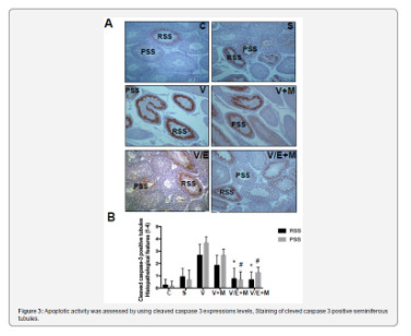
Introduction
Varicocele is an abnormal vascular dilatation of pampiniform plexus, commonly developing at puberty. Although underlying mechanisms remain poorly understood genetic background, anatomical aberrations, incompetence of venous valves, difference between the drainage of left and right testicular veins were suggested in the etiology [1]. As left spermatic vein being longer than the right vein, it is more commonly incurred to increased hydrostatic pressure and dilatation. Compression of the left renal vein between the aorta and the superior mesenteric artery may also contribute to the disturbed intravenous pressure [2].
The prevalence of varicocele varies between 15-20 % in general population and 30-40% in infertile men, and 11-19% of adolescents [3-6]. It was reported that varicocele is a progressive disease and early diagnosis and treatment in youth may enhance fertility potential [7]. Several contributing factors in the pathophysiology of varicocele have been proposed such as higher temperature of testis, the disorder of neuroendocrine system, autoimmunity, accumulation of renal and adrenal metabolites, genetic and epigenetic factors, hypoxia and oxidative stress [8-10].
Varicocele represents a chronic process within the testicle, which is linked to increased reactive oxygen species (ROS) beyond physiologic limits and, subsequently, disrupting sperm membrane fluidity, causing DNA damage and necrosis [11]. Moreover, superoxide dismutase 1, glutathione S-transferase M1 and T1 which are counteracting free superoxide radicals in cells have been reported to be decreased in men with varicocele, that may be important on disturbed sperm parameters [12]. Apoptosis of germ cells was also demonstrated in the pathogenesis of varicocele-related infertility [13]. Clinical findings suggest that surgical repair of varicocele may decrease seminal oxidative stress levels and sperm DNA fragmentation and, thus, may improve sperm quality [14]. Therefore, surgical intervention seems to be a reliable option in the treatment of varicocele-related male infertility, although some controversial reports exist.
Additionally, anti-oxidant medications such as kallikrein, L-carnitine with L-acetyl carnitine, pentoxifylline, coenzyme Q10 have been used to improve the milieu in the testis in men with varicocele [15]. Metformin is a major therapeutic agent in the treatment of type 2 diabetes mellitus as an insulin sensitizer, which decreases hepatic glucose output and increases peripheral glucose uptake. Although its action was not fully elucidated, metformin attenuated intracellular reactive oxygen species and apoptosis in aortic endothelial cells, myocardium, renal tubular cells and testicular cells [16-20].
Aim
Potential effects of metformin on varicocele-induced testicular damage have not been studied in neither humans nor in animal models. Thus, we investigated the impact of metformin on spermatogenesis, testicular integrity, and apoptotic activity in the testis of adolescent rats with experimentally-induced varicocele.
Materials and Methods
Thirty-six male adolescent Wistar rats (6-week-old) were randomly and equally divided into six experimental groups. Surgical procedures were carried out under anesthesia with intraperitoneal injection of ketamine (50 mg/kg). The experimental groups were as follows:
• (C) Control group; no surgical procedure was performed, and testis was examined after removal.
• (S) Sham group, a midline incision was performed, and testis was examined 8 weeks later.
• (V) Varicocele - only group: Experimental varicocele was induced by partial ligation of left renal vein with
Silk suture at the area medial to the insertion of the adrenal and spermatic vein into renal vein as described previously [21].
• (V+M) Varicocele + metformin group: All rats were treated with metformin (300 mg/kg per day by oral gavages) for 8 weeks following induced varicocele.
• (V/E) Varicocele + varicocelectomy group: Varicocelectomy was performed 4 weeks and the examination of the testis 8 weeks after the induction of varicocele. No medication was used.
• (V/E+M) Varicocele + varicocelectomy + metformin group: Varicocelectomy was performed 4 weeks after the induced varicocele. Metformin treatment (300 mg/kg per day by oral gavages) was initiated after the induction of varicocele and continued for 8 weeks. Left testes were examined 8 weeks after the induction of varicocele in all varicocele - induced groups. As maximum apoptotic activity initiates approximately 28 days after the induction of varicocele the procedure of varicocelectomy was performed 4 weeks after the formation of varicocele [22].
Histologic preparation and evaluation
The testicular tissue was fixed in Bouin’s solution (75% picric acid, 5% glacial acetic acid, and 25% formaldehyde) and embedded in paraffin blocks. Sections (5 μm) were formed, deparaffinized, and stained with hematoxylin and eosin. Spermatogenesis was examined in each group using Johnsen’s score (a score of 1-10 was assigned to each tubule regarding epithelial maturation) as described previously [23]. Sections were examined in a random order under a standard light microscope with 10x and 40x magnification by a blinded histologist; unaware of which group each rat belonged to. Histological grading was done by examining approximately 80 randomly selected seminiferous tubules per rat. Thus, a total of approximately 480 seminiferous tubules were scored for each group.
Histomorphometry analysis
A total of 103 randomly selected seminiferous tubules stained with hematoxylin-eosin were analyzed in each group. The presence of round spermatid stage (RSS) and primary spermatocyte stages (PSS) were assessed as described previously and compared among the groups [24].
Immunohistochemical staining for cleaved caspase-3 and ImageJ analysis
Cleaved caspase-3 was used for immunohistochemical staining. Testicular tissue samples were immediately fixed in 10% neutral-buffered formalin, embedded in paraffin, and sectioned (5 μm). Sections were deparaffinized and blocked for endogenous peroxidase activity with methanol containing 3% H2O2 for 10 m. Ultra V Block (Lab vision, Freemont, CA) for 7 m at room temperature. Cleaved Caspase-3 (#9664, Cell Signaling, U.S.) was applied at a dilution of 1: 500 and incubated overnight at +4 °C in a humidified chamber for nonspecific binding. The sections were washed in phosphate-buffered saline (PBS) and incubated with biotinylated horse anti-rabbit IgG (3 mg/mL; Vector, Burlingame, CA) at a 1: 500 dilution for 1 h at room temperature.
Antibodies were detected using a VECTASTAIN avidinbiotin complex (Vector PK 4000) for 30 m at room temperature. Antibody complexes were visualized after incubation with 3,3’-diaminobenzidine tetrahydrochloride (DAB, Bio-Genex, San Ramon, CA.) and were mounted under glass coverslips in Entellan (Merck) and then evaluated under a light microscope. Immunohistochemical staining for cleaved caspase-3 was analyzed by counting 100 seminiferous tubule cross-sections in each group and expressed as the apoptotic index. In each photomicrograph, the following parameter was measured with ImageJ software: expression levels of cleaved-caspase-3 in both groups at round spermatid stage (RSS) of testes. Each of this parameter was measured 3 times for each image and the average of the 9 measurements of each sample was used for the statistical analysis. Histopathological features examined in rats with normal testis and with sham, varicocele, varicocele+ metformin in a subjective scoring (0 - not present; 1 - low grade; 2 - moderate grade; 3 - high grade; 4 - very high grade).
Statistical analysis
Histopathological findings (Johnsen’s score) were assessed by nonparametric Kruskal-Wallis test, and the mean Johnsen’s score was used in the comparison of the groups. Multiple comparisons were made using Tukey’s procedure. p<0.05 was considered statistically significant. Analysis of variance was used for statistical analysis of the apoptotic index among the groups.
Results
Assessment of spermatogenesis
Johnsen’s score was significantly lower in V group (4.14±1.25) compared to C group (9.1±0.3) or S group (9.0 ± 0.2) groups (p<0.05). V+M group had significantly higher score (6.9±0.6) than V group (p<0.05). V/E group and V/E+M group had similar Johnsen scores (8.9 ± 1.02 and 9.2 ± 0.6). These findings suggest that the administration of metformin resulted in 40.6% of improvement in spermatogenesis in rats with varicocele. However, this favorable effect was not observed when metformin was used along with varicocelectomy.
Histological and morphological changes in seminiferous tubules
Histological and morphological changes in the testes of rats were compared via hematoxylin and eosin staining and degenerated tubules (DT) were only detected in V and V+ M groups, not in C, S, V/E and V/E+M groups (Figure 1). Visual assessment of the disorganized seminiferous tubules further supported these findings as seen in Figure 2. Seminiferous tubule degeneration scores were used for quantification of data (Figure 2b). V group had significantly higher scores of RSS and PSS compared to C and S group (2.6±0.8 and 3.7±0.4; 0.2±0.4 and 0.2±0.4; 0.9±0.6 and 0.6±0.7, respectively) (p<0.05). V/E group had significantly lower RSS (0.7±0.8) and PSS (0.8±0.7) scores than V group (p<0.05). V+M group had significantly lower RSS and PSS scores (1.8±0.7 and 2.6±0.7, p<0.05) in comparison to V group, implicating beneficial effects of metformin in rats with varicocele. When compared to V/E group, V/E+M group did not exhibit any difference in RSS (0.6±0.6) and PSS (1.4±0.5) scores, suggesting the absence of additive positive effect of metformin in varicocelectomies rats.
Apoptotic activity
Apoptotic activity was assessed by using cleaved caspase 3 expressions levels, staining of cleved caspase 3 positive seminiferous tubules were shown in Figure 3a. Cleaved caspase 3 expressions were significantly higher in V group (3.5 ± 0.5) compared to C (0) and S (0.2 ± 0.4) groups. V+M group had significantly lower cleaved caspase 3 level (3.0 ± 0.7) than V group. V/E group had lower cleaved caspase-3 expression levels (1.0 ± 0.7) compared to V group. Treatment of varicoceleectomy rats with metformin (V/E+M) did not further reduce apoptotic activity in the seminiferous tubules (1.75 ± 0.43) when compared to the varicocelectomy group (V/E) (Figure 3b).
Discussion and Conclusion
The present study demonstrates that metformin can reduce the extent of testicular damage in rats with varicocele, although having no effect in rats following varicocelectomy Spermatogenesis, seminiferous tubule integrity and the degree of apoptosis were improved using metformin in the presence of varicocele although it was not as remarkable as what was obtained through varicocelectomy. A review of the literature revealed that the impact of metformin on varicocele was not investigated in humans or animal models until now.
Although it is a commonly identified abnormality not all men with varicocele present with infertility. Some intrinsic factors may render some men to become susceptible to varicocele, thus, the best candidates who benefit from varicocelectomy yet to be clarified. Since oxidative stress was shown to be important in the pathophysiology of varicocele some agents have been used to improve the milieu in the testis [1]. A number of anti-oxidant medications have been studied to relieve detrimental effects of varicocele in the testis [25]. These agents have been used either alone or as an adjuvant therapy with surgery. However, surgery remains the treatment of choice and there exists insufficient data to recommend medical therapy in men with varicocele. Barekat et al. [26] reported that administration of an antioxidant agent N-acetyl cysytein as an adjunct therapy improved semen quality following varicocelectomy [26]. Tek et al. [21] demonstrated that vascular endothelial growth factor decreased apoptosis in varicoceleinduced rats as evidenced by diminished caspase-3 positive cells [21]. Both studies showed the benefit of these as adjunct therapy following varicocelectomy. However, in the present study metformin did not enhance the effect of varicocelectomy.
Minutoli et al. [13] demonstrated that neuronal apoptosis inhibin factor and surviving expressions were significantly reduced following varicocele induction and polydeoxyribonucleotide, an agonist of adenosine A2A receptor, administration restored testicular function [13]. Several other studies detected increased germ cell apoptosis in rats with varicocele [21,22,27]. However, in another study, apoptosis was found to be decreased in germ cells in the testes of infertile men with varicocele as compared with normal men [28]. It was speculated that the fixation of testis in formaldehyde might have played a role in the different result. In the present study, cleaved caspase 3 expression was used to assess apoptotic activity and it was found that metformin reduced apoptotic activity in rats with varicocele, whereas no additional effect was observed when metformin was administered after varicocelectomy.
Metformin is commonly used in type 2 diabetes mellitus and polycystic ovarian disease as an insulin sensitizer [29]. Also, it is present in various tissues including myocardium, liver, pancreas, thyroid, adipose tissue, hypothalamus, pituitary, and male and female gonads [19,30,31]. It has been reported that metformin is mainly transported into cells by organic cation transporters as passive diffusion is limited [32]. Although the mechanism of action is not yet fully elucidated recent studies suggested that metformin acts through AMP-activated protein kinase (AMPK) pathway, inhibits the activity of the respiratory electron transport chain in mitochondria, induces epigenetic modifications which in part may explain long term effects and decreases oxidative stress and apoptotic activity [16,19, 33-35].
Male reproductive system utilizes all these metabolic pathways and is prone to be affected by metformin administration [20,36,37]. Metformin was found to stimulate lactate production which is important in the development of germ cells and show an anti-apoptotic effect in rat Sertoli cells [38]. It was also reported that metformin reduced the apoptotic cells and caspase-3 level in rat testis [20]. The findings of the present study are consistent with previous studies that metformin reduced apoptosis in testis with varicocele. Yan et al. [37] reported that metformin improved the semen parameters related to its effects on weight loss, increased testicular weight and reduced testicular cell apoptosis [37]. On the other hand, Tartarin et al. [36] reported metformin at concentration 10 times higher than therapeutic levels decreased testosterone secretion and the number of Sertoli cells in rats when it was administered during pregnancy [36]. Faure et al. [39] reduction in testicular weight and testosterone level were observed in 6-week-old chickens treated with metformin for 3 weeks [39].
Several groups demonstrated that post-operative administration of metformin can exert protective effects in male reproductive function in rat models [40]. Bosman et al. [41] demonstrated that infertile hyperinsulinaemic men could benefit from metformin treatment in combination with an enriched antioxidant diet [41]. Besides, metformin was reported to act as a protective compound when used in the media for cryopreservation of spermatozoa [30]. In conclusion, metformin reduces detrimental effects of varicocele, although no additional benefit is expected following varicocelectomy. Further studies are required to apply metformin for this indication in humans.
#open access journals#peer review journals#Juniper Publishers PubMed Indexed Articles#reproductive medicine#GJORM in juniper publishers
1 note
·
View note
Text
Marine Macroalgae Bioextract Changes the Index of Reflectance in Pepper Plants
Abstract
In recent years, marine organisms have received attention for being a source of natural compounds, enabling the creation of a new and promising field of study. Marine macroalgae are a vast source of raw materials and biologically active molecules used in the pharmaceutical, cosmetic, food and agricultural industries. Due to its importance, the present study aimed to assess whether the bioextract of the red macroalgae Kappaphycus alvarezii acts as a biostimulant in the metabolism of plants and changes the reflectance indexes. In particular, the biological activity of the red macroalgae bioextract was focused on stimulating plant growth and photosynthetic activity. A scientific experiment was carried out using a red marine macroalgae bioextract applied on the pepper culture (Capsicum annuum L.). The pepper culture was chosen due to its high sensitivity to stress, generating good responses in the evaluation of the biostimulant and anti-stress capacity of an algal bioextract. The fresh mass of the aerial part and the length of the aerial part were evaluated, and a metabolic evaluation was also performed, represented by the photosynthetic rate produced by the pepper plants.
Read more about this article: https://juniperpublishers.com/ofoaj/OFOAJ.MS.ID.555822.php
Read more Juniper Publishers Google Scholar articles https://scholar.google.com/citations?view_op=view_citation&hl=en&user=V6JxtrUAAAAJ&citation_for_view=V6JxtrUAAAAJ:2P1L_qKh6hAC
#Juniper Publishers Google Scholar#Aquaculture#Fisheries journals#Marine science#coastal zone#Marine Physics
0 notes
Last Seen Blogs

alwaysmarilynmonroe
Always Marilyn Monroe


jocounselingarte
Jo /Art, Counseling and literature

calmlights
mai

modaparamujeres
Moda para mujeres