#Microscopy
Text


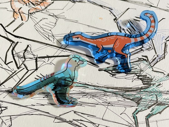

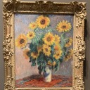
gifts for microbio and paleo enthusiasts! :DD
#science#biology#microbiology#microscopy#cell biology#marine biology#paleontology#paleoblr#dinosaurs#phycology#algae#diatoms#coccolithophore#bacteria#nitrogen fixing bacteria#pins#keychains#small business#artists on etsy#mu's wares
2K notes
·
View notes
Text
Meeting a water bear. 🌱
Basically wishing I could make myself tiny and get lost in some moss. So much beauty in little drops of water.
#water bear#tardigrade#rotifers#cilliates#microscopy#microorganisms#digital painting#digital art#artists on tumblr#animal art#art#painting
6K notes
·
View notes
Text


2024-03-07
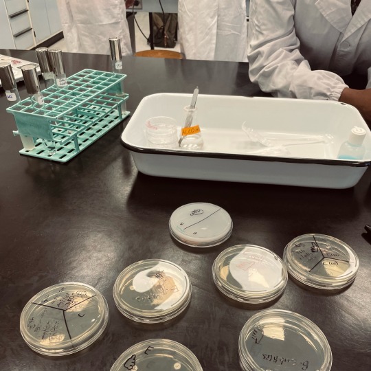
There’s really nothing like being a science student: that feeling of both drowning in work and stress but also somehow enjoying it at the same time.
#are we all masochists or what#everyone I know is so unwell#but we’re all still here#but anyway I guess this is a bit of a lab compilation#went out and swabbed some trash cans today#hopefully we’ll get lots of bacteria#will update once it incubates#studyblr#studyspo#dark academia#study aesthetic#aesthetic#student#stem academia#my posts#chaotic academia#bacteria#microscopy
207 notes
·
View notes
Text
If you ever find yourself playing a game of twenty questions, there's a little-known life form you can pick that is sure to leave your opponent stumped.
It is neither animal, vegetable, nor mineral. It's not even a bacterium or fungi.
It's called a Euglenid – and it's a weird fusion of a bunch of different living things.
Continue Reading.
312 notes
·
View notes
Text
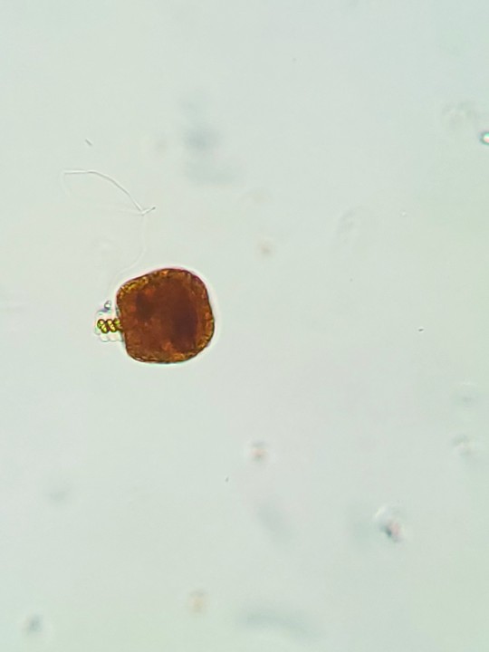
Today's joy is brought to you by things found in 0.01mL of water.


226 notes
·
View notes
Text
Flatworm Dugesiidae sp
Collected from the stem of an emersed iris. The pigmented spots camouflage it with the river mud, and the eyespots help it find dark niches.
246 notes
·
View notes
Text
I FUCKING LOVE MERGING UNETHICAL SCIENCE WITH OCCULTIST MAGIC! I LOVE UNVEILING FORBIDDEN SECRETS MANKIND WAS NOT MEANT TO KNOW! I LOVE SUCCUMBING TO UNENDING TORMENT BROUGHT ON BY OUR OWN HUBRIS!

337 notes
·
View notes
Text
Gametophyte of Pleurocladula albescens.
114 notes
·
View notes
Text
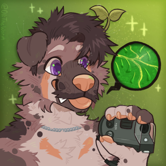
Handheld Microscope🔬-- New Icon for myself, also done live on stream. I literally streamed longer than I normally do because I really wanted to get this done. Worth it🦐
#furry#art#furry art#digital art#fursona#digital drawing#digital artist#furryart#oc#anthro#microscope#microscopy#furries in stem
121 notes
·
View notes
Text
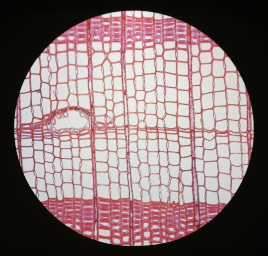
Alan Crivellaro (Italian, b.1990)
Microscopic colour-stained sections of wood - 2007
Installation at "True to Nature: Open-air Painting in Europe 1780-1870" exhibition, Fitzwilliam Museum, Cambridge 2022
Images © Dr. Alan Criverello
722 notes
·
View notes
Note
Hello, Mr. Holmes! How are you?
So, long story short, I ended up with an optical microscope in my room more or less 4 months ago, with 200 previously made slides (secured in a proper box), and lots of new ones too, for me to prepare myself. I love microbiology (it's one of my hyperfixations, curse my neurodivergency) and now I love it even more (my mother has had to drag me away from the microscope - I named it Wesley - in the middle of the night multiple times now).
After much conversation, I finally convinced my mom to buy me the proper equipment to prepare the slides!
So, I'm sending this ask to you, as I know you also have a microscope and that you use it a lot: what kind of equipment do you recommend me buying (gloves, scalpel blades, tints, etc), while still remembering that all of the stuff needs to stay in my room (properly taken cared of by me, of course)?
For example, I'm unsure if different dyes are used for different smears and specimens due to it's affinity (I've noticed that on 'organic matter' slides, images are usually tinted purple or pink, while on plant-based slides, images are usually tinted green and blue, with a few red structures.) Considering that I don't have access to a mortuary, I will mostly make plant slides. There must be a difference in the dyes then, right?
Sorry for the long text! Hope this isn't too much of a bother.
- a 17-year-old :)
Congratulations on your new light microscope. I do hope you get the best out of it. I am overjoyed that someone else appreciates the art of microscopy and microbiology.
However, you need to be careful to not strain your eyes. It is recommended to take breaks every 15 minutes to close your eyes or focus on something in the distance to reaccommodate your eyes. And get up every 40 minutes, stretch and correct your posture. And it is recommended to not use a microscope more than 5 hours per day. John has to chase me away from my microscope sometimes to take a break when I sit there for hours, my posture like a Caridea.
Concerning equipment, you will obviously need a scalpel or other sharp blade to make very thin slices of your specimen, as thin as possible. And forceps to move your samples (best just get a whole dissection kit it has everything). Obviously slides and coverslips, pipettes for the stains or water, maybe some tubes. A pen to label your slides. In many staining procedures ethanol or acetone is also used. A waste jar to safely dispose of any chemicals, but be careful what you mix. A rack for staining and containers. I would recommend nitrile gloves, some people are sensitive to latex.
The dyes you use depend on the specimen. For example in histological slides of tissues hematoxylin and eosin are most commonly used (short HE-stain). That's what you most likely saw on your slides, it's blue, purple and pink.
Hematoxylin is a basic compound extracted and oxidised from the logwood tree (Haematoxylum campechianum), and it stains acidic compounds in the cells (or basophilic because they have an affinity for basic substances). For example nucleic acids like DNA or RNA get stained by hematoxylin because they are basophillic. And where are lots of nucleic acids? In the nucleus and ribosomes, that is why they appear blue to purple in the staining because they bind hematoxylin.
Eosin is an acidic compound, and stains basic or acidophilic compounds red or pinkish, like proteins, collagen, cytoplasm, extracellular matrix.

(Ductus epididymidis with HE-stain)
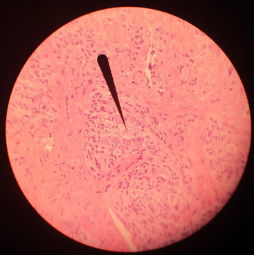
(Tongue HE-stain, pointer marking a ganglion; that is my picture)
Of course there are more specific stains for specific tissues like Golgi's silver staining for neurons.
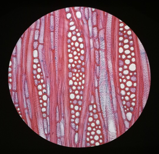
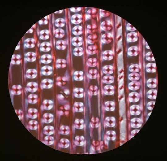
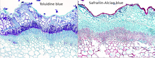
For plants toluidine blue is often used, high affinity for acidic tissues, and can stain blue to green to purple.
It is often combined with safranin, a basic azine, which is probably the red stain you saw. It stains polysaccharides and lignin, woody parts of the plant. Safranin and astrablue is also often combined, astrablue stains non-lignified parts of the plant.
(Ulex europaeus stem; not my pictures I don't have any samples currently, source Atlas of plant and animal histology)
Safranin is also used in bacteriology, in the famous Gram staining. In Gram staining you use crystal violet (blue/purple), Lugol's iodine solution, then wash it with ethanol and add safranin (red) as a counter stain. Bacteria is gram-positive if the crystal violet stays in their thick murein cell wall, can't be washed out with the ethanol and the bacteria stays blue. Gram-negative appear red because of the counterstain.
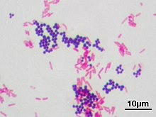
(Staphyloccocus aureus (violet, gram positive) & Escherichia coli (red, gram negative); not my picture, source Wikipedia)
However, I am not sure whether you have access to any of those substances, if they are too expensive for you or if they are too hazardous if used in your own room for a prolongued time. Of course those substances need to be stored properly, and your own room is probably not a good place, especially for ethanol or acetone. The fumes. I would recommend to ask your biology or chemistry teacher whether they can recommend anything further and where to buy said solutions in your area, and if they can't they are idiots. There are also many useful resources and tutorials on Youtube.
Another fascinating experiment for your microscope, that you can perform without buying any chemicals, is a hay infusion. You put hay into a container filled with water, and let it sit undisturbed for a week in a sunny area but not in direct harsh sunlight. During that time the microorganisms in the hay are reproducing in the solution, feeding on the polysaccharides of the hay. Protozoans also flourish in the hay infusion and eat the bacteria. It might get cloudy and a bit foul smelling (best not do it in your own room if you don't want to sleep next to a rotting smell).
When you put a drop of the solution onto a slide and look at it in the microscope, you should see a variety of microorganisms like bacteria (like Bacillus subtilis), amoeba, ciliates, heliozoa, algae et cetera. At different depths of the liquid you should find different kinds of organisms, because of differing oxygen content.
However, pathogens can also occur in the hay infusion so handle it carefully and work sterile, wash your hands properly.
And even if you don't work at a morgue you can still get tissue samples to experiment on, after all meat is sold in supermarkets, basically the same as a human body. And at the butchers they even sell organs like chicken hearts, pig kidney, liver, blood et cetera. Or observe your own hair under the microscope.
Which kind of samples and slides were included in your starter kit? Be careful to not leave them lying around in the sunlight, or the stain might fade. Always store them in the proper box.
#roleplay#rp#sherlock roleplay#sherlock rp#johnlock roleplay#johnlock rp#sherlock#bbc sherlock#sherlock holmes#sherlock holmes rp#sherlock holmes roleplay#science#scientist#research scientist#histology#microscope#microscopy#bacteria#bacteriology#pathology#anatomy#biology#chemistry#scientists#pictures#he stain#specimen#samples#slides#sherlock replies
46 notes
·
View notes
Photo
'A little messy but I like it 🔮', @cosmodernism, 2022
216 notes
·
View notes
Text


microscope life drawings - tardigrade and colony of Vorticella ciliates.
#animal art#microscopy#microscope#ciliate#vorticella#life drawing#sketch#tardigrade#water bear#pencil#drawing#studies#artists on tumblr#traditional art#protist#protists#microorganisms
838 notes
·
View notes
Text


2024-04-13
Incredibly stressful week. Just test upon test, deadline upon deadline. School and my extracurriculars have ramped up, and my to-do list seems suffocating at times.
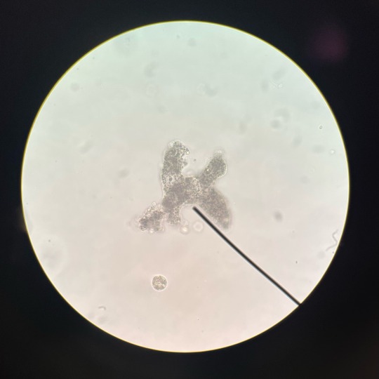
But hey: at least I have good tea and a great eye for finding amoebas.
#is anyone interested in seeing a video of my amoeba#I managed to catch it on camera through the eyepiece#it’s so cute!#just a silly little guy#love protists#also do not be alarmed if at one point I start talking about breast cancer#doing my first research project and it’s pretty freaking cool!#studyblr#studyspo#dark academia#study aesthetic#aesthetic#student#my posts#chaotic academia#study#stemblr#stem academia#microscopy
59 notes
·
View notes
Text
Hey. 108 gigapixel scan of Girl With a Pearl Earring with depth information.
69 notes
·
View notes
Text
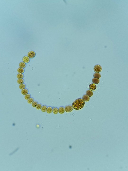
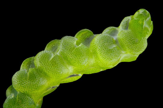
Today's little bit of joy is brought to you by this absolutely perfect filamentous cyanobacteria specimen. This beauty sits perfectly in a single plane, the whole thing in focus, and has both a heteorcysts AND an akinete.
170 notes
·
View notes
Last Seen Blogs