#imaging
Photo

crooked falls
#vancouver#canada#waterfall#imaging#luxlit#lensblr#canonphotography#original photographers#pnw#pws#nature
1K notes
·
View notes
Text

Evening walk #chicago
.
#streetphotography #blackandwhite #blackandwhitephotography #lakemichigan #winter #trees #foggy #fog #rain #reflection #puddles #photo #path #lights #lights #urbanphotography #city #photography #photooftheday #night
#chicago#blackandwhite#streetphotography#lakemichigan#fog#foggy#rain#reflection#trees#path#lights#night#imaging
121 notes
·
View notes
Text
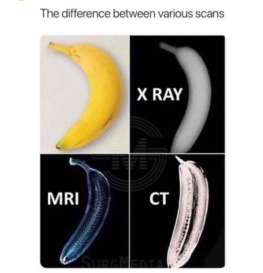
You may or may not learn something new from this meme xD
#biology#medicine#scan#health#hospital#imaging#reddit memes#discord memes#twitter memes#memes#meme#funny#relatable#insidesjoke#humor#humour#science
48 notes
·
View notes
Text

#he's at it again#he's so annoying istg#being shot for dead really does make you jobless huh#I'm blocking his ass rn#can't even spell for shit#how was he supposed to be a songwriter 💀#imaging#all the people#the beatles#john lennon#paul mccartney#george harrison#ringo starr#beatles#memes#broke ass bitch#broke people should never laugh
43 notes
·
View notes
Text
+
39 notes
·
View notes
Text

An x-ray machine operator stands next to a protective lead shield which has a little window for viewing the patient. The electro-therapeutic guide. 1912.
Internet Archive
167 notes
·
View notes
Link
A new non-invasive imaging technique called raster-scan optoacoustic mesoscopy (RSOM) shows promise for detecting diabetes progression and severity by visualizing microscopic changes in the skin’s blood vessels. Recently published research from the Technical University of Munich (TUM) and Helmholtz Munich reveals RSOM’s ability to create detailed three-dimensional maps of the skin’s microvasculature down to capillary dimensions. Machine learning then pinpoints subtle alterations reflecting vascular damage tied to advancing diabetes.
Tracking disease progression in diabetes remains a major challenge. Current blood tests like glycated hemoglobin (HbA1c) analysis lack sensitivity for detecting early vascular complications or monitoring subtle worsening over time. Skin manifestations also tend to appear late and crudely indicate systemic microvascular damage. This often delays interventions until irreversible organ injury sets in.
The new technique leverages the skin’s accessibility to “read out” diabetes’ impacts on tiny vessels in ways not previously possible. It images living tissue without dyes or contrast agents using optical and ultrasonic waves. Powerful AI methods then extract and analyze hundreds of explainable microvascular features predictive of diabetes severity.
Continue Reading
45 notes
·
View notes
Photo
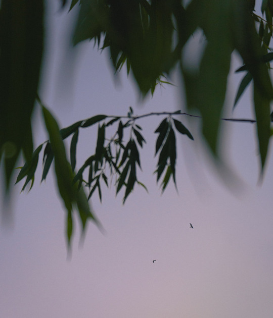
Wanderers
Instagram
#original photographers#photographers on tumblr#photographers on instagram#photographers on flickr#lensblr#lensculture#imaging#nature#plant#leaves#sky#birds#minimal#minimalism#evening#twilight#dusk#Andrei Grigorev
66 notes
·
View notes
Photo

High-resolution images of terminals in water chains.
Shiotari, A., Sugimoto, Y. Ultrahigh-resolution imaging of water networks by atomic force microscopy. Nat Commun 8, 14313 (2017). https://doi.org/10.1038/ncomms14313
153 notes
·
View notes
Text
Reason to Live #8864
Imagining a world where your characters can live in. – Guest Submission
(Please don't add negative comments to these posts.)
#sad#help#hope#reason to live#depressed#depression#empty#alone#mental illness#anxiety#trauma#guest submission#mental health#imagination#imaging#world#creative#creativity#ocs#characters
70 notes
·
View notes
Text
Do y'all think we can Kung pow penis midjourney like a virus
#dunkar rant#Imaging#Thousand of 'art' blog where the art is just kpp#What would that even look like? Don't know but it would be funny
10 notes
·
View notes
Text

Afternoon walk #chicago
.
#blackandwhite #blackandwhitephotography #lakemichigan #winter #trees #sky #clouds #photo #photography #photooftheday
#chicago#blackandwhite#sky#clouds#lakemichigan#landscape#trees#tree#photographer#photography#original photography on tumblr#photo#photographers on tumblr#original phography#imaging#original photographers
24 notes
·
View notes
Text
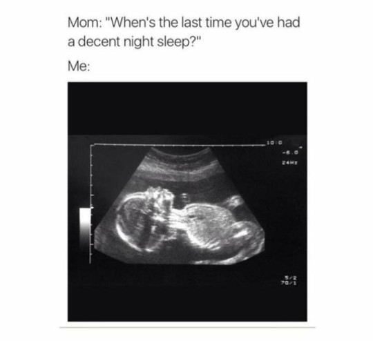
Sooo true
#baby#ultrasound scan#science#imaging#student memes#memes#meme#funny#relatable#insidesjoke#humor#biology
11 notes
·
View notes
Text
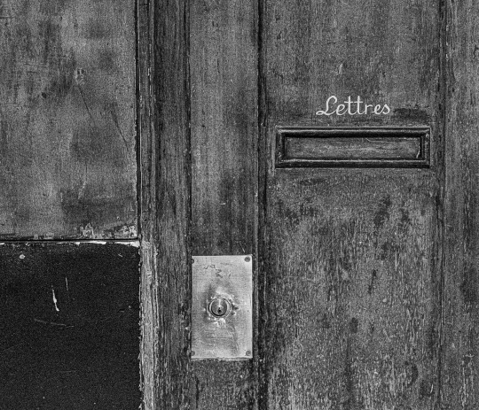
Paris' streets door
©pierre-yves chassaigne
#pierre yves chassaigne photography#pierre yves chassaigne#paris#original photographer#original tumblr photographer#isu#imaging#photozine#photoblog#bnwphotography
10 notes
·
View notes
Photo
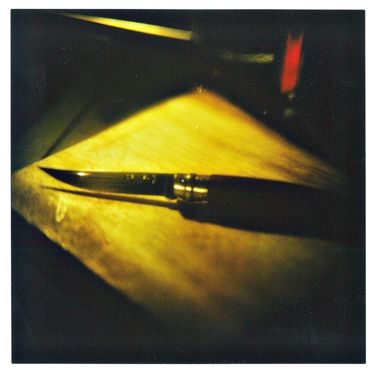
Still Life for Knife
SL660 + Pentax SMC 50mm f1.2 + Instax Square Film
#original photography#original photographers#photographers on tumblr#nonscamera#sl660#polaroid#Instant Film#instaxsquare#instax#analog photography#filmisnotdead#filmisalive#film photography#ishootfilm#believeinfilm#imaging#lensblr
66 notes
·
View notes
Text
New Viewing Platform
A new system that allows trans-scale optical imaging of large field of view and volume so that cells in whole organs and tissues can be mapped dynamically
Read the published research article here
Video from work by Taro Ichimura and colleagues
Transdimensional Life Imaging Division, Institute for Open and Transdisciplinary Research Initiatives, Osaka University, Osaka, Japan
Video originally published with a Creative Commons Attribution 4.0 International (CC BY 4.0)
Published in bioRxiv, February 2024 (not peer reviewed)
You can also follow BPoD on Instagram, Twitter and Facebook
5 notes
·
View notes
Last Seen Blogs

sidsskullshirt
It looked easier on youtube.

dkboutique
DK Boutique & Spa

awoolmonds
S.O.L.

desert-lily
Now @ delilah-briarwood
betawolfpuppy
Beta Wolf