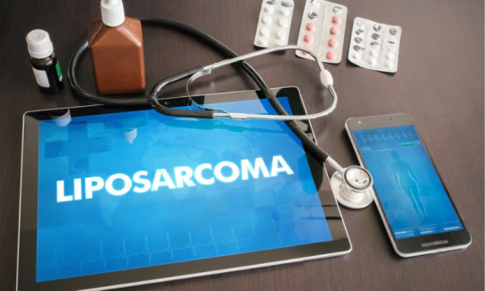
#liposarcoma
Text
Myxoid liposarcoma of the spermatic cord: A rare entity by Emmanuel E. Sadava in Journal of Clinical Case Reports Medical Images and Health Sciences
ABSTRACT
An 81-year-old man consulted at our hospital for evaluation of a long-established left inguinal mass. The patient denied experiencing pain, food intolerance, constipation or urinary tract symptoms in the past. A physical examination revealed a 15x10cm painless mass in the left inguinal region, distinct from the testicle, with no palpable changes during Valsalva´s maneuver. Magnetic resonance imaging (MRI) showed a 79mm heterogeneous lesion of the spermatic cord which projected itself through the inguinal canal into the scrotal sac, displacing the testis inferiorly. Laboratory testings were negative for testicular tumor markers such as α fetoprotein and human chorionic gonadotropin-β. A surgical resection of the inguinal tumor with an “en-bloc” inguinal orchiectomy was performed. The inguinal floor was repaired with a modified Bassini technique without the use of a mesh. The histopathological report confirmed findings were consistent with a myoxid liposarcoma. No further treatment was indicated and the patient continued follow-up with bi-annual MRIs. 18 months later, the patient continues with no signs of recurrence.
Key words: liposarcoma, liposarcoma of the spermatic chord, abdominal wall surgery, inguinal mass.
INTRODUCTION
Sarcomas constitute a heterogeneous group of rare solid tumors of mesenchymal cell origin. Collectively they account for approximately 1% of all adult malignancies with an annual incidence of 2.5 cases per million population[1]. In adults, the most common soft tissue sarcomas are liposarcomas. Overall, they account for approximately 17% of all soft tissue sarcomas. Most cases arise from de novo, therefore, the development from a preexisting benign lipoma is rare. Liposarcomas usually appear as a slowly enlarging, painless mass in a middle-aged person with a slightly higher incidence in men.
These tumors are classified in three main biologic forms: 1) well-differentiated liposarcoma; 2) myxoid and/or round cell; and 3) pleomorphic. The latter being a rare high-grade with a high recurrence rate and poor prognosis. The well-differentiated and myxoid types have favorable prognoses. However these tumors locally recur after incomplete excision[2].
The anatomic site of the primary disease represents an important prognostic factor, influencing treatment and outcome. Extremities (43%), the trunk (10%), visceral (19%), retroperitoneum (15%), or head and neck (9%) are the most common primary sites. Scrotal location is relatively rare, accounting for 3.6% of all liposarcomas. The origin of intra scrotal liposarcomas include the spermatic cord (76%), testicular tunic (20%), and the epididymis (4%).
CASE REPORT
An 81-year-old man with a medical history of follicular cutaneous lymphoma and an open left hemi-colectomy for colon cancer consulted at our hospital for evaluation of a long-established left inguinal mass. The patient denied experiencing pain, food intolerance, constipation or urinary tract symptoms in the past. A physical examination revealed a 15x10cm painless mass in the left inguinal region, distinct from the testicle, with no palpable changes during Valsalva´s maneuver. Magnetic resonance imaging (MRI) showed a 79mm heterogeneous lesion of the spermatic cord which projected itself through the inguinal canal into the scrotal sac, displacing the testis inferiorly (Figure 1). Laboratory testings were negative for testicular tumor markers such as α fetoprotein and human chorionic gonadotropin-β. Ultrasound-guided biopsies of the mass were requested and their histopathology analysis revealed myxoid stroma with fusocelular proliferation.
A radical resection was suggested but, a week prior to the surgical procedure, the patient was diagnosed with COVID infection during which he intercurred with myocardial infarction and ischemic stroke. He underwent a double coronary angioplasty with drug-eluted stents and required anticoagulation and antiplatelet therapy posteriorly. The case was discussed at a multidisciplinary meeting and a conservative management of the inguinal tumor was decided. The patient was reassessed 12 month later with a new MRI, which showed the inguinal mass increased in size (99mm) compared to the previous study, and a computed tomography (CT) with no evidence of metastatic disease. A surgical resection of the inguinal tumor with an “en-bloc” inguinal orchiectomy (Figure 2) was performed. The inguinal floor was repaired with a modified Bassini technique without the use of a mesh. The patient had an uneventful recovery and was discharged from the hospital on postoperative day two.
The histopathological report confirmed a 130x120x120mm low-grade fibro myxoid neoplasm (Figure 3). The surgical margins were negative. Immunohistochemistry showed strong reactivity for S100 and vimentin, whereas SOX10, desmin, CD34 and estrogen receptors were negative. These findings were consistent with a myoxid liposarcoma. No further treatment was indicated and the patient continued follow-up with bi-annual MRIs. 18 months later, the patient continues with no signs of recurrence.
Figure 1: Pelvis MRI T2 axial and coronal images illustrating a left inguinal canal soft tissue density measuring 78 x 68mm.
Figure 2: A Intraoperative image of the liposarcoma. Left inguinal surgical approach with the spermatic cord lesion and left testicle in vivo. B: Intraoperative image of left inguinal mass (a) excision with radical orchiectomy (o).
Figure 3: A Hematoxylin and eosin staining: fusocelular and myxoid infiltrative neoplastic proliferation, made up of ovoid cells and finely granular chromatin. Scarce elongated cytoplasm arranged in fascicles accompanied by elongated, thin, curvilinear blood vessels with zones of perivascular cellular condensation. B: Immunohistochemistry positive for S-100.
DISCUSSION
Liposarcomas invade through local extension and rarely invade through the lymphatic route, making regional lymph node dissection lose its value and having no impact on survival. Nevertheless, high-grade subtypes are associated with high rates of recurrence and hematogenous spread; lungs, liver and peritoneum being the most common sites of metastasis. Surgical resection (with appropriate negative margins: >1cm) is the standard primary treatment in most patients with stromal cell sarcomas. Complete tumor resection is the primary prognostic factor for local recurrence, and liposarcomas are not the exception. Performing an “en-bloc” resection involving a high orchiectomy (including the surrounding tissue) is important to obtain negative margins [1].
Local recurrence rates for sarcomas, including liposarcomas of the spermatic cord, have been reported to be as high as 30-50%. Because of this, and despite the patient’s disease-free status, long term follow-up remains a crucial step in the detection of recurrences that might still be potentially curable. Current controversy arises on the use of adjuvant chemotherapy or radiotherapy. Being a rare and infrequent entity makes it hard for a single institution to accumulate enough cases to perform prospective randomized controlled trials. Extrapolated data from retrospective analyses support the use of adjuvant radiation on selected high-risk situations (tumor recurrence, high-grade tumors or residual disease). Concerning the role of chemotherapy, the use of adjuvant chemotherapy remains controversial and there is no definitive role in the management of localized liposarcomas[3].
In conclusion, myxoid liposarcomas of the spermatic cord are infrequent entities. As most soft tissue sarcomas, they have an indolent course and should be considered as a differential diagnosis of inguinal masses with no palpable changes during Valsalva´s maneuver. Complete surgical resection with high-orchidectomy “en-bloc” is encouraged.
For more information: https://jmedcasereportsimages.org/about-us/
For more submission : https://jmedcasereportsimages.org/
#liposarcoma#liposarcoma of the spermatic chord#abdominal wall surgery#inguinal mass#Magnetic resonance imaging#epididymis#MRI#histopathology#Emmanuel E. Sadava#jcrmhs
0 notes
Text
Understanding Primary Angiosarcoma of the Breast
Understanding Primary Angiosarcoma of the Breast
Primary angiosarcoma of the breast is a rare and aggressive kind of breast cancer. Angiosarcoma is a type of cancer that starts in cells that line blood and lymph vessels. It can occur anywhere in the body, including the breast.
This article takes a closer look at primary angiosarcoma of the breast, including symptoms, risk factors, and treatment.
What is primary angiosarcoma of the…

View On WordPress
0 notes
Text
ultrasound rooms are so dark lol
#cancer mention btw for the rest of these tags#i have never had an ultrasound before so idk what's normal!!!#but . it took both longer and shorter than i expected?#and the tech kept making faces . idk if that was a 'interacting w annoying software' face or a 'this is concerning' face#also she felt the lump and she was like hm. this side is squishy and this side is more lumpy...#like WHAT DOES THAT MEAN?#so now im actually like. way more nervous. lol#bc google is telling me lipomas (the thing my mom said it was and thus the reason i didnt get it checked out for ~4 years) are squishy#and liposarcomas (aka . that but cancer) are like. firm. so uhhhhhhh#even if it is benign i would really like it out of my body. bc it literally looks like i have a rib or smthn popping out of my body when i#wear bikinis/tight tops. anyways .... cant believe i have to work rn. when im freaking out abt having literal cancer
2 notes
·
View notes
Text
Liposarcoma: What You Need To Know | Dr. Haytham El Salhat
Introduction to liposarcoma
L
iposarcomais a type of cancer that develops in the body’s fat cells. It is a rare condition, with an estimated incidence of 1 in 1 million people per year. Liposarcoma can occur at any age, but it is more common in middle-aged and older adults. It can occur in any part of the body but is found in the legs, arms, and trunk. Liposarcoma has several types, depending on how the cancer cells look under a microscope. These types include: well-differentiated, myxoid, pleomorphic, and dedifferentiated liposarcoma.
Liposarcoma is a serious condition that requires prompt diagnosis and treatment. If left untreated, it can grow and spread to other parts of the body, making it more difficult to treat.
Symptoms of liposarcoma
A lump or mass in the soft tissue, which may be painless but can also cause pain and discomfort
Difficulty moving the affected area
Swelling and weakness in the affected area
Weight loss and fatigue
Pain or discomfort in the affected area
A feeling of fullness even after small meals
A change in the shape or size of the lump over time
Changes in skin color or texture over the lump
See a doctor for a proper diagnosis if you have symptoms. It’s important to have regular check-ups if you have risk factors for liposarcoma.
Diagnosis of Liposarcoma
Medical history: A doctor will ask about your symptoms and past health.
Physical examination: A doctor will examine you to look for lumps or masses in the soft tissue.
Imaging tests: Imaging tests like MRI, CT scan, or ultrasound can aid in diagnosis. These tests can help to identify the size and location of the tumor.
Biopsy: A biopsy is usually needed to confirm the diagnosis of liposarcoma. A small sample of the tumor will be taken and examined under a microscope.
Subtype determination: Doctors identify the subtype by examining the cancer cells. The subtype will affect treatment options and prognosis.
Keep in mind that liposarcoma can be hard to diagnose and may resemble other conditions. A biopsy is needed for a definite diagnosis. If you have symptoms of liposarcoma, see a doctor. Follow their instructions for diagnostic tests.
Treatment Options for Liposarcoma
Surgery: Surgery is the main treatment for liposarcoma. The goal of surgery is to remove the tumor and surrounding tissue. Depending on the size and location of the tumor, surgery may be done through a small incision or a larger one. In some cases, surgery may need the amputation of the affected limb. The type of surgery will depend on the subtype of liposarcoma, the size of the tumor, and the location of the tumor.
Radiation Therapy: Radiation therapy shrinks tumors before surgery or kills remaining cancer cells after surgery. It can also ease pain and symptoms for patients who cannot have surgery.
Chemotherapy: Chemotherapy can kill remaining cancer cells before or after surgery. It can also shrink the tumor to make surgery easier.
Clinical Trials: Patients with liposarcoma may also be eligible for clinical trials to test new treatments.
The best way to treat liposarcoma depends on the tumor type, size, and spot and the patient’s health. Talk to a team of specialists for the best treatment options.
Remember, liposarcoma is a complex disease and treatments may have side effects. It’s crucial to check treatment progress and side effects with the healthcare team.
Coping with Liposarcoma
Emotional challenges: Liposarcoma can cause feelings of fear, anxiety, and depression. To cope, have a support system like friends, family, or a counselor.
Practical challenges: Liposarcoma can make daily activities, appearance, and finances difficult. It is important to have a plan in place to manage these challenges.
Maintaining a positive outlook: Stay positive and focus on what can be done, not what can’t. Doing activities that bring happiness and calm can improve well-being.
Coping with treatment side effects: Treatment for liposarcoma can cause side effects such as pain, fatigue, and nausea. It is important to discuss these side effects with the healthcare team and to have a plan in place to manage them.
Coping with recurrence: Liposarcoma can recur even after treatment. It is important to have the plan to monitor and manage any recurrence.
Importance of support system: A support system can be made of friends, family, or a counselor. Joining a support group can also be helpful as it allows connecting with people going through similar experiences.
Conclusion
Liposarcoma is a rare type of cancer that develops in the body’s fat cells. Early detection and quick treatment can improve the outcome. Symptoms include lumps or masses in soft tissue, pain, and difficulty moving. The diagnosis is made with a biopsy and imaging tests. Treatment options include surgery, radiation therapy, and chemotherapy. It’s helpful to have a multidisciplinary team of specialists to create the right treatment plan.
Living with liposarcoma can be difficult. It’s important to have a support system of friends, family, or a professional counselor. Support groups can also be helpful. Liposarcoma can make daily tasks, appearance, and money difficult. Make a plan to manage these challenges.
Having a positive attitude and concentrating on possible actions can enhance well-being. Coping with treatment side effects and recurrence is also important. Everyone’s experience with liposarcoma is unique and coping strategies will vary. It’s important to find what works best for the individual and to seek help when needed.
Liposarcoma is serious, but early detection and quick treatment can help manage it and lead to a good quality of life. Be aware of symptoms, get regular check-ups, and seek help if needed.
0 notes
Text
0 notes
Photo


You are strong! But some days we all need a little more awareness, and a few extra spoons! (in yellow!)
The yellow awareness ribbon represents those with
adenosarcoma, bladder cancer, cancer, bone cancer, carbon monoxide poisoning, craniofacial acceptance, endometriosis, epithelioid sarcoma, ewing sarcoma, microcephaly, myxoid liposarcoma, obesity, osteosarcoma, sarcoma, spina bifida, suicide prevention
And the dark and light versions of the design are available on my Threadless store in a variety of products and background colours!
threadless dark- https://meridiandesigns.threadless.com/designs/yellow-awareness-pocket-dark
threadless light- https://meridiandesigns.threadless.com/designs/yellow-awareness-pocket-light
facebook ★ instagram ★ twitter ★ pinterest
#yellow#spoonie#adenosarcoma#bladder cancer#cancer#bone cancer#carbon monoxide poisoning#craniofacial acceptance#endometriosis#sarcoma#microcephaly#myxoid liposarcoma#osteosarcoma#obesity#spina bifida#suicide prevention
0 notes
Photo

MedsurgeIndia is providing the affordable Liposarcoma Treatment in India.They are also offering the top hospitals with top doctors in India, Turkey & Thailand.
0 notes
Text
☀️ Solar Return | Saturn in the 1st house

Saturn often causes people anxiety because of the difficulties it presents. Since Saturn is currently in the first house of my Solar Return chart, I thought it would be a good idea to write about what I have noticed since turning 28 in February of last year.
Remember that everyone experiences Saturn differently; in my natal chart, this planet is also involved in significant transits that are also connected with the events I’m describing.
Saturn in the 1st house of a Solar Return chart
Saturn's placement in the first house foretells restrictions on one's appearance and social behavior. The body somehow becomes a burden, which can therefore make it difficult for the person to interact with others and conduct daily activities. Additionally, the person may become so self-conscious that leaving the house causes them distress. They could develop increased shyness, reserve, and isolation.
My experience
[TW: body issues, illness]
Being an astrologer, I anticipated having to deal with a physical issue this year. My best guess was having to deal with a negative self-image and growing self-criticism. My self-consciousness is already fairly high, so I assumed it would worsen and keep me from going out even more. It turns out that this is not the case.
The weight gain was the first change I saw. Since I haven't changed my diet or exercise routine much, the change truly caught me off guard (which fits the Saturn in Aquarius theme pretty well). It wasn't until all my clothing stopped fitting me that I even realized I had gained weight. As I can't replace my entire wardrobe at once and must gradually add new pieces, getting ready has been tricky and frustrating.
The extra weight, on the other hand, has not been an issue. In fact, I prefer the way I look right now. Since I grew up in Paris, where the majority of women my age are slim and have smaller frames, and since I have more curves due to my Peruvian ancestry, I've always been self-conscious of my appearance. Knowing that we no longer obsess and despise our bodies 14 years later would make my 14-year-old self quite happy. :)
This is the only beneficial impact of Saturn that I have noticed: developing a more mature perception of my appearance and looks.
The second change is a little bit more serious. A few weeks ago, I discovered a small tumor on my chest. My doctor says it shouldn't be malignant, but given my family history (my dad passed away from an aggressive liposarcoma and another family member has had breast cancer) I need to do some tests to rule out any possibility. We’re quite certain that the growth is not malignant because I'm not currently experiencing any symptoms linked to cancer. According to my doctor, it's also rather typical for small tumors to emerge unexpectedly and then disappear. Even though it's probably not a big issue, it's the first time I've had to deal with something similar and quite a significant occurrence this year.
These are the only changes I've observed since February. However, I'm just 6 months in, so perhaps more events will occur before my next birthday. I’ll keep you posted. :)

#solar return#saturn in the 1st house#1st house#saturn#saturn in aquarius#astro notes#predictive astrology#astrology notes#astrology#astro community#astro observations
72 notes
·
View notes
Text
What are the five various forms of cancer?
Carcinoma: Skin cancers, lung cancers, colon cancers, pancreatic cancers, and ovarian cancers, as well as epithelial, squamous, and basal cell carcinomas, melanomas, papillomas, and adenomas
Sarcoma: Cancer that develops in bone, cartilage, fat, muscle, blood vessels, or other connective or supporting tissue — "bone, soft tissue malignancies" such as osteosarcoma, synovial sarcoma, liposarcoma, angiosarcoma, rhabdomyosarcoma, and fibrosarcoma.
Leukemia is a type of cancer that begins in blood-forming tissue, such as bone marrow, and causes a large number of abnormal blood cells to be produced and enter the blood — "leukemia," lymphoblastic leukemia, myelogenous leukemia, T-cell leukemia, and hairy-cell leukemia are all examples of leukemia.
Lymphoma and myeloma: Cancers that start in immune system cells, including "lymphoma," T-cell lymphomas, B-cell lymphomas, Hodgkin lymphomas, non-Hodgkin lymphomas, and lymphoproliferative lymphomas.
Central nervous system malignancies include gliomas, meningiomas, pituitary adenomas, vestibular schwannomas, primary CNS lymphomas, and primitive neuroectodermal tumors, which originate in the brain and spinal cord tissues.
for more information, consult Dr. Rajinder Kaur Saggu one of the best Breast cancer surgeon in Delhi
3 notes
·
View notes
Photo

El 13 de julio se celebra el Día Internacional del Sarcoma, un tipo de cáncer que es frecuente en la etapa de la niñez y en adultos, que se desarrolla en los huesos y tejidos blandos del organismo.
Se pretende informar y sensibilizar a la población mundial acerca de este tipo de cáncer, que posee más de 150 variedades reconocidas por la Organización Mundial de la Salud (OMS).
¿Qué es el sarcoma?
El sarcoma es un tipo de cáncer poco frecuente, caracterizado por un tumor maligno que se localiza en los huesos y en los tejidos blandos del organismo. Generalmente se forma por cambios o mutaciones en el ADN dentro de las células.
Se clasifican de la siguiente manera:
Sarcoma de partes blandas: se desarrolla en los músculos, tejido adiposo, vasos sanguíneos, nervios y tejido conectivo. En su etapa inicial es difícil de diagnosticar. Entre las clases más frecuentes se destacan: liposarcomas, fibrosarcomas, rabdomiosarcomas, angiosarcomas, linfagiosarcomas, sarcomas sinoviales.
Sarcomas óseos o del esqueleto: se origina en el hueso, clasificado de la siguiente manera: osteosarcoma (tejido óseo), condrosarcoma (cartílago) y fibrosarcoma (componente fibroso de los huesos).
Existen factores de riesgo para la aparición de este tipo de cáncer vinculados al estilo de vida, alimentación y ausencia de actividad física. Otros factores de riesgo son los siguientes:
Patologías hereditarias que incrementan el riesgo de sarcoma, tales como retinoblastoma familiar y la neutofibromatosis tipo 1.
Tratamientos radiológicos para el cáncer.
Inflamación por acumulación de líquido linfático (linfedema), que puede incidir en la aparición de angiosarcomas (sarcoma de partes blandas).
Exposición a algunos productos químicos industriales y herbicidas.
Exposición a ciertos tipos de virus, como herpesvirus humano 8.
Algunos síntomas relacionados con esta patología no son fácilmente palpables o localizables, los cuales deben ser evaluados por parte de un médico especialista. Mencionamos algunos de ellos:
Inflamación y dolor localizado en los huesos.
Nuevas protuberancias o protuberancias existentes que aumentan de tamaño, con o sin dolor.
Dolor abdominal progresivo.
Vómitos.
Heces fecales de color oscuro o negro.
Pérdida de peso.
¿Cuál es el diagnóstico y tratamiento de esta patología?
Para el diagnóstico del sarcoma en pacientes se requiere una evaluación física por parte de un médico especialista, complementado con diagnóstico por imágenes (radiografía, resonancia magnética, tomografía computarizada, ecografía, gammagrafía ósea) y muestras de tejido (biopsia) para su análisis
En el tratamiento del sarcoma interviene un equipo médico multidisciplinar, conformado por oncólogos, radiólogos, traumatólogos, anatomopatólogos y cirujanos plásticos. De acuerdo al grado de complejidad en cada caso se aplicará el tratamiento pertinente:
Intervención quirúrgica para extraer las células cancerosas.
Radioterapia.
Quimioterapia.
Tratamiento farmacológico.
Inmunoterapia.
Comparte información útil e interesante sobre el Día Internacional del Sarcoma. Utiliza el hashtag #sarcoma.
4 notes
·
View notes
Link
0 notes
Text
0 notes
Text
What is the solution for cancer?
Soft tissue sarcoma (STS) refers to a group of malignant tumors derived from non-epithelial extraosseous tissues, mainly from the mesoderm, partly from the neuroectoderm, including muscle, fat, fibrous tissue, blood vessels and peripheral nerves . STS is divided into 12 major categories based on tissue origin. According to different morphologies and biological behaviors, there are more than 50 subtypes. The most common subtypes include: undifferentiated pleomorphic sarcoma (UPS), liposarcoma (LPS), leiomyosarcoma (LMS), synovial sarcoma ( SS). The most common soft tissue sarcoma in children and adolescents is rhabdomyosarcoma (RMS). Soft tissue sarcoma is a group of highly heterogeneous tumors, which are characterized by local invasiveness
, invasive or destructive growth, local recurrence and distant metastasis.
The pathological features of STS that occur in the nasal cavity and sinuses are similar to other parts of the body. However, because it can affect important structures such as the orbit, optic nerve, skull base bone, dura mater, cranial nerve and even brain tissue, the diseased site is deep, the anatomical structure is complex, the treatment is difficult, the range of surgical resection is limited, and the surgical margin Negative is difficult to guarantee, and related treatments may have obvious complications, which affect the survival and prognosis of patients.
Surgical treatment is the most important and most likely effective treatment for STS. With the development of endoscopic skull base anatomy and surgical techniques, the safety and effectiveness of endoscopic sinus surgery for the treatment of nasal cavity and sinus tumors have been fully confirmed, and it has become the main surgical method for nasal cavity and sinus STS. This is also the theoretical and practical basis for the feasibility of this research.
The study intends to conduct a single-arm, prospective, observational study of endoscopic sinus surgery for the treatment of soft tissue sarcoma of the nasal cavity and paranasal sinuses to explore the therapeutic effect and complications of endoscopic surgery for the treatment of soft tissue sarcoma of the nasal cavity and paranasal sinuses, and explore its relationship with chemotherapy and radiotherapy. The model of comprehensive treatment between.
0 notes
Text
Understanding Lipomas: The benign lumps

Lipomas are predominantly innocuous masses, characterized as benign tumors composed of fat tissue. Benign tumors are non-cancerous growths. The transformation of a lipoma into a cancerous form known as "Liposarcoma" is exceedingly rare. Individuals with lipomas may consider medical consultation if they experience pain, discomfort,or related symptoms. Moreover, concerns regarding their aesthetic impact may motivate individuals to contemplate removal.
Connect with us to know more!
📞: : +91-9032802805
📩: : [email protected]
🌐: : https://www.arcusplasticsurgery.com/
Please Follow our SM Pages:
1)Facebook: https://www.facebook.com/ArcusPlasticSurgeryClinic
2)Instagram: https://www.instagram.com/arcusplasticsurgeryclinic/
3)Twitter: https://twitter.com/arcusclinic
4)Pinterest: https://in.pinterest.com/Arcusclinic123/
5)Linkedin:https://www.linkedin.com/company/89536913/admin/feed/posts/
6)Tumblr: https://www.tumblr.com/blog/arcusplasticsurgeryclinic
7)Reddit: https://www.reddit.com/user/arcusplasticsurgery
8)Flickr: https://flickr.com/photos/198305088@N04/
9)Imgur: https://imgur.com/user/Arcusplasticsurgeryclinic
#lipomarcacion#lipomassage#lipoma#lipomatik#lipomanual#lipomas#lipomaremoval#lipomall#trilipomaximus#lipomagnetsiparis#lpglipomassage#lipomassagelpg#lipomaxrf#lipomaticclinic#lipomallkemang#lipomallpuri#lipomarcaje3d#lipomasagge#lipomastia#lipomasculina#lipomasaz#minilipomanaus#lipomasto#vellipomake#lipomassagehouston#postlipomassages#lipomasage#arcus
0 notes
Text
Liposarcoma Treatment Market Size, Reports, Demands, Share - Forecast 2029
Global Liposarcoma Treatment Market By Treatment Type (Chemotherapy, Targeted Therapy, Immunotherapy, Others), Route of Administration (Oral, Parenteral, Others), End-Users (Hospitals, Homecare, Specialty Clinics, Others), Distribution Channel (Hospital Pharmacy, Online Pharmacy, Retail Pharmacy), Country (U.S., Canada, Mexico, Brazil, Argentina, Peru, Rest of South America, Germany, France, U.K., Netherlands, Switzerland, Belgium, Russia, Italy, Spain, Turkey, Hungary, Lithuania, Austria, Ireland, Norway, Poland, Rest of Europe, China, Japan, India, South Korea, Singapore, Malaysia, Australia, Thailand, Indonesia, Philippines, Vietnam, Rest of Asia-Pacific, Saudi Arabia, U.A.E, Egypt, Israel, Kuwait, South Africa, Rest of Middle East and Africa) Industry Trends and Forecast to 2027
In the consistent Liposarcoma Treatment market research report, industry trends are put together on macro level with which clients can figure out market landscape and possible future issues about Liposarcoma Treatment industry. The scope of this market report include but is not limited to latest trends, market segmentation, new market entry, industry forecasting, future directions, opportunity identification, strategic analysis and planning, target market analysis, insights and innovation. The report presents with the CAGR value fluctuations for the specific forecasted period which helps decide costing and investment strategies. An influential Liposarcoma Treatment market report brings precise and exact market research information that drives business into the right direction.
Key Players
Pfizer Inc. (U.S.)
GSK plc (UK)
Novartis AG (Switzerland)
Mylan N.V. (U.S.)
Boehringer Ingelheim International GmbH. (Germany)
AstraZeneca (U.K.)
Johnson & Johnson Private Limited (U.S.)
Bayer AG (Germany)
Merck & Co., Inc. (U.S.)
Bristol-Myers Squibb Company (U.S.)
Cadila Pharmaceuticals (India)
Browse More Info @ https://www.databridgemarketresearch.com/reports/global-liposarcoma-treatment-market
The research studies entailed in the winning Liposarcoma Treatment market report supports to estimate several important aspects that includes but are not limited to investment in a rising market, success of a new product, and expansion of market share. The strategies underlined here mainly consist of new product launches, expansions, agreements, joint ventures, partnerships, acquisitions, and others that boost footprints in this market. Several other factors such as import, export, gross margin, price, cost, and consumption are also analyzed under the section of production, supply, sales and market status.
Key questions answered in the report:
Which product segment will grab a lion’s share?
Which regional market will emerge as a frontrunner in coming years?
Which application segment will grow at a robust rate?
Report provides insights on the following pointers:
Market Penetration: Comprehensive information on the product portfolios of the top players in the Liposarcoma Treatment Market.
Product Development/Innovation: Detailed insights on the upcoming technologies, R&D activities, and product launches in the market.
Competitive Assessment: In-depth assessment of the market strategies, geographic and business segments of the leading players in the market.
Table Of Content
Part 01: Executive Summary
Part 02: Scope Of The Report
Part 03: Global Market
Part 04: Global Market Size
Part 05: Global Market Segmentation By Product
Part 06: Five Forces Analysis
More Reports:
Diuretic Drugs Market
Patient Engagement Technology Market
Healthcare Business Intelligence Market
Chinese Hamster Ovary cells (CHO) Market
Anti-cancer Drug Market
About Us:
Data Bridge Market Research set forth itself as an unconventional and neoteric Market research and consulting firm with unparalleled level of resilience and integrated approaches. We are determined to unearth the best market opportunities and foster efficient information for your business to thrive in the market
0 notes
Text

It is important to consult a physician for an accurate diagnosis even though the majority of lipomas are not harmful. This will help make sure that the appearance of the appearance of a lipoma doesn't mean it's something grave, like the liposarcoma or cyst.
A lipoma looks like the shape of a lump that is soft and doughy which is a bit rubbery to sensation and easily moves with little pressure. The majority of the time, it grows slowly and causes no pain unless it presses on blood vessels or nerves.
There are a variety of reasons to be concern
Lipomas are tumors that grow slowly and originate from fat tissues. They are usually formed between skin and the underlying muscle layer, but they may also develop within soft tissues and muscles all over the body. They are soft and rubbery to the touch, and do not cause pain unless they are pressing against nerves or sensitive tissues. Browse through https://zdrowie.radiozet.pl/choroby/dermatologia/Tluszczak-przyczyny-powstawania-wyglad-usuwanie-tluszczakow site if you need to have details info about lipomas.
A majority of doctors can identify this problem with performing a physical exam. Doctors may prescribe tests such as ultrasound, x-rays, computed tomography (CT) scans, or the magnetic resonance image (MRI) that will provide a more detailed view of the fat tissue.
The reason for lipomas is not understood fully, however they seem to be most prevalent in those suffering from certain conditions or injuries. In particular, trauma to a soft tissue area and a history of inflammatory conditions like Madelung's syndrome may cause you to be more likely to develop these types of tumors. These conditions can also be seen even in people who do not have obvious risks, such as obesity.
Signs and symptoms
They tend to grow slowly and may form within tissues of muscles, or inside organs like stomachs and the intestines. The lumps are usually delicate and crumbly, but they can cause pain when they come into contact with nerves or blood vessel.
Most people with lipomas don't need treatment, but when they're bothersome or get bigger, they can be removed by doctors. them using local anesthesia in one of the procedures performed as an outpatient. An injection of local anesthetics is made over the lipoma. The doctor cuts out the lump and stitches it up.
The lump will be examined by your healthcare provider whom will inform you what it is and what caused it. If they are not certain if the lump is a tumor, you may need a CT scan or an MRI to get better pictures of the soft tissue in your body and determine what the growth is made of. You may also need an aspiration biopsy using a fine needle (a procedure to remove an amount of tissue using a small needle). Tests will identify if the swelling is due to a tumor, or an adipose.
Diagnosis
The majority of lipomas can be identify with a quick examination. Doctors will inspect the lump to determine whether it's painful or moves easily. A biopsy may be recommended to verify the diagnosis and ensure it's not cancerous. The procedure involves anesthesia of the area that is affected and then removing small pieces of tissue that are examined with the magnifying glass.
The growths of lipomas are usually benign, and don't require any treatment. However, it is important to get any bumps or lumps examined out by a doctor to confirm that there isn't a an adnexal tumor. You should seek medical help if your lipoma becomes suddenly uncomfortable, or when it changes in size or shape. Discuss your family's history, since the disease can run within families. If the condition recurs, it may be treated by surgical procedures or injectables. The most common treatment is liposuction.
Treatment
They aren't harmful Therefore, doctors prefer to leave them alone. If they are painful or have changed in appearance or size, doctors may want to look them up more often. Additionally, they might want to check for the rare disorder hereditary Multiple Lipomatosis or Gardner Syndrome (also known as Dercum's Disease or Familial Adenomatous Polyposis). This condition can result in painful lipomas as well as other problems.
Your provider will take an in-person sample from the tumor for analysis to make sure it isn't cancerous (aliposarcoma). In some rare instances doctors may recommend scans for imaging, including ultrasound, magnetic resonance imaging (MRI), or computed tomography (CT) scans.
Conclusion
A lipoma can be surgically removed by your physician through a tiny cut. It's likely that you'll be receiving local anesthesia to ensure that it doesn't hurt. It is possible to undergo an operation called liposuction that will get rid of fat cells by a tube. There is a good chance that a tumor will pop up once it is removed.
0 notes
Last Seen Blogs

saltysaga
Behind the mask of ice

brieffestivalarbiter
Untitled

holosaur
the moons are longer

rude-fem
♀

sysivalkea
pystypäin räntäsateeseen