#medical imaging
Text
We ask your questions so you don’t have to! Submit your questions to have them posted anonymously as polls.
#polls#incognito polls#tumblr polls#anonymous#questions#tumblr users#medical#medical imaging#polls about health
2K notes
·
View notes
Text
Another day, another “your test results look completely normal”
#chronic illness#chronic pain#fibromyalgia#chronic fatigue#disabled#disability#ouchies#medical tests#medical imaging#tired of this grandpa
142 notes
·
View notes
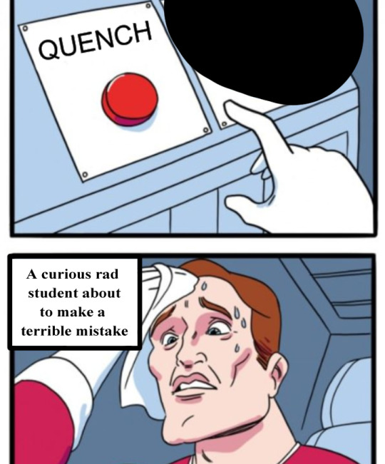
Text
memes for the tired rad student


17 notes
·
View notes
Text


[ID: 6 GIFs of MRI scans.
GIF 1: A bottom to top scan of the brain and tissue.
GIF 2: A bottom to top scan of the bone.
GIF 3: A slower bottom to top scan of the brain and tissue.
GIF 4: A front to back scan of the brain and tissue.
GIF 5: A left to right scan of the brain and tissue.
GIF 6: A scan of a skull rotating on a vertical axis.
End ID]
mri scans :•]
my video, credit this post if you use elsewhere!
#tw medical#stim#stimblr#stimmy#medical#medical stim#mri scan#medical imaging#also the image ids were made with my limited medical knowledge but slight understanding of what i'm looking at;#so my apologies for the ''unprofessional'' terms and possibly incorrect descriptions of the scans '^_^#scopo gifs
82 notes
·
View notes
Text
Revelation Revolution
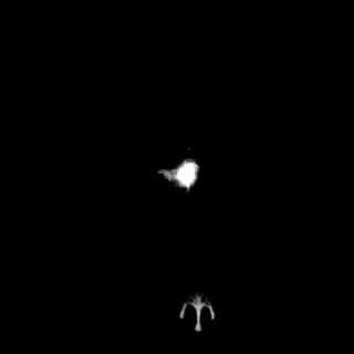
Whole-body positron emission tomography (PET) one hour after injection of radioactive glucose highlights rapidly metabolising tissue such as the liver metastases of a colorectal tumour seen here within the abdomen, along with normal accumulation of the tracer in the heart, bladder, kidneys and brain
Edward J Hoffman – born on this day, January 1st in 1942 – along with Michel Ter-Pogossian and Michael E. Phelps – developed the first human Positron Emission Tomography scanner in 1973
Movie adapted from PET scan GIF created by Jens Maus
Video in the Public Domain
You can also follow BPoD on Instagram, Twitter and Facebook
#science#biomedicine#biology#medical imaging#colorectal cancer#metastasis#liver#PET scan#positron emission tomography#born on this day
17 notes
·
View notes
Photo

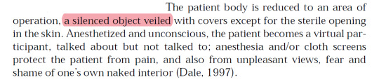
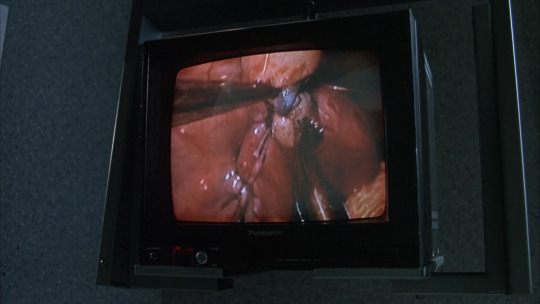
The once private area of the inner body has transformed into a public space of sightseeing
José van Dijck Bodies without borders
Dead Ringers (David Cronenberg | 1988)
#dead ringers#david cronenberg#josé van dijck#bodies without borders#/gore#surgery#medical imaging#biopolitics#insides#eye#knives
64 notes
·
View notes
Text
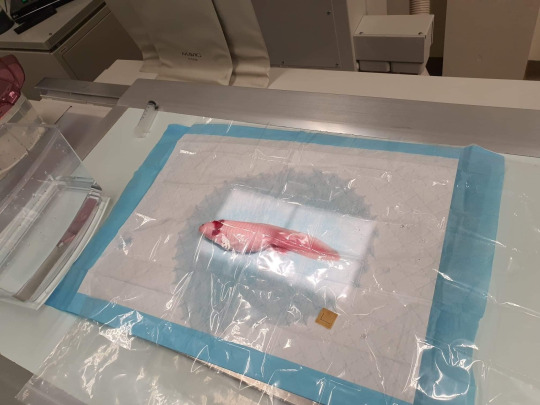

lotl getting a scan, ohto be damp and laminated
#axolotl#photos of animals having medical imaging or procedures done that look silly are far and few between here#medical imaging#animals#animal memes#funny animals#cute
17 notes
·
View notes
Text


mothapparel
121 notes
·
View notes
Note
hi! there’s something i've been wondering about for a long time and hope you can help me out with. i understand that x-rays/ct scans/mri/etc are important to be able to determine the extent of injuries and such but in an emergency situation, is there time for those procedures? or are there other/quicker ways to accurately determine what is going on and how to deal with it? i hope you can answer this for me because it has been on my mind for so long now…
There's generally a lot more time than you think in emergency situations. Or time can be created by giving fluids or intubating a patient, or a number of other interventions.
There are a finite number of situations where a time delay of a minute or two is actually critically important. These are emergencies like an airway blockage (choking, swelling, trauma to the airway, etc..), lack of breathing, extreme blood loss (spurting arterial blood), or the heart not pumping blood.
All of the above can be assessed and supported without imaging. You can intubate someone with an airway problem, intubate and/or provide rescue breathing for someone who is not breathing, put pressure or a tourniquet on someone who is bleeding out, and perform CPR on someone who's heart is not pumping until you can solve the problem that is causing the heart to not pump (or determine that you cannot solve this problem).
Everything else, including heart attacks, strokes, large-scale trauma, and many other things, can spare the time to do EKGs, x-rays, CT scans, and ultrasounds. MRIs are typically not done in emergencies because ain't nobody got time for that, like, ever.
X-rays take literal seconds. You can do them in a brief enough pause in CPR to make them worth it. It's literally "pause CPR-turn the patient-shove the x-ray pad under the patient-roll pt back- snap the pic-roll patient-remove x-ray pad-roll patient-keep doing CPR". The pic can be evaluated right on the machine less than a second later.
You can do ultrasounds literally while CPR is being done. No pauses needed. They can be read right there.
You can set up for a 12-lead EKG while CPR is being done, pause for like 6 seconds while it's being recorded, and go right back at it.
CT scans take minutes, so you're probably not going to do them during CPR, but you would for a stroke or major trauma because it can give a lot of info very quickly that can help make decisions.
Now, modern imaging is really important for a lot of reasons. It's faster and more specific in a lot of cases than other methods, and most modern docs really genuinely don't have the training to do their jobs without them (with the exceptions of very rural or austere practitioners) even when there are technically other ways to get similar information.
HOWEVER, since you happened to ask someone who is a literal physical diagnosis (the practice of diagnosing through history and physical examination) and wilderness first aid instructor, I can say that a lot can *technically* be done without imaging at all.
You can get really close to knowing where a clot in the brain is based on physical exam (something that would generally be done (honestly faster) with a CT scan). You can very accurately tell how bad a pneumothorax or hemothorax is and figure out where to put a chest tube through percussion and physical maneuvers (something you'd generally use a CT or x-ray for). You can locate and treat a life-threatening cardiac tamponade with a long needle and a 3-lead EKG, after diagnosing it with physical exam (something you'd otherwise need ultrasound for). You can tell a bone is broken and what muscles were impacted and whether or not it actually needs surgery with physical exam (usually done with x-ray or CT or even MRI later). You can tell that a problem is appendicitis and even locate where an inflamed appendix is with physical maneuvers (usually done with an ultrasound or CT). These and so many, many more things.
We typically do imaging because it is faster and can (in theory) be more accurate. In the last 40 or so years, practitioners have not been trained to be particularly accurate with physical diagnosis- just trained enough to realize there is a problem that can be further elucidated with imaging. This training takes a LOT of time to be particularly skilled at, and there is already too much information crammed into 4 years of medical school and 3-7 years of residency, and much of what is prioritized is not physical diagnosis (okay, I will get off my dang soap box).
We also do a lot of imaging because insurance expects it and generally feels it is less likely to miss something than physical diagnosis (which I believe is more a problem with training than actual accuracy of the medium). Unfortunately, in prioritizing interpretation of imaging over performance and interpretation of physical exam, we have created an extremely expensive model of diagnosis. The time you'd have to pay a practitioner for to do even a very detailed physical exam costs a hell of a lot less than the price of a single CT scan.
But from the 1910s through 1980s (when computers got good enough to do advanced imaging efficiently), we were pretty dang good at using physical diagnosis exclusively or backed up with more basic (and cheaper) tests like EKGs and x-rays. But like I said above, I'll get off my soap box.
-Ross @macgyvermedical
#whump reference#writing reference#injury#hospital life#medical education#physical diagnosis#medical imaging#medblr#nurblr
128 notes
·
View notes
Text
*activates MRI machine*
“Commence primary ignition!”
#dougie rambles#personal stuff#mri scan#MRI machine#cat scan#magnetic resonance imaging#medical imaging#radiology#biomagnetics#no context#my poor attempt at a joke#what#this sounded funnier in my head#shitpost#weaponised#radiation#this kills the man#star wars#death star
4 notes
·
View notes
Text
“And, remember, if you accidentally press something you didn’t mean to, or are completely lost, the panic button is the 2D mode.”
About ultrasound.
26 notes
·
View notes
Text
Updated ultrasound 🥲 Follow-up next week so they can decide surgery approach.


10 notes
·
View notes
Text
I go to med school and on a daily basis we are given the choice to wear scrubs or business cas and it still amazes me that people would willingly choose to wear business cas over literal pajamas
#funny#joke#med school#medical school#business casual#scrubs#I love scrubs#X-ray#sonography#medical imaging
7 notes
·
View notes
Text
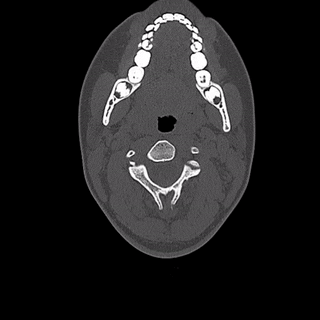
A note on reading Axial 'Transverse' CT slices!
First you gotta imagine your patient standing in front of you (i.e., your left is their right)
Then... you knock 'em over
Typical Axial CT slices look 'up' the patient, cranially, from their feet towards their head!

(disclaimer: please do not actually knock your patients over)
7 notes
·
View notes
Text

Seven Shots at Close Range — Stela Mandel
Painting (mixed media), 36" x 18" x 1.5", 08/23/2020
Presented at The deYoung Open 2023 (ID #50)
Listed for $1800 *
Artist statement:
In my mixed media work, I use medical imaging such as X-rays and MRIs, as a point of departure, sometimes serving as a metaphor for looking under the surface. I combine these intricate, mysterious images with other materials, attempting to reveal or tell a story.
On August 23, 2020, Jacob Blake, a 29-year-old African-American man, was shot and seriously injured by police in Kenosha, Wisconsin, as his children sat in his car. In the midst of a pandemic we watched videos of this shooting and those of other young black men. I was afraid that Jacob Blake’s story would be lost in the noise of the news. "Seven Shots at Close Range" is my attempt to memorialize this tragedy and failure.
Stela Mandel is a scientific illustrator and multimedia artist based in Sausalito, CA. To learn more about her life and work, view this image in greater detail, and view her other creations, visit stelamandel.com . You can also find her on Instagram @/stelamandel or on her Facebook page.
The deYoung Open is a triannual exhibition featuring artwork by California-Bay Area creators. The most recent exhibit was on display at The deYoung Museum in San Francisco from 09/30/2023 to 01/07/2024. To learn more and view a digital gallery of all 883 pieces that were featured, visit deyoungopen.artcall.org . And if you're an artist from Alameda, Contra Costa, Marin, Napa, San Francisco, San Mateo, Santa Clara, Solano, or Sonoma County, you should consider making a submission for 2026! Applications will probably go live in early June 2026, so you have some time to plan :)
I am not an affiliate of The deYoung Museum, Stela Mandel, or any of the artists featured in The deYoung Open 2023. I'm just posting to celebrate some amazing CA artists. If you are the artist and would like me to take this post down or add additional credit, please message me on Tumblr.
* Listing price is shown on the deYoung Open website at time of writing. The artwork may no longer be available for sale.
#The deYoung Open 2023#Seven Shots at Close Range#Stela Mandel#medical#multimedia art#scientific illustration#medical illustration#medical imaging#xray#police brutality#california artist#bay area artist#california#bay area#san francisco#the deYoung museum
2 notes
·
View notes
Text

Next Generation Images
A new 7 Tesla ultra-high resolution MRI scanner takes human neuroscience imaging to the next level
Read the published research article here
Erwin Hahn 7T MRI Laboratory, Henry H. Wheeler Brain Imaging Center, Helen Wills Neuroscience Institute, University of California, Berkeley, Berkeley, CA, USA
Image originally published with a Creative Commons Attribution 4.0 International (CC BY 4.0)
Published in Nature Methods, November 2023
You can also follow BPoD on Instagram, Twitter and Facebook
#science#biomedicine#neuroscience#biology#mri scan#mri#magnetic resonance imaging#medical imaging#next generation#brains#brain scan
7 notes
·
View notes
Last Seen Blogs

cryingtearsofjamjam
Lovergirl Jullia

thyshadowwriter
after hour writer

yedits-blog
Icons
photos-of-everyday-life
Photos of Everyday Life