#ml 5.02
Photo


11K notes
·
View notes
Text

WHY IS NO ONE TALKING ABOUT BUSTIER FINALLY SHOWING?!?!?!? LIKE THIS WOMAN TOOK FORVER TO ACTUALLY SHOW OMGOMFOMF
@thelibraryloser
#miraculous ladybug spoilers#season 5#multiplication spoilers#omg omg omg#it actually happened#i cant believe this#ml 5.02
37 notes
·
View notes
Text
0 notes
Note
so i’m going to my first appointment to start t soon and i know i need to do blood work but i wanna known if the blood work looks for anything other than testosterone levels and things of that nature?? cause i still live with my family and a regular blood test would show i have the in my system and i really can’t have that happening. thank you
Lee says:
They do look for things other than your testosterone levels, but I think they test for general health things. It isn’t the same thing as a drug test, unless your endocrinologist added one of those on to your blood work orders.
Your endocrinologist can order specific things to be tested if they’re worried about something- after one of my blood work results came in they were worried because my thyroid hormone was really low and I didn’t even know they were testing for that.
I’ll tell you the words listed on my most recent blood work results:
MCH 31.3 pg
MCVL 90 fL
Absolute basophils 22 cells/uL
Platelet count 167 thousand/uL
Absolute eosinophils 70 cells/uL
Eosinophils 1.6%
White blood cell count 44 thousand/uL
Basophils 0.5%
MPV 11.3 fL
Absolute neutrophils 24446 cells/uL
Red blood cell count 5.02 million/uL
Hemoglobin 15.7 g/dL
MCHC 34.7 g/dL
Monocytes 6.8%
Hematocrit 45.2%
Absolute lymphocytes 1562 cells/uL
RDW 12.2%
Absolute monocytes 299 cells/uL
HDL cholesterol 47 mg/dL
CHOL/HDLC ratio 3.8 (calc)
ALT 7 U/L
Alkaline phosphatase 87 U/L
Creatinine 1.01 mg/dL
BUN/Creatinine ratio 12 (calc)
Glucose (86 mg/dL)
eGFR African American 93 mL/min/1.73m2
Globulin 2.1 g/dL (calc)
Bilirubin, total 0.7 mg/dL
Calcium 10 mg/dL
Chloride 106 mmol/L
Potassium 4 mmol/L
Albumin 4.8 g/dL
Sodium 140 mmol/L
Albumin/Globulin ratio 3.3 (calc)
Carbon dioxide 27 mmol/L
AST 12 U/L
Protein, total 6.9 g/dL
Urea Nitrogen (BUN) 12 mg/dL
LDL-cholesterol 116 mg/dL (calc)
TSH w/reflex to FT4 1/95 mIU/L
Triglycerides 67 mg/dL
Cholesterol, total 179 mg/dL
Estradiol, ultrasensitive LC/MS 23 pg/mL
Testosterone, total, MS 512 ng/dL
Vitamin D, 25-OH, total, IA 28 ng/mL
Non HDL cholesterol 132 mg/dL
Lymphocytes 35.5%
Neutrophils 55.6%
So that’s 48 different things that they tested for when I went in for my routine blood work for testosterone. I think they do your hormone levels and a standard metabolic panel, but they aren’t testing for drugs.
#Lee says#blood work#bloodwork#testosterone#Anonymous#transgenderteensurvivalguide#trans#transgender
29 notes
·
View notes
Text
Biomed Grid | Nutraceutical Potential of Parka Speciose (Stink Bean): A Current Review
Abstract
Background: Inadequate fruits and vegetables intake contributes to the prevalence of major diseases such as cardiovascular diseases and cancers. Parkia speciosa [stink bean] is a common vegetable consumed in the Southeast Asia. Although it contains various phytochemicals that can help prevent disease development, the effort to develop a specific treatment or food products from Parkia speciosa remains a challenge. Here, we explore research works of the medicinal benefits of P. speciosa that can be used as a guide to develop future clinical studies.
Method: We conducted a database search on PubMed, Google Scholar, and Science Direct using the keywords “medicinal benefits”, “Parkia speciosa” “antioxidant”, “hypoglycemic”, “antitumor”, “antimicrobial” and “cardiovascular effects”. We included clinical trial, in vitro and in vivo studies that were written in English or Malay; and excluded review articles with no time limitations.
Result: We reviewed a total of 28 research articles. No clinical trial was found. The articles were grouped into antioxidative, hypoglycemic, antitumor, antimicrobial and cardiovascular effects. Six articles had combination of the medicinal properties. Seeds and empty pods are the most common plants parts used. Each bioactivities differed depending on the plant parts, extracts, methods, cultivar and plantation site.
Conclusion: P. speciosa demonstrated antioxidative, hypoglycemic, antitumor, antimicrobial and cardiovascular effects that were contributed by its phytochemical compounds. This finding could be used as a database for future clinical studies. We recommended researchers to use the information from the articles reviewed for drug development and clinical trial.
Keywords: Medicinal Benefits; Parkia Speciosa; Antioxidant, Hypoglycemic; Antitumor; Antimicrobial; Cardiovascular Effects.
Background
P. speciosa or stink bean is a classic Malaysian favourite and is commonly grown and cultivated in Southeast Asia e.g. Indonesia, Malaysia, and some parts of North-Eastern India [1]. P. speciosa is also known as ‘petai’ in Malaysia, Singapore, and Indonesia [2], ‘sator’ or ‘sataw’ in Thailand [3], ‘u’pang’ in Philippines [2], and ‘yongchak’ in India [4]. P. speciosa earned its nickname ‘stink bean’ from its strong and pungent odour. The plant belongs to the pea or bean family Fabaceae and placed in Leguminosae and Mimosaceae [4]. P. speciosa contained several phytochemical compounds such as polyphenols and flavonoids [5,6], terpenoids [5,7,8], Alkaloids [5,7-10], saponins [7-10], steroids [7,8,10], tannins [7,9] and phytosterol [11,12]. Globally, the World Health Organization [WHO] reported that about 80% of the world population relies on traditional med icine to cure ailments [13]. In Malaysia, P. speciosa has been used traditionally to treat various diseases and ailments such as hypertension [4] and kidney disorders [14]. There is a limited data of medicinal benefits of P. speciosa especially on the cardiovascular and antioxidant effects. From the previous review articles, only two [15,16] research experiments were included for cardiovascular effects; and only seven [15] and nine [16] research experiments were included for antioxidant effects. The findings of the current review article could be used as a database for future clinical studies. This review aimed to explore the medicinal benefits of P. speciosa as to provide the foundational knowledge on the topic; so that the preclinical and clinical studies can be conducted for future drug development.
Methods
We conducted a literature search for medicinal benefits of P. speciosa using PubMed, Google Scholar, and Science Direct using the keywords “medicinal benefits”, “Parkia speciosa” “antioxidant”, “hypoglycemic”, “antitumor”, “antimicrobial” and “cardiovascular effects”. We included clinical trials, in vitro and in vivo studies; and exclude review articles. The literature search was limited to articles published in Malay and English without time limitations.
Results and Discussion
We found a total of 28 eligible articles: 23 in vitro and 5 in vivo studies. No clinical trial was found on this topic. Of the articles reviewed, 14 articles reported on antioxidant activity, 5 articles on hypoglycemic activity, 5 articles on antitumor activity, 6 articles on antimicrobial activity and 4 articles on cardiovascular effects. There were combinations of bioactivities studied in six articles reviewed: one article documented on the antioxidant, hypoglycemic and antimicrobial activities [6], one article performed research on antioxidant and antitumor effects [17], one article reported on antioxidant and antimicrobial effects [18], two studies described both the antioxidant and antimicrobial activities [5,18] and one study combined antioxidant and cardiovascular effects [19].
Antioxidant activity
A total of 13 in vitro and 1 in vivo study on the antioxidative property of P. speciosa were reviewed. In most of the studies cited, the seeds of P. speciosa were used [5,19-22] (Table 1). Other studies analysed empty pods [6,17,23,24], pods [9,18] and seed coat and pericarp of the P. speciosa bean [25]. The commonly used tests for antioxidative activity are 1,1-diphenyl-2-picrylhydrazyl free radical [DPPH] scavenging and reducing ferric ion antioxidant potential [FRAP] assay. Other assays used are anti-lipid peroxidation, superoxide radical scavenging activity, 2,2’-azino-bis [3-ethylbenthiazoline- 6-sulfonic acid] [ABTS] radical scavenging activity and metal chelating activity. The relationship between total phenolic and flavonoid contents with antioxidant level was evaluated in four studies [5,20,22,26]. Two studies examined the antioxidant level indirectly [21,25], whereby the antioxidant level was related to hydrogen sulphide [H4S] in one study, and the other to Heinz body inhibition [aggregation of denatured hemoglobin in the red blood cell resulting from oxidative process]. Four studies compared the antioxidant level between P. speciosa and other plants [20,22,26]. Generally, the antioxidant level varies depending on the part of the plant, the extraction variables [solvent-extract ratio, time, temperature and solvent type] and the plantation location.
In vitro studies
Table 1: List of Abbreviations.
In a study by Balaji and co-workers [9], the flavonoids and phenolic content, as well as the antioxidant activity were evaluated. The flavonoids content estimation was carried out using the calorimetric method, while the phenolic content was carried out using the Folin-Ciocalteu reagent method. Reducing power assay and DPPH were used to examine the antioxidant activity. The study found that P. speciosa pod powder extract had 14.16±0.02 mg gallic acid equivalents per gram [GAE/g] dry weight total phenolic content and 5.28±0.03 mg rutin equivalents per gram [RE/g] dry weight total flavonoid content. Generally, the phenolic and flavonoid compounds are related to antioxidant activity of the plant. The extract also showed potent antioxidant activity as demonstrated by IC50 values of the extract in DPPH, 74.37μg/ml compared to the standard butylatedhydroxy toluene [BHT] 35.40 μg/ml. IC50 indicates the concentration of test extracts or positive controls that inhibit or scavenge the radical formation by 50% [23].
Although the DPPH radical scavenging activity was lower than BHT, it was evident that the extract contained a substance/s with proton-donating ability and was capable of inhibiting free radicals. Ko and coleague [23] evaluated the antioxidant activities of aqueous and ethanolic extracts of P. speciosa empty pods using several assays namely: anti-lipid peroxidation, superoxide radical scavenging activity, DPPH radical scavenging activity, ABTS radical scavenging, metal chelating and reducing power. It showed that the ethanol extracts possessed stronger antioxidant activity via all the assays except for superoxide radical scavenging activity. This finding is influenced by a higher level of polyphenols [phenols and flavonoids] in the ethanolic extract. The anti-lipid peroxidation IC50 was 5.02±1.06 μg/ml, DPPH radical scavenging activity was 64.2±3.46 μg/ml, ABTS radical scavenging 19.6±0.44 μg/ml, metal chelating activity 319 ± 26.3μg/ml and reducing power activity 274±16.1μg/ml. The extracts contained several polyphenolic constituents, the most abundant of which were gallic acid, catechin, ellagic acid and quercetin. Two types of P. speciosa in Thailand [‘Sataw- Khao’ and ‘Sataw-Dan’] were studied by Wonghirundecha and co-workers [18] for their total phenolic content, antioxidant and antimicrobial activities. It was found that the extraction yield, total phenolic and total flavonoid contents of ‘Sataw-Dan’ pod extracts were higher than that of ‘Sataw-Khao’ pod extracts. In contrast, ‘Sataw- Khao’ pod extracts showed higher DPPH [1218.07± 8.72 μmol Trolox equivalent/g dry weight [TE/g] vs 920.32±6.15 μmol TE/g dry weight], ABTS radical scavenging activity [1610.67±11.88μmol TE/g dry weight vs 1261.14±17.44μmol TE/g dry weight] and metal ion chelating activity [9.76±0.03 Ethylenediaminetetraacetic acid equivalent/g dry weight [EDTAE/g] vs. 5.86±0.02 EDTAE/g dry weight] compared to Sataw-Dan pod extracts.
Thus, the authors concluded that there was no relationship between total phenolic content and antioxidant activity in the PS extract, suggesting that other phytochemicals apart from polyphenols may contribute to the antioxidant activity. In addition, both extracts showed antimicrobial effect in the form of inhibition zone formation through the agar well diffusion assay. In a study by Aisha and co-workers [17], eight empty P. speciosa pod extracts were examined for phenolic content and antioxidant, cytotoxic and antiangiogenic activities. The results of the study showed that the methanolic sub-extract had the highest total phenolic content, 25.55± 1.57 GAE/100 mg. In the DPPH scavenging assay, antioxidant activity was also highest in the methanolic sub-extract, [IC50 26±3.0μg/ ml]. It can be visualized from all the extracts used that; the antioxidant level corresponded to the total phenols content.
Sonia and co-workers [6] further explored the antioxidant, anti- inflammatory, anti- diabetic and anti-microbial activity of P speciosa. The FRAP value of the hydromethanolic extract was higher than of the ascorbic acid controls (1.9mM Ferrous sulfate (FESO4)). Its strong antioxidant activity was supported by DPPH study, as it significantly decreased in vitro DPPH radical concentration (64.52±2.4 % inhibition, IC50315.75 μg/ml), and hydrogen peroxide (H4O2) assay [78.06 ± 5.7 % inhibition, IC50 166.3μg/ml]. The extract exhibited anti-inflammatory properties via inhibition of lipooxygenase activity (38.6±10.2 % inhibition, IC50 493.34 μg/ml; control 56.9 ± 11.4% inhibition , IC50 280.71 μg/ml], proteinase inhibitory activity (22.78±3.6 % inhibition, IC50 1142.3μg/ml; control 29.9±5.9 % inhibition, IC50 53.75 μg/ml ) and RBC membrane stabilization activity (99.21±12.6% inhibition, IC50 67.01μg/ml ;control 99.36±6.7 % inhibition, IC50 53.75μg/ml).
There is increasing demand for new ingredients from natural sources in the food industry. Moreover, there are suggestions to use agrowaste materials [in this case, P. speciosa empty pods] as functional food ingredients. Gan and Aishah [24] studied the physicochemical properties’ characterization, looking at the antioxidant property of P. speciosa as potential functional flour. Between two method of drying [freeze dried and oven dried], different functional properties can be seen. Higher antioxidative properties were shown in freeze dried P. speciosa pod (FDPSP) which consist total phenolic content (TPC) of 110.0mg GAE/g sample and total flavonoid content (TFC) of 8.5 mg pyrocatechol equivalents/g sample. These extracts [pre-diluted 50x] gave %DPPHsc, %ABTSsc and FRAP values of 65.3%, 77.4% and 1.9mM FESO4, respectively. Therefore, P. speciosa powder can be used as a substitute for commercial flour. Five Malay raw salads namely: leaves of Cosmos caudatus (‘Ulam Raja’), Oenanthe javanica (‘Selom’), Murraya koenigii [curry leaf], Centella asiatica (‘Pegaga) and the seeds of P. speciosa (‘Petai’) were studied by Reihani and Azhar [20] for their total phenolic content and antioxidant activities. The total phenolic content [mg GAE/g of plant on dry basis] were highest in Murraya koenigii (33.18) and lowest in P. speciosa (6.45). Pertaining to antioxidant property, Cosmos caudatus had the highest DPPH free radical scavenging activity (212.8) and Centella asiatica the lowest (32.4).
Oenanthe javanica and Cosmos caudatus demonstrated the highest ferric reducing activities: 199.96 μmol TE/g and 183.11μmol TE/g, respectively; while P. speciosa showed the lowest (44.67 μmol TE/g). It was found in this study that there was no significant correlation between antioxidant activity and total phenolic content. This may be due to steric hindrance and presence of other reducing agents in the extracts studied. Siow and Gan [19] examined the antioxidative bioactive peptides from P. speciosa seeds using alcalase. Prior to extraction, the P. speciosa seeds protein hydrolysate was analysed for amino acid composition. Glutamine and aspartic acid were found in the highest amounts, followed by cysteine (154, 132, 84.8 per 1000 amino acid residuals, respectively). Glutamine and aspartic acid are strong antioxidants as they act as electron donors [27]. In this study, the effects of temperature, substrate-to-enzyme ratio and incubation time were taken into consideration.
The highest DPPH free radical scavenging activity and FRAP activity (2.9 mg GAE/g and 11.7mM FeSO4, respectively) were demonstrated in a condition of 50℃, substrate/enzyme (S/E) ratio of 50 and 2hours incubation time. The high temperature causes unfolding of the protein molecules thus making the protein active site more accessible to the enzyme and exposes the protein donating residues. Partial hydrolysis of the proteins is essential to give higher DPPH value, compared to extensive protein hydrolysis caused by higher enzyme concentration. The DPPH values increased along with the incubation time. Upon fractionation of the protein hydrolysates that had the highest DPPH free radical scavenging activity and FRAP activity, the peptide fraction of <10k Da showed the strongest bioactivities. Using advanced mass spectrometry, 29 peptide sequences were identified to be responsible for the potent bioactivities, that could be developed into novel nutraceuticals.
The production of phytochemicals in plants depends on the variety or species and external variables such as environmental conditions, agricultural practices and post-harvest handling [5]. A study by Ghasemzadeh and co-workers [5] compared the phytochemical constituents and biological activities of P. speciosa collected from 3 regions of Malaysia [Perak, Negeri Sembilan and Johor]. The result showed that the seeds (ethanol extract) collected from Perak contained highest phytochemical content at concentration of 100 μg/ mL, DPPH (66.29%) and FRAP (522.1 μM of Fe (II)/g) scavenging activity, followed by Negeri Sembilan and Johor. The authors analyzed the correlation between the parameters studied and found that the antioxidative activity was significantly correlated with flavonoid content. In another study, Maisuthisakul and co-workers
[26] analyzed the correlation between parameters [yield, radical scavenging activity 1/EC50, phenolic compounds, flavonoids, moisture, ash, protein, fat, carbohydrate, dietary fibre, calcium, iron and vitamin C] of 28 Thai plants. EC50 is the concentration of extract necessary to decrease DPPH radical scavenging by 50%. Generally, the seeds of the plants studied had high energy, and P. speciosa seeds were among the plants with the highest energy content (441.5 Kcal/100g edible portion, db). It showed antiradical activity of 1.5 (1/EC50), total phenolics of 51.9 mg GAE/g db and total phenolics of 20.3mg RE/g db.
The yield of extractable compounds ranged from 0.6% to 4.5%. The yields from berries, fruit and seeds were higher than from other parts of the plants. In terms of correlation analysis, plants with high yield had high carbohydrate content and low fibre content. A high yield did not correspond to high content of phenolic compounds and high antioxidant activity. The study also revealed that plants with strong antioxidant activity had high total phenolic and flavonoid contents. Interestingly, Liang and co-workers [21] investigated the hydrogen sulphide [H4S] releasing capacity of ten organosulphur rich fruits and vegetables [garlic, red onion, yellow onion, scallion, shallot, leek, spring onion, Chinese chives, durian and stinky beans]. H4S is a gaseous signaling molecule that has several effects on human health including antioxidant effect [28]. To demonstrate the importance of this gas, the authors quoted previous studies to investigate how garlic has been used as an herbal remedy for thousands of years and having cardioprotective effect. Benavides and co-workers [29] from their work in rats proposed that the two major components of garlic extract, namely diallyl trisulphide [DATS] and diallyl disulphide [DADS] are converted to H4S resulting in relaxation of the rat aorta ring.
Similarly, Chuah and co-workers [30] showed that through H4S-dependent mechanism, S-allyl cysteine, the major compound of aged garlic was able to protect against myocardial infection. In the study by Dong Liang and colleagues [21], the H4S releasing capacity of plant essential oils was evaluated by fluorescent method using BCu [H4S selective and sensitive turn- on fluorescent probe] as probe and human breast cancer MCF- 7 cells as the medium. MCF-7 cells incubated with BCu for 3 hours were treated with H4S donors to produce H4S. Fluorescence produced by H4S captured by the probe was measured by a microplate reader. The results showed that P. speciosa had the highest DATS equivalent (DATS-E) value (158 mmol DATS/kg of raw material), followed by garlic (18.5mmol DATS/kg of raw material) and yellow onion (4.59 mmol DATS/kg of raw material).
DATS-E value is a concept introduced in which each plate was divided into two zones; one zone loaded with different concentrations of samples, and the other zone loaded with different concentration of DATS. This method was performed to enable data comparison among different groups. Among the reasons P. speciosa had the highest DATS- E value was because of its high sulphur content, and presumably its cyclic sulphur compounds could be converted to H4S more efficiently than linear compounds. This study indirectly relates the presence of H4S in P. speciosa to antioxidant bioactivities. Twentyfive edible tropical plants were examined by Wong and co-workers [22] for their antioxidant properties. The total polyphenol contents, free radical scavenging, ferric ion reducing (expressed as TEAC-Trolox equivalent antioxidant capacity) and cupric ion chelating capabilities (CCA) were determined. The result showed that P. speciosa leaves extract had the highest TPC but showed low TEACDPPH and TEACFRAP. The non-correspondence between TPC and antioxidant activities is due to the fact that P. speciosa may contain other compounds that can oxidize the Folin-Ciocalteu, other than polyphenols. In addition, P. speciosa leaf extract chelated the cupric ions the most. This finding indicated that the extract had potential secondary compounds. TPC showed satisfactory correlation with TEACDPPH and TEACFRAP, indicating that polyphenols in the extracts were partly responsible for the antioxidant activities. Conversely, TPC correlated poorly with CCA, indicating that polyphenols might not be the main cupric ion chelators.
Indirect relationship between inhibitory activity of Heinz body induction and antioxidant activity was studied by Tunsaringkarn and co-workers [25]. Hemoglobin chains undergo denaturation process through oxidative damage by reactive oxygen species and produced Heinz body, an aggregation of denatured and precipitated hemoglobin within red blood cells [31]. Hence it can be a biomarker for oxidative damage in the body. In this study, P. speciosa seed coat extracts showed the highest activity of Heinz body inhibition (39.98%, IC25 2.68 mg/ml), followed by X. xylocarpa bark (42.75%, IC25 15.71 mg/ml), P. speciosa Hassk. pericarp (44.89%, IC25 28.14 mg/ml) and E. rheedii Spreng.seed coat (55.12%, IC25 33.20 mg/ ml) X. xylocarpa bark, E. rheedii seed coat, Hassk seed coat, and X. xylocarpa stem contained high tannin concentration. It has been shown that the percentage of Heinz body inhibition was correlated to tannin concentration. The previous study suggested that the antioxidant activity of tannin was mainly due to iron chelation rather than hydroxide radical scavenging [32]. This finding also supported the results of the study by Wong and co-workers [22] discussed before.
In vivo study
Al Batran and co-workers [1] studied antioxidant and antiulcer activity of P. speciosa ethanolic leaf extract in ethanol-induced gastric ulcer in rats. Sprague Dawley rats were divided into 7 groups: Groups 1 and 2 received 0.5% carboxymethylcellulose (CMC) as vehicle; Group 3 received 20mg/kg omeprazole and groups 4-7 received ethanolic leaves extract of P. speciosa at doses of 50, 100, 200 or 400 mg/kg. After 1 hour, CMC or absolute ethanol was administered to groups 2-7. The rats were euthanized after 1 hour and the gastric mucosa was examined for gastric juice acidity, gastric wall mucus, macroscopic gastric lesion evaluation, antioxidant activity, histological examination and immunohistochemical staining. Regarding the antioxidant activity, gastric tissue homogenate prepared from the groups that were treated with plant extract displayed significant antioxidant activity, with decreased levels of malondialdehyde (MDA) and elevated levels of total glutathione (GSH) and superoxide dismutase (SOD), in response to oxidative stress due to ethanol treatment.
MDA is the final product of lipid peroxidation which causes a loss of membrane fluidity, impaired ion transport and membrane integrity resulting in loss of cellular function [33]. It was found that the groups treated with the plant extract reduced this process. GSH plays a role in determining ulcer severity and acts as tissue protective agent [34,35]. SOD converts superoxide to hydrogen peroxide (H4O2), which is transformed into water by catalase in the lysosomes or by glutathione peroxidase in the mitochondria [36].
Hypoglycemic Activity
There were 5 articles that demonstrated the hypoglycemic property of P. speciosa, 2 in vitro and another 3 in vivo studies. One of the in vitro hypoglycemic studies has been discussed earlier by Sonia and co-workers [6] as part of their study. Two studies examined empty pods and seeds separately [37,38], one study examined seeds and pericarp separately [39] and two studies examined empty pod [6,11]. Among the assays used were α-glucosidase inhibition activity, alpha amylase inhibition activity, and porcine pancreatic lipase (PPL) inhibition assay and glucose oxidase method. All five studies exhibited a strong hypoglycemic activity of the plant. Two studies explored the compounds responsible for the hypoglycemic activity [11,38]. There was some inconsistency on parts of the plant that gives greater hypoglycemic effect. Tunsaringkarn and colleagues [39] found that the P. speciosa pericarp had a higher hypoglycemic activity than the seeds, but Jamaluddin and colleagues [37,38] had opposite findings. This inconsistency could be due to the cultivar, harvesting time and method, types of extract and assays performed.
In vitro study
A study by Tunsaringkarn and colleagues [39] evaluated twenty species of Thai plants of Mimosaceae family. α- glucosidase is the enzyme responsible for digestion of polysaccharide and oligosaccharide to monosaccharides [40]. Using α-glucosidase inhibition assay, P. speciosa pericarp was one of the plants that showed high inhibition activity; IC50 0.0581 mg/ml (89.46%). As a comparison, other plants had α-glucosidase inhibition of: Entada rheedii seed coat IC50 0.0043, Archidendron jiringa seed coat IC50 0.0054, Albizia lebbeck branch bark IC50 0.0397 and Albizia lebbeckoides bark IC50 0.0702mg/ml. P. speciosa seed showed lower percentage of α- glucosidase inhibition activity (45.72%).
Sonia and co-workers [6] studied the anti- diabetic activity of P. speciosa empty pods via two assays: alpha amylase inhibition and porcine pancreatic lipase (PPL) inhibitory assay. The alpha amylase acts to hydrolyze dietary starch [maltose] to glucose. In this study, the researchers explored how the P. speciosa pod extract can inhibit alpha amylase resulting in reduction of post prandial hyperglycemia. It was found that P. speciosa showed a maximum inhibition of 79.2% at 500ug/ml. The extract exhibited anti- diabetic property as the IC50 of P. speciosa were found to be 199.29μg/ml compared to standard acarbose (324.18μg/ml). From another aspect, pancreatic lipase functions to digest dietary fat, specifically hydrolyzing triacylglycerol to 2-monoacylglycerol and fatty acids. Through PPL inhibitory assay, the maximum inhibitory activity of P. speciosa extract at 500ug/ml was 89.5%; IC50196.61 μg/ml compared to Orlistat, 76.3%; IC50227.27μg/ml.
In vivo studies
Jamaludin & Mohamed [37] studied the hypoglycemic effect of P. speciosa extracts using glucose oxidase method. In this study, healthy Sprague Dawley rats were induced to be diabetic via intravenous injection of 60mg/kg alloxan. The two groups of normal and alloxan-induced diabetic rats were given the P. speciosa extract in the range of 25-500mg extract/ kg body weight (BW), together with 1g glucose/kg BW of rat. The result showed that only the chloroform extracts from both the empty pods and seeds had a strong hypoglycemic activity on diabetic rats. The seed had a higher hypoglycemic activity than the pod. The reduction of blood glucose in alloxan diabetic rats given 0.4g/ kg P. speciosa pod and seed were 36% and 57%, respectively. The hypoglycemic action took place as early as within an hour, best after 2 hours and lasted for at least 24 hours. The study also revealed the dose-response relationship of P. speciosa seed on blood glucose level. Maximum reduction of blood glucose was seen with 500mg/ kg BW [77% decrement]. Optimum dosage seen was 200mg/kg BW.
There was insignificant raise of blood glucose levels of normal rats fed with 0.4 g seed or pod extracts. As an extension of the above study, Jamaludin and co-workers [38] explored further on the actual compound contributing to the hypoglycemic effect of P. speciosa seeds. The hypoglycemic fraction S-9.4 was identified through chromatographic separation. The fraction showed 83% reduction of blood glucose at 100mg/kg BW, compared to 111% reduction of blood glucose at 5mg glibenclamide/kg BW. The minimum effective dose was 25mg/ kg BW. The fraction was found to contain mixture of β-sitosterol (66%) and stigmasterol (34%). Interestingly, the two sterols exhibited synergistic effect. No hypoglycemic effect produced when the sterols were tested individually. The similar author, Jamaludin and co-workers [11] identified component P-7.1 (stigmast- 4-en-3-one) as the compound responsible in producing hypoglycemic activity of pod extract. Compared to 111% blood glucose reduction by 5mg/ kg BW glibenclamide, 100mg/ kg BW of P-7.1 reduced the glucose levels by 84%. The minimum effective dose was 50mg empty pods/ kg. The extract at 400mg/ kg BW did not significantly change the blood glucose levels in normally fed rats.
Antitumor/Antimutagenicity
We found five articles highlighting the antitumor activity of the plant. Several terms including antimutagenicity, antiproliferation and antiangiogenic were combined as to describe antitumor activity. All of the articles were in vitro studies. One of the studies by Aisha and colleagues [17] has been mentioned earlier in antioxidant activity. Three articles used P. speciosa seed [41-43], one article used empty pods [17] and one article used pericarp and seed coat [44]. Various techniques have been used to demonstrate the antitumor activity such as Epstein-Barr virus (EBV) inhibitory assay [41], Ames preincubation method against Trp-P-1 in Salmonella typhimurium TA98 [42], thioproline determination [43], Peripheral Blood Mononuclear Cell (PBMC) culture and 2,3-bis-(2-methoxy- 4-nitro-5-sulfophenyl)-2H-tetrazolium-5-carboxanilide (XTT) cell proliferation test [44]; and using rat aortic rings, matrigel matrix with Human Umbilical Vein Endothelial Cells [HUVEC] and Vascular Endothelial Growth Factor (VEGF) level [17]. Effect of thermal processing to the antitumor activity was assessed in two studies [42,43]. All articles demonstrated positive antitumor activity of P. speciosa.
In vitro studies
Murakami and colleagues [41] studied the antitumor activities of 114 methanol extracts of edible Malaysia plants for their antitumor activity towards tumor promoter HPA-induced Epstein Barr Virus (EBV)activation in Raji cells. EBV activation was evaluated by detecting early antigen (EA) which was stained with high titre EA- positive sera from nasopharyngeal carcinoma patients. The results were ranked into 4 categories, based on inhibitory rate (IR) toward EBV activation and cell viability (CV): +++ strongly active (IR ≥70% and /or 50% >CV); ++ moderately active (70% > IR ≥ 50% and CV ≥ 50%); + weakly active (50% > IR ≥ 30% and CV ≥ 50%) and - inactive (30% > IR and CV ≥ 50%) P. speciosa showed weakly active inhibitory rate towards tumor promoter HPA-induced Epstein Barr Virus (EBV) activation, with 45% IR and>90% CV. Of the samples studied, 32% [37 samples] showed strongly active, 25% (28 samples) moderately active and 17% (19 samples) weakly active inhibitory action. Research study done by Tangkanakul and co-workers [42] examined 10 foods in central and southern Thailand containing local Thai vegetables. Methanol extracts of both the raw ingredients and homogenized foods were evaluated for antioxidant activity using DPPH scavenging assay and antimutagenic assay using Ames preincubation method (against Trp-P-1 in Salmonella typhimurium TA98).
Total phenolic content was determined using the Folin- Ciocalteau reagent. Raw P. speciosa showed antioxidant activity of 0.04 g vitamin C equivalent / 100g [VCE/100 g], total phenolic content of 0.13 g GAE/100 g and 31% inhibition against Trp-P-1. A strong antimutagenicity was demonstrated by galangal, lemongrass, wild betal, turmeric and kaffir lime leaf (87-97%). Both antioxidant activity and antimutagenicity increased after the plant was cooked. The antioxidant activity increased from 0.04 g VCE/100g to 0.14gVCE/100 g, and the the antimutagenic activity increased from 31%-69%. This result showed benefit of cooking to enhance both bioactivities. Thioproline or thiazolidine-4-carboxylic acid (TCA) is a sulphur-containing amino acid, produced from condensation of formaldehyde and cysteine. It was documented as an effective nitrite- trapping agent in human body, thus inhibiting the endogenous formation of carcinogenic N-nitroso compounds [45].
In this experiment, Suvachittanont and colleague [43] evaluated the formaldehyde, thiol and TCA level in various edible leguminous seeds in Thailand. The uncooked P. speciosa was found to have the highest level of formaldehyde [0.77 ± 0.07 mmol/100 g], which decreased upon boiling [0.25±0.18 mmol/100 g]. This observation could be due to volatilization of formaldehyde and formation of TCA upon boiling. Consistently, the TCA content of uncooked P. speciosa was <0.001 mmol/100 g dry beans and it increased to 0.14 ± 0.02 mmol/100 g dry beans after boiling. The highest level of TCA was found in Archidendron clypearia [‘Niang Nok’] both in uncooked and cooked status. In subsequent study, Tunsaringkarn and colleague [44] evaluated the antiproliferation activity of human white blood cells, Peripheral Blood Mononuclear Cell (PBMC) with 21 Thai Mimosaceous plant extracts. P. speciosa pericarp and seed coat were among plants with high inhibitory cell proliferation (17.17% and 12.16%, respectively).
The researchers correlated the high inhibitory effect with tannin level, which was also consistently high, 250mg/g in Parkia speciosa pericarp and 350mg/g in Parkia speciosa seed coat. This finding suggested tannin as potential cancer therapeutic agent. In the assessments of antiangiogenic activity, the methanolic extract and all its sub-extracts showed more than 50% inhibition of rat aortic microvessel outgrowth [17]. P speciosa extracts also inhibited tube formation on matrigel matrix involving HUVECs (Human Umbilical Vein Endothelial cells). Under light microscopy, the HUVECs treated with P. speciosa extracts showed formation of cytoplasmic vacuoles, which are markers of autophagy as a result of nutritional deprivation which is essential to maintain cell viability [46]. The vascular endothelial growth factor (VEGF) concentration of treated HUVECs was also reduced, (36± 2.2pg/ml) compared to 51±1.6 pg/ml in untreated cells. The extracts did not show acute toxicity.
Antimicrobial activity
We found six articles on this bioactivity, and all were in vitro studies. Three studies have also experimented on the antioxidant studies described earlier [5,6,18]. Main methods used in the experiments were agar-well diffusion assay and disc diffusion method.
In vitro studies
Uyub and co-workers [47] reported that the extracts of P. speciosa seeds in petroleum ether, chloroform, and methanol demonstrated antibacterial activity against Helicobacter pylori but none was found in the water extract. The activity was found highest in the chloroform extract followed by methanol and petroleum ether. In comparison, the chloroform extract of P. speciosa showed a moderate inhibition zone diameter to mg extract ratio (25.0), compared to other plant extracts where the ratios were in the range of 1.5- 117.5. In the study of Sakunpak and Panichayupakaranant [48], amongst the forty-four extracts of twenty-two Thai edible plants that were investigated for antibacterial activity using the disc diffusion method, the methanolic P. speciosa seed extract was found be able to inhibit Helicobacter pylori growth, while the ethyl acetate extract was effective against Escherichia coli.
These extracts, however, had no inhibitory effect on Salmonella typhimurium, Salmonella typhi, and Shigella sonnei growth. According to Gmelin and co-worker [49], two cyclic polysulfide compounds, hexathionine and trithiolane in the P. speciosa seeds are contributing to its antibacterial property. In Thailand, the ‘Sataw’ or P. speciosa pods extract could be potentially used as a natural preservative [18]. Both ‘Sataw-Khao’ and ‘Sataw-Dan’ pod extracts under investigated by the same group of researchers showed antimicrobial activity against food-borne pathogenic bacteria (Bacillus cereus, Listeria monocytogenes, Staphylococcus aureus, Escherichia coli, Salmonella typhimurium, Vibrio cholerae non O1/ non O139) and food spoilage bacteria (Aeromonas hydrophila, Pseudomonas aeruginosa, Serratia marcescens). Although there were no significant differences between ‘Sataw-Khao’ and ‘Sataw-Dan’ pod extracts against all the tested bacteria, but for Gram negative bacteria, the extracts exhibited a lower range of inhibition zone than Gram positive bacteria.
However, the result is not in favour for Vibrio cholerae which was the most susceptible strain in comparison with other tested Gram-negative bacteria. These results are in agreement with the findings of Musa and co-workers [50] who observed that P. speciosa extract could be effective against all Gram-positive bacteria [Streptococcus agalatiae, S. aeruginosa and S. aureus] and some Gram-negative bacteria (A. hydrophila and V. parahaemolyticus). However, the Gram-negative bacteria including Citrobacter freundii, Edwardsiella tarda, E. coli and V. alginolyticus were resisted to the extract. Similar findings were reported by Sonia and co-workers [6], whereby the methanolic extract of P. speciosa pod showed the inhibition against common pathogens [Staphylococcus aureus, Escherichia coli, Pseudomonas aeruginosa and Klebsiella pneumoniae]. The highest zone of inhibition was demonstrated for the gram-positive Staphylococcus aureus at 10 mm. The bioactive compounds are responsible to effectively inhibit and/or stop microbial growth via disruption of the synthesis of microbial nucleic acids, proteins and cell walls [51]. According to the previous studies, P. speciosa pod extract could inhibit all tested pathogenic and spoilage bacteria compared to rambutan peel, mangosteen peel, palmyra peel and coconut husk [52], rambutan peel and seed [53], pomelo peel [54] and banana peel [55] which could only inhibit certain bacteria and their inhibition zones were reported to be inferior to the P. speciosa extract. Fatimah [56] studied the utilization of P. speciosa pod extract as reducing agent in silver nanoparticles (Ag NPs) synthesis and its potential as antimicrobial agent.
She reported that the microwave-assisted synthesis of Ag NPs using P. speciosa pod extract demonstrated antibacterial activity against Escherichia coli, Staphylococcus aureus, and Pseudomonas aeruginosa. The specific compounds that act as reducing agents and support the antimicrobial activity of Ag NPs are generally flavonoids and polyphenol compounds. From the comparison study done by Ghasemzadeh and co-workers [5], P. speciosa extract from Perak had a strong inhibitory effect towards both Gram positive and Gram-negative bacteria. The bacterial strains that were most susceptible to P. speciosa extract were Staphylococcus aureus [7.2±0.346 mm inhibition zone, minimal inhibitory concentration of 40.0 μg/ml] and Bacillus subtilis [8.4±0.320mm inhibition zone, minimal inhibitory concentration of 40.0 μg/ml]. The Gram-negative bacteria were less sensitive to the extracts compared to Gram positive, due to the presence of a cell wall that prevents permeation of the extract into the cell. The antibacterial activity was correlated to the effect of gallic acid.
Effects on Cardiovascular system
Four articles were reviewed for the effects of P. speciosa on cardiovascular system. Three research studies done in vitro involving human umbilical vein endothelial cells [HUVECs], cardiomyocytes (cells of the heart) and Angiotensin-converting enzyme (ACE) [19,57,58]. The other study was done in vivo [59]. One study also experimented antioxidant activity [described in the antioxidant section] [19]. Three of the articles used empty pod extracts [57-59] and one article used seed [19]. The chemical agents used apart from P. speciosa extract were tumor necrosis factor-α (TNF-α), quercetin, nicardipine, N(G)-nitro-L-arginine methyl ester (L-NAME) and ACE solution. Among the outcomes observed are the inflammatory protein expressions: nuclear factor kappa B cell (NFκB) p65, p38 mitogen-activated protein kinase [p38 MAPK) , inducible nitric oxide synthase (iNOS), cyclooxygenase-2 (COX-2) and vascular cell adhesion molecule-1 (VCAM-1) [57,58]; blood pressure level, plasma nitric oxide level, cardiac angiotensin converting enzyme (ACE) inhibitory activity, NADPH oxidase activity and cardiac lipid peroxidation content [59]. The outcome proved cardioprotective effect of the P. speciosa extract.
In vitrom studies
As a basis of understanding, TNF- α is a proinflammatory cytokine that has been used in many in vitro studies to induce inflammation [57]. It stimulates inflammatory markers, such as iNOS, nitric oxide [NO], COX-2 and VCAM-1, through [NFκB] pathway activation [60]. NFκB induces cardiomyocyte hypertrophy [61]. In addition, p38 MAPK was also described to be involved in the inflammatory process by triggering the synthesis of inflammatory regulators such as TNF- α and COX-2 [62]. Elevated p38 MAPK activity resulted in augmented inflammatory, hypertrophic and fibrotic processes in patients with end-stage heart failure and ischemic heart disease [63]. Mustafa and colleague [57] studied the anti-inflammatory activity of P. speciosa empty pod extract in human umbilical vein endothelial cells [HUVECs]. HUVECs were divided into four groups: HUVECs exposed to TNF- α [10 ng/ml] in the presence [25ug/ml] or absence of P. speciosa extract. Quercetin act as positive control while HUVECs without TNF- α served as negative control. The concentration of the P. speciosa extract was chosen at 25ug/ml because of its highest cell viability in HUVECs co-incubated with TNF- α. Quercetin was used as the positive control as it was present in the extract. All the inflammatory protein expressions were reduced in HUVECs with P. speciosa extract, namely NFκB p65, iNOS, COX-2 and VCAM-1, as determined with Western blot analysis.
The nitric oxide [NO] and reactive oxygen species [ROS] level were also decreased [10.24uM and 120%, respectively]. These effects were comparable to that of quercetin. This observation was demonstrated as P. speciosa extract attenuates TNF- α -induced inflammatory responses by blocking the activation of NFκB p65 and thus reduces the iNOS, COX-2 and VCAM-1 expressions as well as ROS and NO production. A relatively similar study design was performed by Gui and colleague [58], in which inflammation-induced cardiomyocytes were used to determine the anti-inflammatory property of P. speciosa extract. The researchers proposed that the anti-inflammatory effect of P. speciosa were due to modulation of NFκB and MAPK pathways. The cardiomyocytes were divided into four groups: negative control, cardiomyocytes exposed to TNF- α, cardiomyocytes exposed to P. speciosa extract and TNF- α and cardiomyocytes exposed to quercetin and TNF- α. The P. speciosa extract [500ug/ml] and quercetin [1000uM] used were different in quantity compared to the previous study. The NFκB p65 and p38 MAPK expression were reduced in cardiomyocytes pre-treated with P. speciosa extract or quercetin.
Similarly, the iNOS, COX-2 and VCAM-1 expression as well as NO and ROS levels were also reduced. This effect confirmed the postulation and could be attributable to the polyphenol content of P. speciosa, specifically quercetin. Using alcalase, Siow and Gan [19] examined the antihypertensive bioactive peptides from P. speciosa seeds. Amino acids such as isoleucine, valine, phenylalanine and tyrosine contributed to the angiotensin converting enzyme [ACE]- inhibitory activity. A comparison of the ACE-inhibitory activity was made between different temperature, substrate-to-enzyme [S/E] ratio and incubation time. The highest percentage of ACE- inhibitory activities [80.2%] were demonstrated in a condition of 50℃, S/E ratio of 50 and 2hours incubation time.
In vivo studies
Kamisah and colleague [59] experimented the effects of P. speciosa empty pod extract in rats given N(G)-nitro-L-arginine methyl ester (L-NAME). L-NAME is an inhibitor of nitric oxide synthase. It reduces plasma nitric oxide and increases systolic blood pressure [due to relaxation of blood vessel], ACE and NADPH oxidase activities, as well as lipid peroxidation in the heart [59]. Twenty-four male Sprague Dawley rats were divided into 4 groups: Group 1 was given L-NAME (25mg/kg, intraperitoneally), Group 2 was given L-NAME and P. speciosa empty pods methanolic extract (800mg/ kg, orally), Group 3 was given L-NAME and nicardipine (3mg/kg, orally) and Group 4 served as negative control. The plasma nitric oxide reduction was inhibited in Group treated with P. speciosa (12.33%), but not with nicardipine (-29.29%). The blood pressure increase was prevented in groups given P. speciosa and nicardipine. However, no difference was seen between the two groups. Consistently, the cardiac ACE, NADPH oxidase activity, as well as cardiac lipid peroxidation content were reduced in groups given P. speciosa and nicardipine. These effects were the result of high polyphenol content in P. speciosa. As being reported in previous studies, quercetin exerted hypertensive effect by enhancing nitric oxide production via induction of endothelial nitric oxide synthase [eNOS] phosphorylation [64]. In addition, quercetin also was shown to reduce ACE protein level in endothelial cells [65] and inhibit myocardial NADPH oxidase-dependent superoxide anion generation in hypertensive rats [64].
Conclusion
P. speciosa or known as stinky bean, is a plant found abundantly in Southeast Asia. Despite of its odorous smell, it has been used both as culinary ingredients and indirectly as a medicine to treat various diseases such as hypertension and urinary tract infection. Various parts of the plant were used, including seed, pod and seed coat. The health benefits of P. speciosa are mainly contributed by the phenolic and flavonoid content. The antioxidant property is due to the presence of phenols, flavonoids, hydrogen sulphide and tannin, although the levels differ based on the types of extracts, parts of the plant, plantation and post-harvest handling. P. speciosa also has hypoglycemic effect by inhibiting α-glucosidase, α-amylase and pancreatic lipase. The effect was due to the synergistic effect of β-sitosterol and stigmasterol, and by a compound known as stigmast- 4-en-3-one. The antitumor activity was exhibited more in pericarp, seed coat and empty pods, compared to the seed of the plant. P. speciosa demonstrated antimicrobial activity against both Gram positive and negative pathogen, although the effect was less potent in the latter. P. speciosa inhibit inflammatory markers by blocking the NFκB and MAPK pathways, prevent plasma nitric oxide loss, as well as inhibiting heart angiotensin-converting enzyme. We found that limited animal study and absent of clinical trial was done in evaluating the medicinal effects of Parkia speciosa. Thus, future animal and human studies are needed to strengthen evidence of its medicinal effect.
Acknowledgement
We would like to thank the Director General of Health Malaysia for his permission to publish this article. We are also grateful to the Director of the Institute for Medical Research (IMR), Kuala Lumpur, for the continuous support and encouragement. We thank Ms Sumarni Mohd Ghazali from Epidemiology and Bio-Statistic Unit, IMR for editing, proofreading and reviewing the manuscript.

Read More About this Article: https://biomedgrid.com/fulltext/volume4/nutraceutical-potential-of-parka-speciose-stink-bean-a-current-review.000842.php
For more about: Journals on Biomedical Science :Biomed Grid | Current Issue
#biomedgrid#American Journal of Biomedical Science & Research#Open access Journals of Biomedical Science#Journals on vaccination
0 notes
Photo

#GStilesRealty This is your chance to own a cleared south facing 5.02 acres --with awesome views -- nestled on the back side of Jade Dr. Only a few minutes from Roseburg with tons of potential to build your dream home in the highly desirable Diamond Heights Development or if you want place your mobile home. Site has standard septic approval, Umpqua Basin Water and electricity available at road. Let your imagination go wild. Take a look before it's gone. Buyer to do own due diligence. MLS#21479091 For more information please call 541-672-1616 or click the link below #GSR #RoseburgRealEstate #GregJohnsonPrincipalBroker #RoseburgOregon http://www.idxhome.com/homes/113526/54/0-JADE-DR-ROSEBURG-OR-97471/21479091 http://www.idxhome.com/homes/113526/54/0-JADE-DR-ROSEBURG-OR-97471/21479091
0 notes
Photo
# Enthaarungswachs rica 400 ML Wasserlöslich Honig Alta Qualität’ Preis : 5.02 € jetzt kaufen # Enthaarungswachs rica 400 ML Wasserlöslich Honig Alta Qualität' Preis : 5.02 € endet in : 2020-08-11 06:45:10 jetzt kaufen
#body cream#body creme#body pflegecreme#Bodylotion#bodylotion männer#enthaarungscreme#enthaarungspads#enthaarungswachs#Körpercreme#Körperlotion#Körperpflege
0 notes
Photo


#miraculous ladybug#mledit#adrienette#marinette dupain-cheng#adrien agreste#alya cesaire#marinette dupain cheng#ml 5.02#gifs#*#ml#ml spoilers
6K notes
·
View notes
Photo
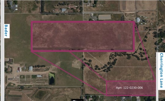
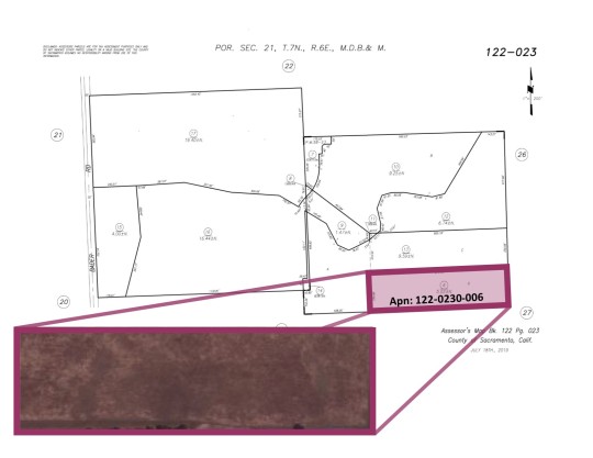

0 Cherrington Ln!!! MLS #: 19076276
Our New Listing! Calling all buyers!! This is a vacant lot that is 5.02 Acres and is zoned for Agricultural residential. This property is perfect to build your new home.
hashtag#vacant hashtag#acres hashtag#newlisting hashtag#buyers hashtag#lot hashtag#property hashtag#residential hashtag#agricultural hashtag#realestate
0 notes
Text
Minor earthquake 3 mag, 12 km NNW of Midland, Texas
Earthquake News on http://www.earthquakenewstoday.com/2020/09/17/minor-earthquake-3-mag-12-km-nnw-of-midland-texas/
Minor earthquake 3 mag, 12 km NNW of Midland, Texas
A minor earthquake magnitude 3 (ml/mb) has occurred on Thursday near Midland, Odessa, Big Spring, West Odessa, Andrews, United States. The temblor has occurred at 03:49:22/3:49 am (UTC/GMT) at a depth of 5.02 km (3 miles). Did you feel it? Leave a comment!
0 notes
Text
Green tea extract and its major component epigallocatechin gallate inhibits hepatitis B virus in vitro.
PMID: Antiviral Res. 2008 Jun ;78(3):242-9. Epub 2008 Feb 8. PMID: 18313149 Abstract Title: Green tea extract and its major component epigallocatechin gallate inhibits hepatitis B virus in vitro. Abstract: Hepatitis B virus (HBV) infection is endemic in Asia and causes major public health problems worldwide. Present treatment strategies for HBV infections are not satisfactory and the clinical limitation of current antiviral drugs for HBV, such as lamivudine, is causing rapid emergence of drug-resistant viral strains during the prolonged therapeutic treatment. In this research, the efficacy of a natural green tea extract (GTE) against HBV in a stably expressed HBV cell line HepG2-N10 is examined. The expression of viral antigens, HBsAg and HBeAg, were determined by using enzyme linked immuno-absorbent assay (ELISA). Quantitative real-time-PCR (Q-PCR) was used for the determination of extracellular HBV DNA and intracellular replicative intermediates and nuclear covalent closed circular DNA (cccDNA). HBV mRNAs were also analyzed by reverse transcription PCR (RT-PCR). Results showed that the 50% effective concentration (EC50) of GTE on HBsAg, HBeAg, extracellular HBV DNA and intracellular HBV DNA were 5.02, 5.681, 19.81, and 10.76 microg/ml, respectively. While the concentration of GTE with the inhibition percentage of 50% on proliferating cells (CC50) was 171.8 microg/ml. Similar analysis of the principal component of GTE, epigallocatechin gallate (EGCG), revealed it has relative weaker efficacy compared to GTE.
read more
0 notes
Text
Can Color Doppler Ultrasonography Differentiate Thyrotoxicoisis in Graves Disease from Subacute Thyroiditis?- Juniper Publishers
Abstract
Background and objectives: Color flow Doppler (CFD) can determine thyroid vascularity. It can be used for confirmatory diagnosis of thyroiditis and Graves' instead of radioactive iodine uptake (RAIU). This study analysed thyroid blood flow, peak systolic velocity (PSV) by CFD and thyroid volume by Gray scale sonography in thyrotoxicosis.
Methods and materials: Thyrotoxic cases (N=78; Graves: M/F=21/27, age: 35.73±1.38; subacute thyroiditis: M/F=09/21, age: 35.37±1.61 (M±SEM, yrs)) were recruited and divided into Graves' and subacute thyroiditis holding RAIU as gold standard. CFD and Gray scale sonography were done for all.
Results: Goiter was tender in 33% of subacute thyroiditis. Neither free thyroxine (FT4: 3.38±0.22 vs. 2.78±0.23ngm/dl, P=0.549) nor thyroid stimulating hormone (TSH: 0.05±0.01 vs. 0.06±0.2^IU/ml, P=0.084) were statistically different between the two groups. Echo pattern was predominantly homogeneous in Graves and heterogeneous in subacute thyroiditis (60.4% vs. 43.3%, P=0.007). Gray scale sonography revealed increased thyroid volume in 73%, normal in 20.8% and decreased in 6.3% of Graves which were 33.3%, 56.7% and 10.3% respectively in subacute thyroiditis (p=0.002). All Graves' had increased flow while 76.7% of the subacute thyroiditis had decreased flow by CFD (P<0.001). Mean (±SEM)PSV of inferior thyroid artery was significantly higher in Graves' than that of subacute thyroiditis (40.70±2.54 vs. 26.71±4.05cm/sec, P=0.003) and correlated with RAIU at 2h (r=0.330, P= 0.003) and 24hrs (r=0.325, P=0.004) as well as blood flow (r=0.663, P<0.001). Sensitivity and specificity of CFD were 93.7% & 83.3% respectively.
Conclusion: It is concluded that CFD may be a cost effective, convenient, safe and noninvasive technique with high sensitivity, specificity and diagnostic accuracy.
Keywords: Color doppler ultrasonography; Graves' disease; Subacute thyroiditis
Go to
Introduction
Thyroid disease is common and the prevalence increases with age. It is one of the major health hazards in many countries including Bangladesh [1]. Thyrotoxicosis refers to the classic physiological manifestations of excessive quantities of thyroid hormone. Differentiation between hyperthyroid Graves' disease and destructive thyrotoxicosis in thyroiditis is very important for management purpose.
Radioactive iodine uptake (RAIU) is recognized as the most accurate test, but iodine-containing foods or drugs can interfere with the results. In addition, RAIU is expensive, not easily accessible and contraindicated during pregnancy and lactation [2]. On the other hand, color flow Doppler (CFD) is a useful, inexpensive, noninvasive and widely available. Its evaluation can be both qualitative (visual assessment of thyroid vascularity) and quantitative [measuring peak systolic velocity (PSV), end diastolic and mean velocities in the inferior thyroid arteries]. Normal PSV in intrathyroidal arteries ranges between 15 to 30cm/sec but it can rise to over 100cm/sec in certain pathologic conditions like Graves' disease [3]. CFD reveals a spectacular thyroid inferno with marked hypervascularity both in systole and diastole. Return of normal thyroid appearance may be possible during remission. Contrary to Graves' disease, vascularity is markedly diminished in subacute thyroiditis. In Graves disease, gray scale ultrasound reveals diffusely enlarged thyroid (2-3 times its normal size) which is hypoechoic and heterogeneous whereas in subacute thyroiditis it shows characteristic focal hypoechoic areas (map like) and enlargement of one or more lobes.
The aim of the study was to evaluate the utility of CFD in differentiation of hyperthyroid Graves' disease from subacute thyroiditis as well as comparing its sensitivity and specificity in light of RAIU test.
Go to
Materials And Methods
This study included 78 patients with thyrotoxicosis (Graves disease: n=48, M/F=21/27, age (M±SEM, yrs): 35.73±1.38; subacute thyroiditis: n=30, M/F=09/21, age (M±SEM, yrs): 35.37±1.61) recruited from May 2014 to April 2015 in the department of Endocrinology, Bangabandhu Sheikh Mujib Medical University (BSMMU). Informed written consent was taken from all participants. Free thyroxine (FT4), thyroid stimulating hormone (TSH) and RAIU and complete blood count with ESR were done for each subject. Thyroiditis and Graves' disease were discriminated on the basis of low versus high uptake. A color Doppler ultrasound scanner (Toshiba Ultrasonography system and color Doppler, Model no: SSA-550A, Toshiba medical system, Tokyo, Japan) equipped with a 7.5MHz broadband linear array high resolution transducer was used and both CFD and Gray scale ultrasound were performed at National Institute of Nuclear Medicine and Allied sciences (NINMAS), BSMMU campus. Thyroid blood flow was seen by color Doppler power mode. PSV of the inferior thyroid artery of >30cm/sec was considered significantly high and suggestive of Graves disease and <30cm/sec for thyroiditis [3]. RAIU was considered as the gold standard. High RAIU was suggestive of Graves' disease and low RAIU considered for thyroiditis. All data were analyzed by SPSS (version 22.0).
Go to
Results
Though few clinical manifestations were statistically different between the two entities, altogether they were not conclusive (Table 1). Virtually all goiter of both the groups were diffuse (p=NS). However, consistency of goiter was mostly soft in Graves' and firm in subacute thyroiditis (P<0.001). Similarly, tenderness was absent in Graves' disease except one case whereas 33% of subacute thyroiditis had tender goiter (P<0.001). FT4 in both Graves' and subacute thyroiditis were above the upper normal limit (3.38±0.22 and 2.78±0.23ngm/ dl respectively, m±SEM; P=0.549) and TSH less than the lower limit (0.05±0.01 and 0.06±0.02|iIU/ml respectively, P=0.084). RAIU at 2hrs (21.35±1.43 vs. 5.02±0.30, mean±SEM; P<0.001) and at 24hrs (37.81±1.72 vs. 2.94±0.35, mean±SEM; P<0.001) were higher in Graves' disease than that of subacute thyroiditis (Table 2).
With in parenthesis are percentages over column total.
*by student's t-test.
RAIU: Radioactive Iodine Uptake.
Echo pattern of thyroid was mostly homogeneous in Graves (60.4%) but heterogenous in subacute thyroiditis (30.0%; p=0.007). As observed by gray scale sonography, increased thyroid volume was found in 72.9% patients of Graves' disease whereas 6.3% had decreased and 20.8% had normal volume. On the contrary, in subacute thyroiditis 56.7% had normal, 33.3% increased, and 10.0% decreased volume (p=0.002) (Table 3).
Within parenthesis are percentages over column total.
Cent percent of the Graves' disease subjects had increased blood flow by CFD. On the contrary, 76.7% of the subacute thyroiditis subjects had decreased flow, 16.7% had increased flow and 6.7% had normal flow (x2 = 58.87, P<0.001) (Table 4). Mean (±SEM) PSV of inferior thyroid artery was significantly higher in Graves' disease than that of subacute thyroiditis (40.70±2.54 vs. 26.71±4.05, P=0.003). Considering 30cm/sec as cut off value, 93.8% of the Graves' subjects had PSV >30cm/ sec; frequency of which was only 16.7% in subacute thyroiditis subjects (p <0.001) (Table 5). Doppler PSV strongly correlate with RAIU in both 2h (r=0.330, p=0.003) and 24hrs (r=0.325, p=0.004; Figure 1 & 2). PSV correlated with thyroid blood flow (r=0.663, P<0.001) as well as thyroid volume (r=0.219, p=0.054). Doppler thyroid blood flow was a strong predictor for diagnosis of Graves' disease and subacute thyroiditis (P<0.001); but none of the others were independently capable as predictor (Table 6).
Within parenthesis are percentages over column total.
by yχ 2 and student's t-test.
Within parenthesis are percentages over column total.
FT4: Free Thyroxine
TSH: Thyroid Stimulating Hormone
Holding the RAIU as the gold standard for diagnosis of Graves' disease and subacute thyroiditis, sensitivity and specificity of CFD were 93.7% & 83.3% respectively. Positive predictive value was 90.0%, negative predictive value was 89.2% and diagnostic accuracy was 89.7%.
Go to
Discussion
In the present study, importance of relatively easily practicable, convenient and less hazardous modules like blood flow and PSV of inferior thyroid artery by CFD sonography and thyroid volume as well as echo pattern by gray scale sonography were investigated in the light of 2h and 24h RAIU as gold standard. It was clearly observed that cent percent of Graves' disease patient had increased thyroid blood flow in comparison to subacute thyroiditis in which more than three quarters showed decreased flow. Cut off value of 30cm/sec was considered in differentiation between Graves' disease from thyroiditis based on a review of relevant literature [3,4]. Our findings were in agreement with the findings of those investigators revealing more than 90% of the Graves' patient above the mentioned cut off while 80% of subacute cases were below.
Although echo pattern was found to be different, yet it was not enough to demarcate the two diseases- it was homogeneous in around 60% of Graves and 30% of subacute thyroiditis and by gray scale sonography 70% of Graves patient had increased volume while only 33% of subacute thyroiditis had so. Thus, neither echo pattern nor thyroid volume could be considered for diagnostic differentiation of the two disease status. Rather PSV was found to be free from performance related error and relatively better in distinguishing between the two diseases as well as less hazardous and easy practicable. Holding RAIU as the gold standard, sensitivity and specificity of PSV were 93.75% and 83.33% respectively with 89.7% diagnostic accuracy The diagnostic performance is very satisfactory and in much agreement with other investigators' findings [5-8].
Though there are similarities of clinical manifestations between Graves' disease and thyroiditis, it was observed in this study that some clinical manifestations like palpitation, weight loss, heat intolerance, hyperdefaecation, excessive sweating, tremor were more prominent in Graves' than subacute thyroiditis whereas neck pain/ tenderness, fever, sore throat were more prominent in subacute thyroiditis as was expected. 75% of the goiter was soft in Graves' disease while in subacute thyroiditis 63% were firm in consistency. Bruit (12.5%) and eye sign (14.6%) were exclusively seen in patients with Graves'. None of the abovementioned features could clearly distinguish between these two groups as features of thyrotoxicosis overlapped in both the groups.
CFD is a cheap and simple technique with no ionizing radiation exposure and is cost effective in the diagnosis of thyrotoxicosis. A typical CFD thyroid examination takes about 10 minutes [9]. Inferior thyroid artery blood flow could be considered as an acceptable alternative to RAIU test especially when there is a contraindication to nuclear imaging of the thyroid. Radioactive iodine should not be used in situations with recent use of iodinated contrast which interfere with the accuracy of radioactive iodine tests [9]. Phillips et al. found that 45% of patient with newly diagnosed thyrotoxicosis had received iodinated contrast within 2 week before endocrine evaluation for other purposes heralding diagnostic difficulty [10]. Therefore, in these scenario if some diagnostic module avoiding the use of radioisotope can be utilized diagnostic difficulties can be overcome. In such context, we might propose and would fully agree with other investigators [11,12] supporting diagnostic modules like PSV by CFD to discriminate between thyrotoxicosis due to Graves' and subacute thyroiditis.
In summary, the thyroid blood flow is a useful marker in diagnosis of Graves' disease and subacute thyroiditis in initial phase. The high correlation between RAIU and PSV by CFD established this modality as an acceptable alternative. Its role in pregnancy and lactation where nuclear imaging is contraindicated needs to be emphasized. Data of present study suggest that CFD may be a cost effective, quick, safe and noninvasive technique in contrast to RAIU with high sensitivity,specificity and diagnostic accuracy.
For more Journals in Juniper Publishers please click on https://juniperpublishers.com/journals.php
For more articles in Journal of Thyroid Research please click on: https://juniperpublishers.com/jetr/index.php
0 notes
Text
Despite price cut to $12.9M, West Palm Beach mansion would set record
Hide caption
The current price tag for the newly built estate would shatter the record price for a single-family home in West Palm Beach, which now stands at $5.02 million.
Even after a $600,000 price cut, a West Palm Beach mansion is on the market with a list price that would far surpass the city’s record sale.
The newly built estate at 2914 Washington Road went on the market in 2017 for $13.5 million, while it was still under construction. The mansion was completed in September, and in late March, the seller cut the price to $12.9 million.
If the house along the Intracoastal Waterway fetches anywhere near that amount, it will shatter the record price for a single-family home in West Palm Beach. The current high-water mark is $5.02 million, the sum paid in October for a house at 5615 S. Flagler Drive.
The 9,634-square-foot manse on Washington Road was developed by The Aquantis Group. It features six bedrooms, a two-story living room, a library, a wine vault, an elevator and, in something of a head scratcher, seven fireplaces.
The house is listed by Burt Minkoff and Ashley McIntosh of Douglas Elliman.
Palm Beach County’s luxury real estate market is sending mixed signals.
The condo market is shattering records. Last month, a full floor at The Bristol, the waterfront condo tower in West Palm Beach, sold for $42.6 million. That sale far eclipsed The Bristol’s previous high-water market of $17.6 million.
The Bristol’s total sales have reached $100 million. The 291-foot-tall tower boasts private elevators and other perks.
The VistaBlue condo on Singer Island in Riviera Beach also has sold well, attracting buyers such as billionaire Joe Mansueto, who founded Morningstar, Inc., and football coach Bret Bielema, who is currently a consultant with the New England Patriots.
However, many high-dollar mansions have been languishing. In one high-profile example, the Ziff estate in Manalapan has been for sale since 2015. It was first marketed off the multiple listing service, then went on the MLS for $195 million in early 2016. It’s now on the market for $137.5 million.
In Palm Beach, a never-lived in mansion at 1071 N. Ocean Blvd. has been for sale since 2015. It went on the market for $84 million. It’s now priced at $59.9 million.
Source Article
The post Despite price cut to $12.9M, West Palm Beach mansion would set record appeared first on NICOLES BOOK MUSINGS.
More Info At: http://www.nicolesbookmusings.com/despite-price-cut-to-12-9m-west-palm-beach-mansion-would-set-record/
0 notes
Last Seen Blogs

ravequeenn-blog-blog
west coast

fortheloveofexy
pipe dream

mighty-sevenzeroseven
DEFENDER OF JUSTICE


onlyfairytail
Fairy Tail!