#radiograph
Text

“Till the end of forever do us part” 💙💚
#my art#en abime#william yew#henry bicknell#drawing#artists on tumblr#fanart#digital art#procreate#illustration#art#radiograph
21 notes
·
View notes
Text
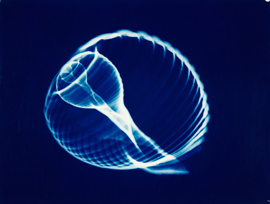
Hilja Raviniemi ~ Shell design, 1970. Suomen valokuvataiteen museon kokoelma. | src Fall 2023 exhibition
view & read more on wordPress
After her more traditional early work, Raviniemi explored the infinite creative possibilities offered by the darkroom, especially in the 1960s. Her recognizable blue era, which differed from the stark black-and-white art photography of the time, began in the late 1960s. Chemist by profession, Raviniemi was an ingenious artist in the darkroom.
In addition to blue-tinted prints, she also created completely abstract photographic artworks using different techniques. Raviniemi’s workplace at the University of Helsinki photography department laboratory also allowed her to make the first artistic radiographic images in Finland. Hundreds of Raviniemi’s radiographic works have been preserved and make up an exceptional ensemble of works in the history of Finnish art photography. read more on wordPress
#Hilja Raviniemi#x-ray#x-ray photography#radiogram#1960s#nature photography#seashell#shelldesign#shell structure#women artists#women photographers#finnish photographer#valokuva#blue tinted#radiographic image#radiograph#Finnish museum of photography
45 notes
·
View notes
Text
snake x-rays anyone?
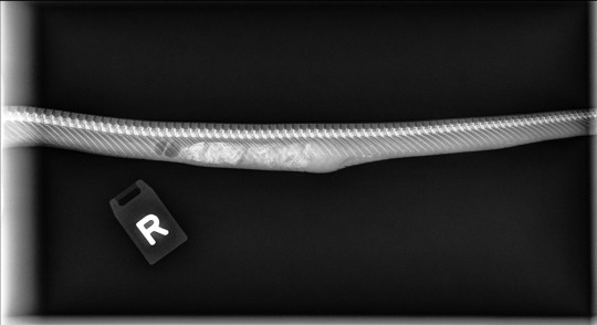
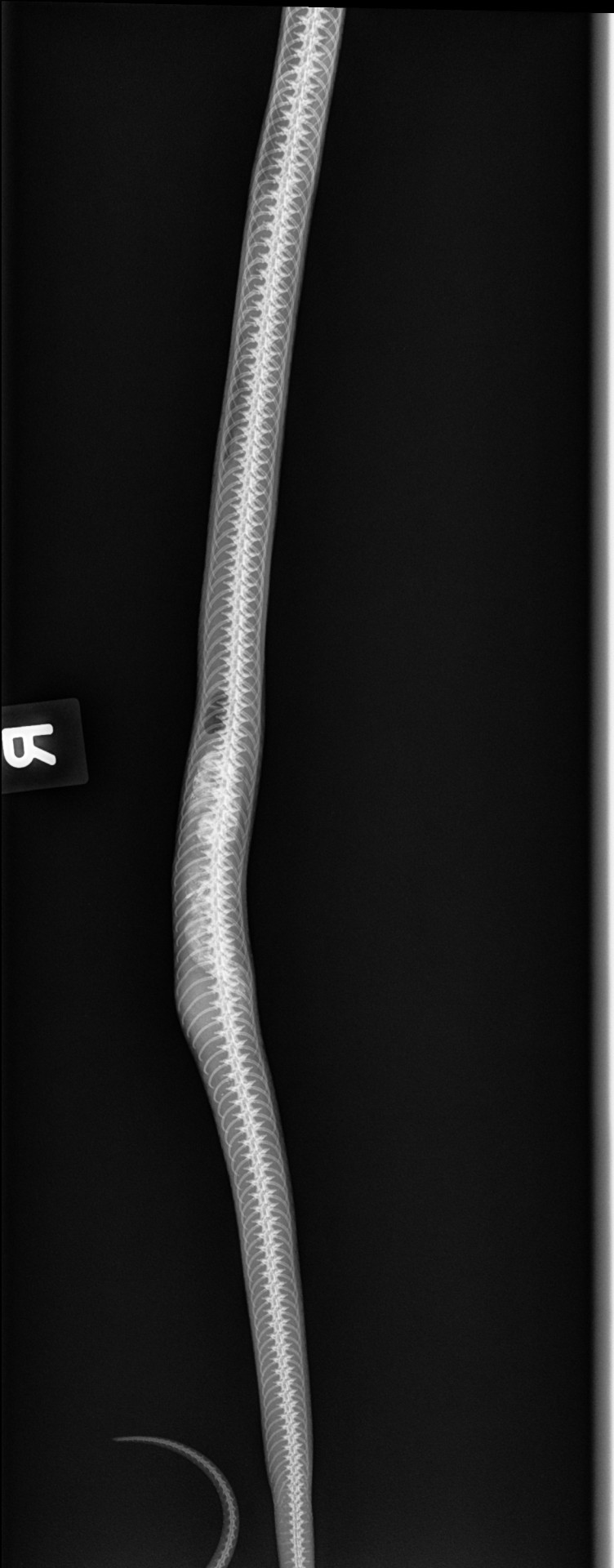
Say hello to Noodle, my almost 19-year-old lavender zigzag corn snake. Last fed 3 weeks ago, and in the last four days or so started getting a firm lump 4" or so cranial to his cloaca. Getting bloated cranial to the lump, yadda yadda, only able to pass a couple dime-sized urates, and only after effort. He's easily exhausted and generally just not feeling good. Been getting warm water soaks nightly to no avail. He has an appointment with his vet on Wednesday. I did these rads today at the clinic I work at, but we work with cats so we have no idea wtf we're seeing or even really how to x-ray a snake :D. Yes he's backed up but the question is whyyyyy and I won't get an answer until Wednesday. Bleh.
#snakeblr#snake#corn snake#x-ray#radiograph#vet#vet tech#veterinary nurse#veterinary medicine#exotic pets#reptiles
13 notes
·
View notes
Text

Today's case is a 70-year-old woman with bilateral thigh pain. Radiograph of the femurs reveals cortical irregularities of the lateral femurs, the classic location and appearance of bisphosphonate-related stress fracture. This is an atypical location for stress fractures in a patient who is not taking bisphosphonates.
Case courtesy of Matt Skalski, Radiopaedia.org, rID: 46147
5 notes
·
View notes
Text
This arg is so gay dear god—
6 notes
·
View notes
Text

Time to play, GUESS THE SPECIES?
🍆🙊🤣
4 notes
·
View notes
Text
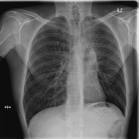
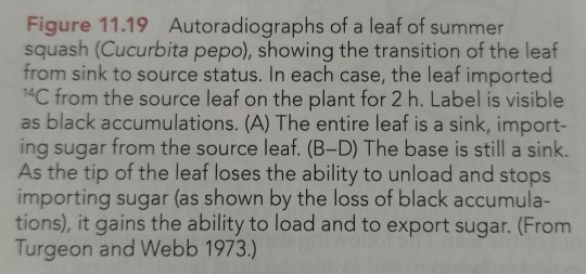
This week we will be showing a case series of chest radiographs. Take a minute to look at the image before you read on. Can you spot the problem?
...
...
...
...
This case demonstrates a mass at the left hilum. In addition, the left hemidiaphragm is elevated, indicating volume loss, and there is air outlining the aortic arch and proximal descending aorta (the so-called "Luftsichel sign," or "air sickle" due to its shape). Findings are classic for left upper lobe collapse (in this case post-obstructive due to the mass). The hyperinflated superior segment of the left lower lobe is what creates the air sickle, as it is situated betwixt the collapsed lung and the aorta.
Case courtesy of Dr Charlie Chia-Tsong Hsu, Radiopaedia.org, rID: 35263
3 notes
·
View notes
Text
World Radiography Day
"Happy World Radiography Day to everyone. Cheers to radiography which makes it easier for doctors to discover many health issues"
#radiography#health#doctor#radiograph#radiographer#special#claretworld#radiographyday#paramountuniversalpvtltd
0 notes
Text

trying to take a lateral image and the cat looked up the second i hit the pedal
1 note
·
View note
Text
During the transition period, the tip exports sugar, while the base imports it from the other source leaves (Figure 11.19).
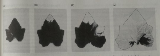
"Plant Physiology and Development" int'l 6e - Taiz, L., Zeiger, E., Møller, I.M., Murphy, A.
#book quotes#plant physiology and development#nonfiction#textbook#radiograph#summer squash#cucurbita pepo#cucurbita#sink to source#sugars#export#import#leaves
0 notes
Text
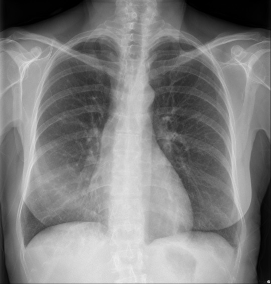
Can you spot the finding that gives this case away?
...
...
...
...
...
...
Indistinct right heart border is right middle lobe pneumonia / right middle lobe collapse until proven otherwise. This is easily confirmed on the lateral view, where the finding is usually obvious. (lateral view posted in the comments).
Case courtesy of A/Prof Stefan Heinze, Radiopaedia.org, rID: 37865
4 notes
·
View notes
Note
Good luck for Duncan's appointment tomorrow! I hope everything goes as smoothly as possible. Our thoughts are with you and the little man.
Thank you!! I am so very excited and if I told him what’s happening tmrw he would be so very NOT excited! His appointment is at 10:00am and then we’ve got an hour and a half drive back home.
I will make sure to update you guys as soon as I can with how the consult went! I’m also going to make sure I specifically request his scans when he has them done tmrw, not just their finding/notes
#idk how the neurology department at Madison is but a lot of times when other clinics send us records they skip things like radiographs#and A) I want to see them B) Doc wants to see them#and C) I want to show you guys#my post#Duncan#ask
103 notes
·
View notes
Text
Ssbine had a really bad wheezing fit last night but I'm trying to remain optimistic 🤧
#it always sounds she's trying to pass a hairball but more breathy#the fact she CLEARLY has something but radiographs and sample tests cant detect it is driving me insane dude#I have one last idea (getting an autovac to keep my home totally dust free) before I do yet another vet visit#if its not internal its GOTTA be external and thats the only thing I haven't tried yet
14 notes
·
View notes
Last Seen Blogs

smileselectdentaloffice
Smile Select Dental

funlovelydesign
Fun Lovely Design

thebrokenheelsdiary-blog
Sans titre

spawnnfrog
spawnn moment ribbit ribbit
seapandora
Seapandora writings