#tumor biology
Text


this isn't THRIVING... this is FALLING APART... with STYLE
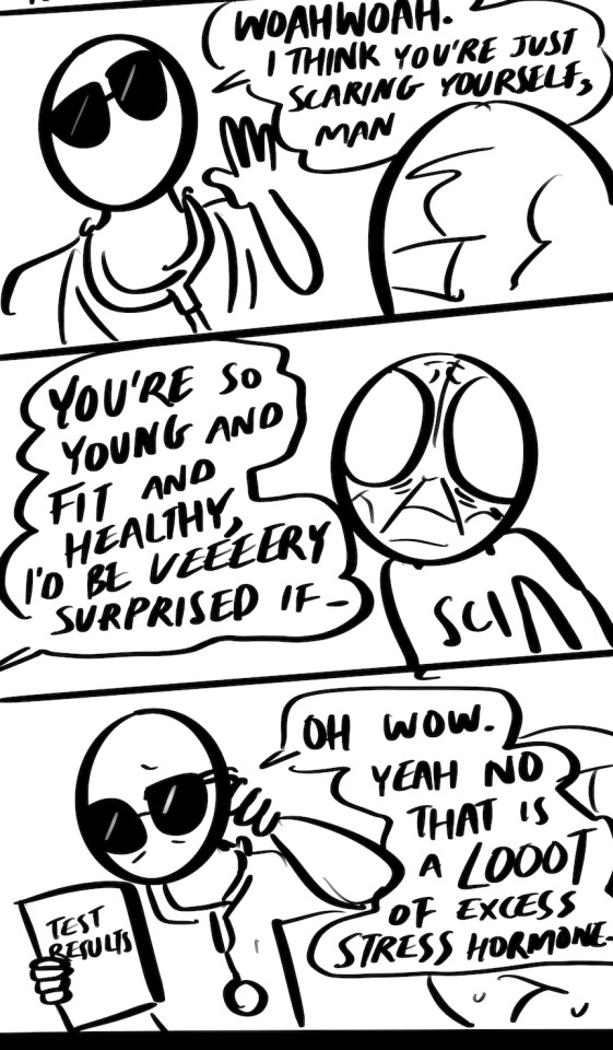
#sci speaks#the sci files#I’ve been chasing up a lot of strange symptoms I’ve been having that . haven’t been causing me pain but#you know . unanswered questions .#turns out I’ve been fighting a battle with my biology all this time and didn’t Know because I was fighting so well.#keep fighting sci .. keep fighting ..#I will WIN… Lord watch me I will win..#I have a tumor. his name is Lamar.#I’m gonna miss him when he’s gone. But he Has To Go.#the burden of being so good at coping . when something is actually Wrong nobody believes you
446 notes
·
View notes
Text
What's the point of teaching calor rubor dolor tumor anymore???
(Latin for heat, redness, pain, swelling;)

Which apparently has been taught to med and immunology students since first century AD and invented by the Roman scholar Celsus?!?
MNEMONICS IN DEAD LANGUAES DON'T HELP PEOPLE REMEMBER THINGS! Now I need to come up with a mnemonic for the mnemonic
please give me ideas for the mnemonic mnemonic, funnier the better
21 notes
·
View notes
Text
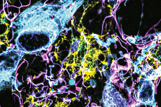
Signs in Surroundings
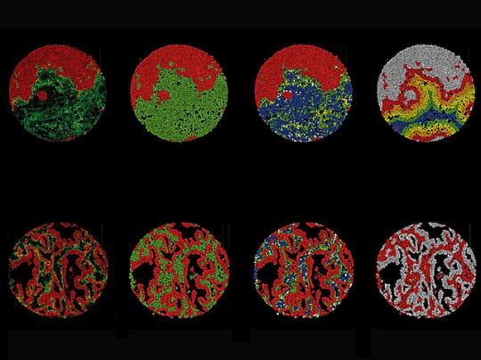
Single-cell mapping of lung cancer samples reveals presence of a distinct subset of connective tissue cells called fibroblasts in the tumour surroundings (microenvironment) is associated with different outcomes for the patient – tumour-like fibroblasts with poor prognosis and inflammatory type with longer survival
Read the published research article here
Image from work by Lena Cords and colleagues
Department of Quantitative Biomedicine, University of Zurich, Zurich, Switzerland
Image originally published with a Creative Commons Attribution – NonCommercial – NoDerivs (CC BY-NC-ND 4.0)
Published in Cancer Cell, January 2024
You can also follow BPoD on Instagram, Twitter and Facebook
8 notes
·
View notes
Text
I was talking on Discord a bit ago (as one does) about whether Space Marines are a separate species from humans (not really), but then thinking about it later, about what the fuck they are biologically -- they’re parasites, of course.
Or at least, their gene-seed is.
Each Legion’s gene-seed is an asexually reproducing lineage that uses a combination of social and biological manipulation to force its human hosts to reproduce and spread it, both inside their human bodies and in the labs of the Apothecarion.
“Parasite” is a bit of of a value judgement, of course. The Space Marines themselves, if they didn’t find the whole discussion heretical and blasphemous, would probably describe the relationship as mutualistic. They benefit immensely! They’re bigger, stronger, more durable, and live longer (if no one kills them)!
(And if they don’t die in the implantation process, of course. A roughly 90% death rate is a helluva fitness cost on both the human hosts and the space marine gene-seed.)
But... the gene-seed also seems to sterilize them[1], and that caries a lot more weight in biology than physical strength or longevity on its own. Space marines’ enhanced bodies don’t work to reproduce human children; instead they grow new gene-seed to implant into more humans.
That’s a little reductive for a social species, of course. Sure, humans with gene-seed implanted into them don’t reproduce directly, but surely they increase the survival and reproduction of other humans sufficiently to make up for it!
Which may be true in canon [2] and is more defensible as a biological argument than as a moral one. [3]
But given that (a) space marine gene-seed carries its own genetic code which (b) it requires the help of its human host and his community to reproduce (c) at the direct expense of the human host’s own life (most of the time) and reproduction (nearly always)... yeah, I think parasite is accurate.
[1] Canon’s not entirely consistent here, but certainly Space Marines rarely if ever have biological children.
[2] Hard to measure that sort of thing in a fictional universe with wildly varying made-up population numbers.
[3] Do the numbers add up? Who knows, maybe. Does this justify anything the Imperium is doing? Whole different question, and no.
#space marine biology#warhammer 40k meta#gene-seed the Canine transmissible venereal tumor of the 30th/40th millennium#parasites#yeah still kinking on these guys
38 notes
·
View notes
Text
Weird question but are tumors edible
#tw tumor#tw tumor mention#like if you ate a cancerous tumor would you get cancer? because that’s probably not how it works but also I don’t know biology#science side please explain
14 notes
·
View notes
Text
I have to talk about something because I realize I haven't yet (it's about biology)
Oxyphil cells are super weird little guys that I learned about while looking at histology slides for anatomy (this was back when I thought I wanted to do nursing). We don't know their function so they're kind of a mystery!
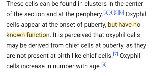
They mostly start to appear after reaching puberty and then increase in number as we age. They're also known as oncocytes (onco meaning mass or bulk + cytes meaning mature cell) but they go by a bunch of more specific names depending on their location. Plants don't have them btw but we do and so do other animals. They have bigger mitochondria that act like gymrats on steroids and more of them compared to the average cell. Apparently they even have more mitochondria than the cells that make up your muscle tissues, which is insane since you can imagine how many powerhouses it would take to fuel muscle activity. I tried to find a vid to show what they look like and I think this youtuber does a good job at that
youtube
Sometimes benign (as in unlikely or slow spread, not cancerous), sometimes malignant (as in spreads like wildfire, cancerous). They're literally just tumor cells, which would make sense since oncology is the study of cancer.
The reason they have so many overpowered powerhouses (still talking about mitochondria) is because they require a LOT of energy breakdown. Your body's cells love love love carbs (like sugar) so much that it prioritizes using them over other nutrients to make stored energy readily available for uses like muscle contractions.
The inefficient way to break down sugar (caused by oxygen-deprived conditions) is kinnnnd ooooof like an Italian grandma making tomato sauce from tomatoes. When Grandma's tomatoes enter into the sauce-ification machine, it has two exits. One exit spews tomato sauce and the other is where all the separated seeds and skin go. Sometimes Grandma pops the seedy skin bits back into the top of the machine to get a little extra out. The oxyphil's mitochondria work like Grandma's machine! Stored sugar (the tomato) goes in and some usable energy called ATP (tomato sauce) comes out while also making this other byproduct (the seeds and skin). That waste/byproduct, pyruvate, can be reabsorbed by the mitochondria to make even more energy in oxygen-deprived environments, producing lactic acid. This localized lactic acid literally makes your body more acidic but your body is usually pretty good at flushing it out (fyi drinking water helps with this process... This is also why they tell people to drink lots of water after getting a swedish massage).
In the case that it isn't clearing itself out enough for whatever reason, like say maybe you consistently produce so fucking much lactic acid because you have a tumor with MANY overpowered af mitochondria.... Well... You might be pretty acidic there. Good news is you can raise your body's pH to be more alkaline via deep breathing (this would make your body oxygen-rich so that you can break down sugars more efficiently). You can also eat more alkaline foods like red cabbage and leafy greens.
Anyways that's all I know about oxyphils and I wish we knew why they exist. It feels so cynical to think they exist just to cause death and I don't really believe that anyways since not everyone gets cancer. Are oxyphils how the body adapts to make more energy readily available in oxygen-deprived conditions, resulting in acidosis? Does that acidity break down neighboring cell walls? Does that mean they permeate the cell membrane and get that healthy neighboring cell to start dysfunctioning too and the cycle continues? Also why aren't they present at birth??? If it's true that they come from chief cells then they wouldn't really have a chance to form since chief cells are also not rly present at birth. Food for thought! I just want to know their function and have been for like two years blaaaahhhhh
Also here is a video of how a grandma makes her tomato sauce. She doesn't put the seedy skin bits back into the machine but some people do. I hope it helps with the analogy
youtube
#oxyphils#oncocytes#oncology#cancer#tumors#not into biology as much anymore but i still nerd out over science stuffs#Youtube#medical
3 notes
·
View notes
Note
Lmao I’m dying over your tags about fruit fly gene names. I listened to an interview with a geneticist who works with C. elegans a while ago and he had major beef with fruit fly geneticists over naming conventions. 😂
you know, I’m beginning to wonder if this isn’t just a normal thing for them. my professor for the developmental biology part of this course is a big drosophila guy and two weeks ago he waxed lyrical about gene naming conventions for it, which was somehow still better than the solid twenty minutes he spent today trying to convince us of drosophila’s superiority as a means of studying development and cancer (presumably as a preface to Thursday’s lesson, where he actually has to talk about c elegans organogeneis experiments). at one point he had to admit that c elegans has a shorter generation time, but he quickly alleviated his own distress by following up that harrowing admission with three minutes of describing how wonderful the Nüsslein-Volhard and Wieschaus experiments were for the study of embryonic development
#i have zero stake in the argument I’m just so so tired of human oncogene and tumor suppressor names#i can’t remember a single pathway except for sonic hedgehog and frankly that one I’d rather forget#asks#biology#anakinsthot#willow's life
13 notes
·
View notes
Text
JUST FOUND OUT WHAT A TERATOMA IS WHAT THE FUCK
WHAT THE FUCKING FUCK OH MY GOD AAAAAAAAAAAAAAAAAAAAAAAAAAAAAAAAAAAAAAAAAAAAAAAAAA
1 note
·
View note
Text
me on a reddit thread: hey it might not be a good idea to make estrogen combo pills OTC (even though progesterone would probably be fine) cause clotting disorder based strokes aren't in the numbers of side effects stats cuz america is dumb about things that are "preexisting"....and also my mom's stroke at 27 definitely isn't counted in the numbers (nor would hailey beibers btw....cuz she had the perfect storm of combo pill hormonal bc, covid, plane ride, and an undiagnosed heart issue so they can't pin it on the estrogen in a suit to a pharma company)
some twat: wellllllll if you donated blood twice monthly you'd neeeeeever have a clot
Like source please....cuz the lieden v thrombophillia foundation (which is my family's mutation I was lucky enough not to get (even though my rheum thinks that's the side my likely CTD comes from....great.....)) has NOTHING about it. I am SURE my aunt and mom would have preferred extra blood draws done to 3x daily self injectable heparin shots when having my cousins and i......when pregnant you can't take the pills, it's gotta be the shots (and this is with them both as single gene carriers lololol)
don't talk out of your ass about health conditions wtf....But only 1/300 women a year will develop a blood clot on OTC estrogen menopause meds.... ONLY 1/300.....ONLY.
#personal#to the tune of happy and you know it: *external factors dont change your genes* 👏👏*external factors dont change your genes* 👏 👏#*unless its in cancer and you raised the chancer* *external factors dont change your genes* 👏 👏#HIGH SCHOOL LEVEL BIOLOGY JESUS FUCKING CHRIST KILL ME#i cant live on a planet in a country with people this WILLINGLY stupid#your entire body genetic makeup isnt changed by a little NEEDLE pulling a few pints of blood out..............#(nor did a medication change your whole body's genetics...nor did a mRNA vax......nor did covid.......)#even fucking cancer is localized gene mutations in specific areas which is how they can tell where metastisized cancer starts#my moms on anastrozole as preventive care and like thats the one also used for metastisized BREAST cancer but its not for brain cancer or#blood cancer or bone marrow cancer or gallbladder cancer or.... or..... or...........#cause even CANCER isnt changing all your genes........which is why my aunt works on *gasp* gene therapy#where they isolate the genetic makeup of the tumor and use mRNA injections to have your immune system target the mutant parts of you#so it has YOU kill the tumor....so cool!!! isnt that cool!!! (her family is brca+ so she went into boobie cancer research)#((looks at my moms family like.....my aunt by marriage has two genetic disorders floating around in there...#and my moms blood relatives most likely also have two genetic disorders floating around in there oooooooof OOF))
3 notes
·
View notes
Text
Paintings Part 11 | Stomach Invaders
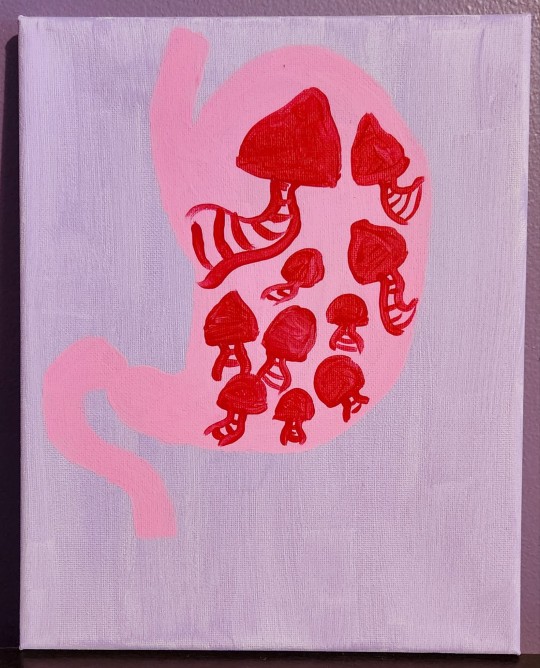
Follow on TikTok @crypticpaw.official
This is a painting I made when a family member discovered they were dealing with polyps in their stomach. We joked and called them their "stomach invaders." I thought they looked a lot like little mushrooms.
What do you want to see next?
#crypticpaw#art#artists of tumblr#artist#stomach#invaders#polyps#science#anatomy#biology#pathology#tumor#painting#painter#organ#cool art#mushrooms#mushroom art#commissions open#weird art#weird
0 notes
Text
Imaging method reveals new cells and structures in human brain tissue
New Post has been published on https://thedigitalinsider.com/imaging-method-reveals-new-cells-and-structures-in-human-brain-tissue/
Imaging method reveals new cells and structures in human brain tissue
Using a novel microscopy technique, MIT and Brigham and Women’s Hospital/Harvard Medical School researchers have imaged human brain tissue in greater detail than ever before, revealing cells and structures that were not previously visible.
Among their findings, the researchers discovered that some “low-grade” brain tumors contain more putative aggressive tumor cells than expected, suggesting that some of these tumors may be more aggressive than previously thought.
The researchers hope that this technique could eventually be deployed to diagnose tumors, generate more accurate prognoses, and help doctors choose treatments.
“We’re starting to see how important the interactions of neurons and synapses with the surrounding brain are to the growth and progression of tumors. A lot of those things we really couldn’t see with conventional tools, but now we have a tool to look at those tissues at the nanoscale and try to understand these interactions,” says Pablo Valdes, a former MIT postdoc who is now an assistant professor of neuroscience at the University of Texas Medical Branch and the lead author of the study.
Edward Boyden, the Y. Eva Tan Professor in Neurotechnology at MIT; a professor of biological engineering, media arts and sciences, and brain and cognitive sciences; a Howard Hughes Medical Institute investigator; and a member of MIT’s McGovern Institute for Brain Research and Koch Institute for Integrative Cancer Research; and E. Antonio Chiocca, a professor of neurosurgery at Harvard Medical School and chair of neurosurgery at Brigham and Women’s Hospital, are the senior authors of the study, which appears today in Science Translational Medicine.
Making molecules visible
The new imaging method is based on expansion microscopy, a technique developed in Boyden’s lab in 2015 based on a simple premise: Instead of using powerful, expensive microscopes to obtain high-resolution images, the researchers devised a way to expand the tissue itself, allowing it to be imaged at very high resolution with a regular light microscope.
The technique works by embedding the tissue into a polymer that swells when water is added, and then softening up and breaking apart the proteins that normally hold tissue together. Then, adding water swells the polymer, pulling all the proteins apart from each other. This tissue enlargement allows researchers to obtain images with a resolution of around 70 nanometers, which was previously possible only with very specialized and expensive microscopes such as scanning electron microscopes.
In 2017, the Boyden lab developed a way to expand preserved human tissue specimens, but the chemical reagents that they used also destroyed the proteins that the researchers were interested in labeling. By labeling the proteins with fluorescent antibodies before expansion, the proteins’ location and identity could be visualized after the expansion process was complete. However, the antibodies typically used for this kind of labeling can’t easily squeeze through densely packed tissue before it’s expanded.
So, for this study, the authors devised a different tissue-softening protocol that breaks up the tissue but preserves proteins in the sample. After the tissue is expanded, proteins can be labelled with commercially available fluorescent antibodies. The researchers then can perform several rounds of imaging, with three or four different proteins labeled in each round. This labelling of proteins enables many more structures to be imaged, because once the tissue is expanded, antibodies can squeeze through and label proteins they couldn’t previously reach.
“We open up the space between the proteins so that we can get antibodies into crowded spaces that we couldn’t otherwise,” Valdes says. “We saw that we could expand the tissue, we could decrowd the proteins, and we could image many, many proteins in the same tissue by doing multiple rounds of staining.”
Working with MIT Assistant Professor Deblina Sarkar, the researchers demonstrated a form of this “decrowding” in 2022 using mouse tissue.
The new study resulted in a decrowding technique for use with human brain tissue samples that are used in clinical settings for pathological diagnosis and to guide treatment decisions. These samples can be more difficult to work with because they are usually embedded in paraffin and treated with other chemicals that need to be broken down before the tissue can be expanded.
In this study, the researchers labeled up to 16 different molecules per tissue sample. The molecules they targeted include markers for a variety of structures, including axons and synapses, as well as markers that identify cell types such as astrocytes and cells that form blood vessels. They also labeled molecules linked to tumor aggressiveness and neurodegeneration.
Using this approach, the researchers analyzed healthy brain tissue, along with samples from patients with two types of glioma — high-grade glioblastoma, which is the most aggressive primary brain tumor, with a poor prognosis, and low-grade gliomas, which are considered less aggressive.
“We wanted to look at brain tumors so that we can understand them better at the nanoscale level, and by doing that, to be able to develop better treatments and diagnoses in the future. At this point, it was more developing a tool to be able to understand them better, because currently in neuro-oncology, people haven’t done much in terms of super-resolution imaging,” Valdes says.
A diagnostic tool
To identify aggressive tumor cells in gliomas they studied, the researchers labeled vimentin, a protein that is found in highly aggressive glioblastomas. To their surprise, they found many more vimentin-expressing tumor cells in low-grade gliomas than had been seen using any other method.
“This tells us something about the biology of these tumors, specifically, how some of them probably have a more aggressive nature than you would suspect by doing standard staining techniques,” Valdes says.
When glioma patients undergo surgery, tumor samples are preserved and analyzed using immunohistochemistry staining, which can reveal certain markers of aggressiveness, including some of the markers analyzed in this study.
“These are incurable brain cancers, and this type of discovery will allow us to figure out which cancer molecules to target so we can design better treatments. It also proves the profound impact of having clinicians like us at the Brigham and Women’s interacting with basic scientists such as Ed Boyden at MIT to discover new technologies that can improve patient lives,” Chiocca says.
The researchers hope their expansion microscopy technique could allow doctors to learn much more about patients’ tumors, helping them to determine how aggressive the tumor is and guiding treatment choices. Valdes now plans to do a larger study of tumor types to try to establish diagnostic guidelines based on the tumor traits that can be revealed using this technique.
“Our hope is that this is going to be a diagnostic tool to pick up marker cells, interactions, and so on, that we couldn’t before,” he says. “It’s a practical tool that will help the clinical world of neuro-oncology and neuropathology look at neurological diseases at the nanoscale like never before, because fundamentally it’s a very simple tool to use.”
Boyden’s lab also plans to use this technique to study other aspects of brain function, in healthy and diseased tissue.
“Being able to do nanoimaging is important because biology is about nanoscale things — genes, gene products, biomolecules — and they interact over nanoscale distances,” Boyden says. “We can study all sorts of nanoscale interactions, including synaptic changes, immune interactions, and changes that occur during cancer and aging.”
The research was funded by K. Lisa Yang, the Howard Hughes Medical Institute, John Doerr, Open Philanthropy, the Bill and Melinda Gates Foundation, the Koch Institute Frontier Research Program, the National Institutes of Health, and the Neurosurgery Research and Education Foundation.
#2022#aging#antibodies#approach#Arts#Biological engineering#Biology#Biomolecules#blood#blood vessels#Brain#Brain and cognitive sciences#brain research#brain tumors#Cancer#cell#cell types#Cells#chemical#chemicals#Design#diagnostics#Diseases#education#electron#engineering#form#Foundation#Future#genes
0 notes
Text
How Dog Cancer Became a New Species (CTVT)
You probably think of cancer as being not infectious, right? One of your cells made a pro gamer move and started dividing uncontrollably, but it’s not like that could become someone else’s problem. I mean, I can’t even get a kidney transplant, gaining superhuman urine production with the power of three kidneys, without needing drugs to stop my body from rejecting it.
But it turns out there actually are some types of cancer that are not only infectious, but become plagues in their own right! Lets talk about that.
View On WordPress

12 notes
·
View notes
Text
Explore cellular intricacies through systems biology. Osbaldo Resendis-Antonio's team integrates diverse biological data to understand metabolic alterations in diseases like cancer. Their predictive models offer insights into tumor heterogeneity, paving the way for personalized treatments.
#systems biology#biological data#metabolic alterations#cancer#predictive models#tumor#personalized treatments
0 notes
Text
Study reports first mathematical approach to measure total tumor-specific mRNA from mixed tumor samples -- ScienceDaily
Study reports first mathematical approach to measure total tumor-specific mRNA from mixed tumor samples — ScienceDaily
Researchers at The University of Texas MD Anderson Cancer Center have developed a new approach to quantify tumor-specific total mRNA levels from patient tumor samples, which contain both cancer and non-cancer cells. Using this technique on tumors from more than 6,500 patients across 15 cancer types, the researchers demonstrated that higher mRNA levels in cancer cells were associated with reduced…

View On WordPress
#approach#Cancer; Brain Tumor; Lung Cancer; Colon Cancer; Computer Modeling; Computational Biology; Hacking; Information Technology#Mathematical#Measure#Mixed#mRNA#reports#Samples#ScienceDaily#study#Total#tumor#tumorspecific
0 notes
Text

oh god ok I just caught up on this and WOW

so, thought #1: yeah these are awful lol
thought #2: I don't think AI is going to create a uniquely bad atmosphere for fraud in academic articles, because we've already established the incentives and conditions for fraud to be a substantial problem in the first place
most people heard about the foundational Alzheimer's research that was found to be fabricated, and that was probably the most high profile case in recent years
institutions basically lock PI's in an eternal grant-writing grind where they have to produce interesting, relevant, and novel research continually in order for them and everyone under them to keep their jobs. it de-incentivizes any type of research that seeks to verify or reproduce previous studies (journals generally won't publish this) and if you end up NOT finding something ground-breaking, then it's seen as a scientitic failure rather than a naturally possible outcome of investigating the world, in which things sometimes aren't interesting
this obviously lays incentives for fudging data in the whole field of science itself, but there's been high amount of fraud coming from institutions in china which is why I was disappointed but not especially surprised by this
I'm prefacing this by saying that scientists based in china have been asking for solutions to this and rightly pointing the blame to the uniquely weird government funding scheme as the cause. I'm not going to get too much into it, but there have been issues with fake paper mills for years, and "citation circles" (iirc you can get a cash kickback for how many times you're cited) of people agreeing to cite each other's irrelevant papers.
it's gotten to the point where, and this is really bad, PI's over here will ignore papers that come from china-based institutions under the presumption that the data is unreliable.
excerpt from the article:
For example, in 2017, Tumor Biology published by Springer retracted 107 papers, all of which are by Chinese authors, because authors provided made-up contact information of potential reviewers, and the review processes ended up being manipulated by third-party agencies that make profit from “faking” the review processes (Chen, 2017). Also, certain journals have in recent years witnessed a concentration of works by authors from China. We suspect that this is because of closely knit networks of editors, reviewers and authors, which results in superficial peer-review, easy acceptance, and deliberate self-citing from the same journals to boost impact factors (see Guglielmi, 2019 for similar patterns of behaviors occurring to Italian scientists).
re: the last sentence, it does happen to a lesser degree elsewhere, which is why this is a field-wide problem
the chinese government has I think in recent years realized that the way everything is structured is de-legitimizing their research, and there have been huge efforts to crack down on fraud and basically place sanctions on these scientists. but that's sort of just handling the symptoms and not the cause - I'm not sure how far along they are on rectifying the index by which they ascribe merit and funding potential to PI's (we do this in other ways too) but yeah, the whole system of academia itself has to change.
59 notes
·
View notes
Last Seen Blogs

fbleta69
Untitled

lunleil
lacepockets


marvel-universe-of-masterlists
MARVEL MASTERLIST

pulsar-ray
& i'm a transistorosexual