#DiagnosticAccuracy
Text
https://globalbizoutlook.com/2024/04/11/generative-ai-will-transform-healthcare-in-6-ways/
#GenerativeAI#HealthcareTransformation#PersonalizedMedicine#DiagnosticAccuracy#DrugDevelopment#AdministrativeAutomation#FutureHealthcare
0 notes
Text
0 notes
Text

Attend as a speaker/poster/delegate
The CME/CPD accredited 11th World Digital Pathology & AI UCGCongress from December 15–17, 2023 in Holiday Inn Dubai, Al Barsha, UAE & Virtual.
WhatsApp us: https://wa.me/442033222718?text=
In-person Registration details: https://digitalpathology.ucgconferences.com/registration/
Register virtually here: https://digitalpathology.ucgconferences.com/online-registration/
#RadiologyTech#DiagnosticAccuracy#MedicalImaging#DigitalPathology#ImagingPathology#RadiologyPathology#PathologyImaging#ImagingDiagnostics#DigitalHealthcare#PathologyInnovation#DigitalMedicine
0 notes
Text
#AIClinicalTechnology#HealthcareInnovation#DiagnosticAccuracy#PersonalizedMedicine#EfficientWorkflows#PatientEngagement#HealthcareAutomation#CostReduction#PredictiveAnalytics#RemotePatientMonitoring
0 notes
Text
A DIGITAL REVOLUTION IN PATHOLOGY

Laboratory medicine is continuously evolving to provide the much needed support for disease monitoring, treatment decisions and patient safety. With a rise in the adoption of machine learning and artificial intelligence to improve turnover and optimise efficiency, pathology laboratories are transforming into predictive rather than reactive environments.
Diagnostics is the critical first step in the healthcare delivery chain, as 70 per cent of the treatment decisions are based on lab results. The market for diagnostics devices is growing steadily to meet the increasing demand from hospitals and laboratories. The onus is thus on the manufacturer to provide a holistic solution that integrates automation with analytics, training, updates, and troubleshooting. Further, the use of QR codes and RFID chips enables automated tracking of samples and laboratory materials such as reagents, chemicals and equipment.
The role of AI and ML
Today, digitalisation, automation, and laboratory information software (LIS) have found a place in clinical laboratories. However, many lab processes continue to be performed manually or are partially digitised. Efforts are also being taken to improve the pre-analytic, analytic, and postanalytic processes of the clinical laboratories with the AI-support.
The COVID-19 outbreak accelerated the adoption of digital technologies in laboratories. According to a recently published report, several examples of applications of Artificial Intelligence (AI) to COVID-19 are already reported such as AI-enabled outbreak tracking apps, chatbots for diagnostics, AI-powered analysis of scientific publications and triage using natural language processing (NLP) for screening potential patients and prognosis prediction tools, and using radiology CT scans to manage system capacities, among others.
Also available on record are multiple examples of the role played by AI in the management of non-communicable chronic diseases such as cancer and cardiovascular disorders. AI tools aid in augmenting the accuracy of clinical decision and improving patient care.
Studies reveal that AI-supported systems can predict patient waiting time in the phlebotomy unit and organise the entire blood collection process or the autoverification of test results using a Machine Learning (ML) approach. ML can also be used to predict out-ofcontrol events in internal quality control studies, detecting instrument failures before even they occur, or determining compatibility between analysers in central laboratories where several instruments running the same parameters are tested.
Automation of analysers
It is very interesting that the basic manual laboratory techniques and procedures have generally been adopted in principle to automated, or mechanised, fashion in automated analysers, while a wide variety of configurations can be observed in modern instrumentation in clinical laboratory. Step-by-step innovations created these ultimate configurations.
There are several approaches to automated instrumentation historically. For instance, multiple samples are tested in a series in batch analysers. In contrast, in sequential analysers, the samples are tested sequentially, and the results are reported at that order. Continuousflow analysis means a form of sequential analysis with a continuous stream. In random-access discrete analysers, the most common configuration, analyses are performed sequentially on a set of specimens, and each sample can be analysed for a different test selection. In this type of analysis, an interruption can occur in routine working for spanning a stat sample in-between, permitting measurement of a number of and a variety of analytes in each specimen. Discrete term in this type of analysers means that each test of a patient (or a sample) is analysed in a distinct chamber (in a well, cuvette, reaction vessel)
The use of automated analysers has many advantages including reduction of workload, less time consumption per sample analysis, more number of tests done in less time, use of minute amount of sample, decreased chances of human errors, and high accuracy and reproducibility.
Further, Total Laboratory Automation (TLA) systems use conveyor belts to connect pre-analytical specimen processing and other functions directly to an analyser. Such systems also include functions such as post-analytical storage or sorting of specimens or aliquots to be transported to low-volume testing areas—or to be sent to reference laboratories.
At Transasia-Erba Group, we are looking forward to launching exciting new products by 2023. Our R&D labs in France, UK, USA, Austria and India will launch over 10 state-of-the-art analysers in CLIA, molecular, high-end haematology, AI and LIS. Also in the offing is a TLA solution aimed at midand large size laboratories. All these will make Transasia-Erba among the top-five companies globally to have ‘Total solutions in Laboratory Diagnostics.
Digital data management
Modern-day cutting edge smart technologies are giving a much-needed renovation to laboratories and making them ‘smart’. A smart laboratory is nothing but an amalgam of different hardware tools and software that helps in data management, which is one of its integral parts. And when it comes to data management, one cannot ignore the concept of digital data management.
The concept of digital data management generally revolves around utilising tools to store, document, and share, analyse and manage the experimental data. Among all the other software available, Laboratory Information Management System (LIMS) is extremely helpful. LIMS is a software solution that helps in addressing data management, regulatory challenges, and automation. This software is extremely useful in handling laboratory samples and the data. As a result, it helps maintain tests, workflows and reporting techniques that assist in standardising operations.
The most significant part of the LIMS is to enhance the operational efficiency of a lab by streamlining and automating workflows. Also, it eliminates the purpose of manually maintaining information and complying with regulations. An efficient LIMS will easily be able to keep records and report them. As a result, it eliminates the possibility of any human errors.
Benefits of using LIMS
The key advantage of using LIMS is that it provides sample management. It means that with the help of LIMS, it is possible to manage accurate and detailed records of every sample and securely store them. As a result, the data is not lost while it moves from one laboratory to another.
Secondly, the inventory management functionality of LIMS makes it possible to manage reagents and stock supplies. In addition, it also helps in generating automatic recorder alerts in time of depletion of the stock.
Test Management and reporting are the other two benefits of using the Laboratory Information Management System. It is possible to achieve standardised testing structures with the help of LIMS that too with the bonus of providing accurate and complete testing process control.
The Internet of Things
With an increasing focus on offering value-added services, manufacturers are adopting rapidly evolving technologies, such as the Internet of Things (IoT), to meet the huge demand for fully automated products and services.
IoT applications are being used across all industries and healthcare is no exception. In fact, IoT has brought in a lot of ‘convenience’ to the healthcare ecosystem, becoming especially useful for medical device providers. While it offers a host of solutions, it is left to the companies to leverage the potential of IoT to the fullest.
Predictive maintenance
For laboratories, one of the biggest concerns is downtime. Planning for upgrading via cloud-based lab management systems and sophisticated software is a good way to future proof products and reduce the cost of maintaining the instrument. The days of preventive maintenance are long forgotten, now is the time for predictive maintenance. Devices with IoT capability can sense when components are exhibiting faults or when they near their expected end of life and communicate this back to technical support and initiate actions to resolve them – from ordering replacement parts to requesting a completely new device.
Read More: https://www.europeanhhm.com/information-technology/a-digital-revolution-in-pathology
#DigitalPathology#PathologyRevolution#HealthTechInnovation#MedicalDiagnosis#PathologyAutomation#DiagnosticAccuracy#DigitalHealthcare#MedicalTechnology
0 notes
Text
Exploring Pathology Laboratories: Where Science Meets Medical Diagnostics
The global pathology laboratories market is expected to reach USD 612.2 billion by 2030. The major factors driving the industry include increasing life expectancy, the development of healthcare infrastructure in developing nations, favoring reimbursement, and rising demand for routine medical check-ups.

Gain deeper insights on the market and receive your free copy with TOC now @: Pathology Laboratories Market Report
In addition, the advancement of technology is altering the dynamics of diagnosis. Innovative technologies have made Point of Care Testing(POCT) devices portable and improved sampling techniques, making diagnostic services more accessible and efficient. Several advancements are expected to transform the industry by steering patient care through quality, speed, efficiency, and scalability. The healthcare system is progressively emphasizing the importance of pathological & diagnostic labs in becoming a greater clinical decision-making engine, assisting physicians in making inferences and diagnosing as well as monitoring patients more quickly and accurately.
The COVID-19 pandemic has resulted in higher demand for pathology/diagnostic labs as the testing for COVID-19 was necessary during the pandemic. The rapid rise of COVID-19 infections has resulted in increased funding and testing, which has boosted the overall market growth. For instance, the (NIH) National Institutes of Health in the United States announced in September 2020 that it would grant USD 129.3 million in funding to nine organizations including, Maxim Biomedical Inc., MatMaCorp, MicroGEM International, & others, for scaling-up manufacturing assistance to a new set of COVID-19 testing technologies as component of its initiative to boost diagnostics in the region.
#PathologyLaboratories#MedicalDiagnosis#DiagnosticExcellence#HealthcareInnovation#MedicalTesting#LabMedicine#PathologyInsights#DiagnosticAccuracy#HealthTech#PersonalizedMedicine#HealthcareIndustry#LaboratoryTesting#MedicalTechnology
0 notes
Text
MDT Acceptance in Physical Therapy with Dr. Ron Schenk | PT Pro Talk Podcast

Join Dr. Ron Schenk, renowned expert in Mechanical Diagnosis and Therapy (MDT), as he discusses the acceptance of MDT in the field of physical therapy on the PT Pro Talk Podcast. In this captivating episode, Dr. Schenk sheds light on the growing recognition and utilization of MDT among physical therapists. Explore the evidence-based principles and techniques that make MDT an effective approach for assessing and treating musculoskeletal conditions. Gain valuable insights into the benefits of incorporating MDT into clinical practice, including improved patient outcomes and enhanced diagnostic accuracy. Don't miss this informative conversation on the acceptance and relevance of MDT in the field of physical therapy!
#MDTAcceptance#DrRonSchenk#PTProTalkPodcast#PhysicalTherapy#EvidenceBasedPractice#MusculoskeletalHealth#PatientOutcomes#DiagnosticAccuracy#ClinicalPractice#TherapeuticApproach
0 notes
Text
Molecular Diagnostics: A New Frontier in Anatomic Pathology
Anatomic pathology serves as the cornerstone of diagnostic medicine, providing invaluable insights into the nature and progression of diseases through the examination of tissues and cells under a microscope.
This specialized field encompasses a wide range of techniques and methodologies aimed at identifying abnormalities, characterizing diseases, and guiding treatment decisions. From biopsies to surgical specimens, anatomic pathologists play a crucial role in interpreting the microscopic features of tissues, helping clinicians make accurate diagnoses and develop personalized treatment plans. Advances in technology, such as digital pathology and molecular diagnostics, have further expanded the capabilities of anatomic pathology, enabling more precise and comprehensive analysis of specimens. With digital pathology, images of tissue samples can be digitized and analyzed remotely, facilitating collaboration among pathologists and enhancing diagnostic accuracy. Molecular diagnostics, on the other hand, allow for the detection of specific genetic mutations and biomarkers associated with various diseases, paving the way for targeted therapies and personalized medicine. As the field continues to evolve, anatomic pathology remains at the forefront of medical innovation, driving advancements in diagnosis, prognostication, and therapeutic decision-making, ultimately improving patient outcomes and shaping the future of healthcare.#AnatomicPathology #DiagnosticMedicine #PrecisionMedicine #DigitalPathology #MolecularDiagnostics #HealthcareInnovation #MedicalTechnology #Pathology #DiseaseDiagnosis #PersonalizedMedicine #HealthcareAdvancements #MedicalResearch #HealthcareQuality #DiseaseCharacterization #DiagnosticAccuracy
0 notes
Photo
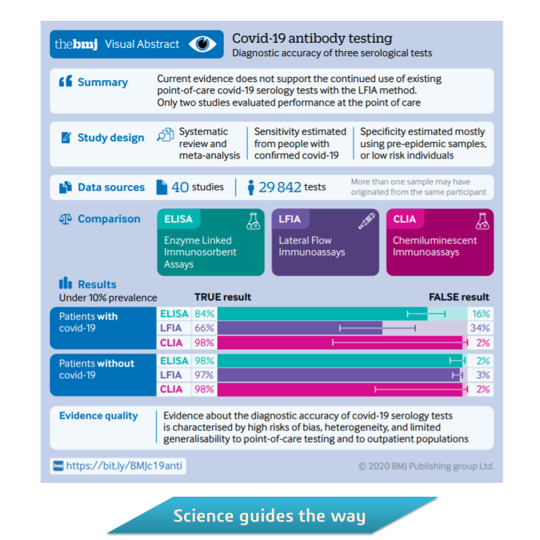
Diagnostic accuracy of serological tests for covid-19: systematic review and meta-analysis
https://inpst.net/diagnostic-accuracy-of-serological-tests-for-covid-19-systematic-review-and-meta-analysis/
#SerologicalTests #Diagnostic #DiagnosticAccuracy
Other social media channels: https://inpst.net/social-media-channels/
1 note
·
View note
Text
Diagnostic Value of High Resolution Neckultrasongraghy, Fine Needle Aspiration Cytology and BRAFV600E Mutation in Diagnosis of Malignant Thyroid Nodules- Juniper Publishers
Abstract
Background: Thyroid malignancy is rapidly raising, nearly 9-15% of the thyroid nodules are malignant nodules. Finding an optimal mean to diagnose malignant thyroid nodule preoperative is a challenge. Combination of molecular analysis, imaging and cytopathology may be helpful. This study aimed to evaluate the value of adding BRAFV600E analysis to nodules with suspicious ultra-sonographic criteria.
Patients and methodology: 50 patients from Kasr alainy endocrine outpatient clinic with solitary or multiple thyroid nodules are included, All patients subjected to full history and examination, thyroid profile, high resolution neck High resolution neck ultrasound (U/S), Fine needle Aspiration Cytology (FNAC), U/S guided FNAC and BRAFV600E analysis on FNAC using DNA sequencing then high resolution melt curve analysis (HRMA) for confirmation.
Results: The incidence of BRAFV600E mutation among papillary thyroid cancer (PTC) patients was 55.6%. it’s sensitivity 42.9%, specificity was 100% in diagnosing PTC. Sensitivity of high resolution neck ultrasonograghy in detecting malignancy was 88.2%, specificity 72.7%. Addition of ultrasonography to BRAFV600E analysis increased ultrasound sensitivity in detecting PTC preoperative to 92%. There was a positive correlation between most suspicious ultrasonography findings and presence of BRAFV600E mutation (increased AP/T diameter, Increase intra-nodular blood flow, cervical lymphadenopathy, absent or incomplete halo (all p value<0.001), irregular border p value 0.004, micro calcifications p value 0.007.
Conclusion: Adding BRAF-V600E analysis to work up of suspicious thyroid nodule would increase the sensitivity of preoperative diagnosis of PTC especially in cases of indefinite pathological findings. HRMA is simple, low cost tool for BRAF-V600E analysis in comparison to DNA sequencing method.
Keywords:Thyroid nodule; Thyroid cancer; High resolution neck ultrasound (U/S); Fine needle aspiration cytology (FNAC); BRAFV600E mutation
Abbreviation:(U/S): High Resolution Neck Ultrasound; FNAC: Fine Needle Aspiration Cytology, PTC: Papillary Thyroid Cancer; HRMA: High Resolution Melt Curve Analysis
Go to
Introduction
Thyroid nodules are a common problem their prevalence reaching 35% by ultrasound examination in some studies [1] and 8-65% in autopsy data [2] There is an increasing rate ofthyroid cancer incidence worldwide, There is an increasing rate of thyroid cancer incidence worldwide. 9~15 % of all thyroid nodules are malignant [3,4]. Accordingly the initial evaluation of thyroid nodule should focus on exclusion of malignancy [3].
High resolution U/S is the most sensitive diagnostic imagingtechnique for the detection of the thyroid nodules [4], it cancharacterize suspicious nodules by some sonographic [5], but itdepends on the skills and accuracy of the operator [6].
FNAC is the most important step in the work up of thethyroid nodule; Diagnostic accuracy has improved using USneedle localization due to a decreased number of inadequatespecimens and false negative results [7]. The pathology reportfrom FNAB according to Bethesda may be read as non-diagnostic,benign, indeterminate lesion (atypia or follicular lesion ofundetermined significance), Follicular neoplasm or suspiciousfor follicular neoplasm, Suspicious for malignancy and malignant[8,9]. Benign lesions on FNAB have an approximate 3% risk ofmalignancy (vary with patient population), and may be followedclinically with ultrasound or with a repeat FNAB [9], which, ifalso benign, decreases the risk of a false negative to 1.3% [10],the only malignant pathology reliably diagnosed through FNABis papillary thyroid carcinoma, as features such as ‘OrphanAnnie’ nuclei, nuclear grooves, intra-nuclear inclusions, andpsammoma bodies can be sufficient for a diagnosis [11]. Benignand malignant follicular neoplasms and oncocytic (Hurthle cell)adenomas and carcinomas cannot be distinguished on the basisof cytology alone, as tissue architecture is required to make thediagnosis of malignancy through observation of capsular orangio-lymphatic invasion [12].
Indeterminate cytology findings vary from 15% to 25%[13]. Indeterminate FNAC results and cyto-diagnostic errors areunavoidable due to overlapping cytological features particularlyamong hyperplastic adenomatous nodules, follicular neoplasmsand follicular variants of papillary carcinoma [14]. Thecorrelation of U/S features with FNAB results helps to overcomethe limitations of FNAB alone [15] even when cytological resultswere the same, the malignancy rate was higher when noduleshad suspicious U/S findings [16], So, U/S features and FNABresults are complementary to each other [17].
From that comes the importance of a new diagnostic toolto solve the problem, to overcome the limitations of FNA and toavoid unnecessary surgery. Several molecular factors have beenadded to improve the diagnostic accuracy of U/S-guided FNAB[2,18] one of the promising genetic factors is the analysis of theV600E mutation [19,20].
Go to
Aim Of The Work
To detect an accurate and useful tool for early preoperativedetermination of thyroid malignancy in the patients with thyroidnodule and assess its value in addition to the previous modalities(ultrasonography, and FNAC) that may increase the diagnosticaccuracy and prevent unnecessary surgery
Go to
Patients And Methods
This single group comparative descriptive study included50 patients from Kasr Alainy endocrine outpatient clinic, CairoUniversity.The thyroid examined contained either solitaryor multiple nodules. Four patients with four nodules werehypothyroid (8%), and two nodules were in hyperthyroidpatients (4%), and the remaining part 44 nodules were ineuthyroid patients (88%).18 examined nodules were solitarynodule and 32 were a nodule in MNG. Nodules’ size ranges from0.6-28mm3, Mean 6.6mm3±5.8mm3 SD, Median 6.0mm3. Patients’ages ranged from 22-65 years old median 43.0±9.6 SD, Median43.5. five males (10%) and 45 females (90%). Demographic dataare included in Table 1.
All patients were subjected to
Full clinical history including age, sex, past historyof head and neck irradiation and positive family history ofthyroid cancer.
Thyroid examination.
Thyroid profile (FT3, FT4, and TSH).
High resolution neck ultrasound.
U/S guided FNAC.
BRAFV600E analysis.
Ethical considerations
All patients were informed about these types of investigationsand for confidentiality their names were omitted and replacedby numerical codes.
Sonographic criteria for thyroid nodules include
Echogenicity, Presence or absence of calcification and itstypes, A-P to Transverse diameter ratio, Blood flow in the noduleand its patterns whether central or peripheral or both, Solitaryor dominant nodules, Presence or absence of halo, Solid, cystic orpartially solid and partially cystic, Sharpness of border.
Signs of increase risk of malignancy in U/S include
Hypo-echogenicity. Micro- or interrupted rimcalcifications, Irregular Margins.
Absence of Halo or incomplete halo.
Increased Intra-nodular flow.
Increase antro- posterior to transverse diameter(AP/T) diameter.
Significant increase in size over time.
Invasion of anterior strap muscles
Presence of abnormal cervical lymphadenopathy.
All cytology specimens were obtained under ultrasoundguidance using a 21-gauge needle attached to a plastic syringe,part of the smear obtained were wet fixed in 95% ethyl alcoholand stained with hematoxylin and eosin stain for routinecytological evaluation, and the other part put in test tube withsaline for genetic examination.
Molecular genetic testing
Specimen collection: Samples were collected from eachsubject in sterile containers for the genotyping technique.Samples were either processed fresh or were stored at 2-8 °C.
DNA extraction: This was performed using ThermoScientific Gene JET Whole Blood Genomic DNA Purification MiniKit supplied by Thermo Scientific Fermentas. The extractedDNA then subjected to the algorithm for molecular detectionof BRAFV600E mutation. The aim of the current study is thedetection of the most common mutation in the BRAF gene.V600E in DNA samples collected from thyroid nodules.
We used DNA sequencing method for BRAF V600E mutationdetection, unfortunately all samples were BRAF V600E mutationnegative, so we tried more accurate method (HRMA), then DNAsequencing for validation of the mutant BRAF results had done.
The extracted DNA were conducted to the work asfollows
Extracted DNA DNA sequencing of exon 15 HRMA DNAsequencing for validation of the mutant BRAF results
DNA amplification using the polymerase chainreaction
DNA amplification using the PCR: Enzymatic amplificationwas performed by PCR using HotStarTaq® Master Mix Kit (250units) supplied by QIAGEN® and BIO RAD T100TM ThermalCycler*.
Detection of PCR amplification products: Fluorescentstained DNA was detected by using Agarose Gel Electrophoresisand Ultra-Violet Light Trans-illumination.
Purification of PCR product: This was performed usingQIAquick® PCR Purification Kit supplied by QIAGEN® (Table 2).
Sequencing coding sequence and intronic boundaries ofExon 2 and Exon 3 in INS gene Cycle Sequencing: Using theApplied Biosystems* (ABI) PRISM® BigDye® Cycle SequencingReady Reaction Kit.
Second purification: Removal of excess Dye DeoxyTMterminators from sequencing reactions by using Centri-Sepcolumns (Princeton separations) Terminator v3.1 CompletedDNA long Read capillary electrophoresis on the ABI 3500.
Analysis of data
Sequences were compared to the published Referencesequence of Homo sapiens (BRAF), NCBI (National Centre ofBiotechnology Information): (NG_007873.3, NM_004333.4,and NP_004324.2) Analysis was done by BLAST (Basic LocalAlignment Search Tool) (www.ncbi.nlm.nih.gov) and the CLCBIOsequence viewer 6 program (www.clcbio.com ).
High resolution melt curve analysis: The HRMA reactionwas done according to the protocol of the kit MeltDoctor TMHRM Master Mix Applied Biosystems, USA.
DNA sequencing for validation of the BRAF mutantresults: The samples with mutant results were processed afterHRM analysis by pipetting the reaction from the reaction tubesfollowed by purification of the products.
Statistical analysis: Pre-coded data was entered on thecomputer using “Microsoft Office Excel Software” program(2010) for windows. Data was then transferred to the StatisticalPackage of Social Science Software program, version 21 (SPSS) tobe statistically analyzed.
Data was summarized using range, mean, standard deviationand median for quantitative variables and frequency andpercentage for qualitative ones. Comparison between groups wasperformed using independent sample t-test or Mann Whitneytest for quantitative variables and Chi square or Fisher’s exacttest for qualitative ones. P values less than 0.05 were consideredstatistically significant, and less than 0.01 were consideredhighly significant.
Go to
Results
There is no statistically significant association betweendifferent sex, nodular site, count and thyroid functions withmalignancy or BRAFv600E gene mutation (Table 3). Pathologyresults showed 28 benign nodules, 17 malignant nodule (12papillary -5 follicular neoplasm) and 5 cases with indefinitenodules. The incidence of BRAF V600E gene mutation in ourpatients with PTC was 55.6%
50 nodules were included but unfortunately because ofrepetition of genetic analysis for BRAF V600E gene mutations byDNA sequencing then again by HRMA methods that leads to that3 samples were not enough to complete genetic analysis so BRAFV600E analysis completed only in 47 samples.
We found a positive correlation with significant p valuebetween some suspicious sonographic criteria and malignancyand some suspicious sonographic criteria and BRAF V600Emutation as increased (AP/T) diameter, absent or interruptedhalo, micro-calcifications, increase intra-nodular blood flow,presence of suspicious LNs and irregular border with increasedboth sensitivity and specificity (Table 4 & 5).
Six malignant nodules were found to harbor BRAF V600Egene mutation, five of them were papillary thyroid carcinomaand one was follicular neoplasm by FNAC with P value 0.001 andnothing was found in benign nodules so there was significantcorrelation between BRAF V600E gene mutation and papillarythyroid cancer. Overall, sensitivity of ultrasound in detectingmalignancy was found to be 88.2%, specificity 72.7%, Sensitivityof US and BRAF V600E mutation combined was 92% (Table 6).
Go to
Discussion
It is noticed that thyroid nodules more common finding infemales more than males, Framingham survey for thyroid nodulesshows that prevalence in females 6.4% and 1.5% in males, andthe Whickham study displays (6.6:1 ratio females /males)[21], This might explain why most of the patients in our studywere females representing 90%. The incidence of malignancyin our study was 34% this result is biased because 12 nodulesdiagnosed as PTC are chosen retrograde after diagnosis of PTCto assess BRAF v600E mutation status.
The incidence of BRAF-V600E gene mutation in our patientswith PTC was 55.6%; previous meta-analyses have publishedvalues for the overall prevalence of the BRAF-V600E mutationin PTC ranging from 29 to 83%. [10,22,23], this wide rangemay be due to variations in PTC subtype, subjects’ geographicalbackgrounds, and research methodology.
No difference in BRAFV600E gene mutation incidencebetween different age groups and this shows agreement withAh Young Park et al. [24], but regarding our result of absenceof significant association of BRAFV600E gene mutation withspecial sex that shows disagreement with Ah Young Park et al.study that shows increase mutation in male sex, this may be dueto the small number of cases involved in our study, also 90% ofour cases are females that cannot reflect the actual prevalence[24].
Our study contained five nodules (10%) with undeterminedcytology, four of them have suspicious US features and all werenegative regarding BRAF-V600E gene mutation, we can’t excludemalignancy in these cases by absence of BRAF-V600E genemutation alone as BRAF-V600E mutation is found only in nearly40% of PTC cases [25].
The presence of BRAF-V600E mutation in one case offollicular neoplasm out of five cases shows agreement with onlyone case report Pennelli et al. [26], this conflicting result maybe due to inadequate cytological diagnosis and interpretationas follicular variant of papillary cancer may be interpreted as amicro follicular lesion in FNA [27], so this case need follow upand pathological confirmation post thyroidectomy to make sureof this result.
In this present study BRAF-V600E mutation was negativein benign cases, this result shows agreement with other studies[10,11] and BRAF-V600E mutation is believed to be specific forPTC because no report has been issued of this mutation in benignnodules [28].
The sensitivity of BRAF-V600E gene mutation in detectingPTC was 42.9%, specificity was 100%, positive predictive value100%, and accuracy 83%, this result shows agreement with Kimet al that shows BRAF-V600E mutation alone has a specificityof 100% and a positive predictive value of 100% for diagnosingmalignancy in PTC [9,29].
Nam et al. [30] reported significant improvements in thesensitivity, negative predictive value, and diagnostic accuracy ofFNA cytology for diagnosing malignancy by adding BRAF-V600Eanalysis to both US and FNAC [30].
Several studies have found a positive correlation betweenthe BRAF-V600E mutation and suspicious sonographic featuresof thyroid nodules [7], other studies show inconsistent results;Kwak, et al. found that only marked hypo echogenicity wasassociated with BRAF-V600E positivity, Hwang et al. found thatonly calcification was associated with BRAF-V600E positivity[29,31] both these studies predominantly evaluated smallpapillary thyroid micro carcinomas, with mean tumor sizesof 6-9mm3, the small size was likely a significant confoundingfactor in characterizing these lesions by preoperative US.
Our study shows positive correlation between mostsuspicious US findings and BRAF-V600E gene mutation andshows that AP/T≥1, absent or incomplete halo, irregular border,micro-calcifications and Increase intra-nodular blood flow havepositive correlation with positivity of BRAF-V600E mutationthese results show agreement with Kabaker et al. [32] thatshowed association of BRAF-V600E positivity with most knownsuspicious U/S finding including taller than wide shape, illdefinedmargin, hypo-echogenicity, micro-calcifications andabsent halo (p value<0.001) [32].
This study also showed that BRAF-V600E gene mutation issignificantly present in cases with cervical lymphadenopathythis result shows agreement with many studies that correlateBRAFV600E gene mutation with the frequency of lymph nodemetastases [10,22] Park et al. [24] study concluded that theBRAF-V600E mutation was associated extra-thyroidal extension,central and lateral lymph node metastasis, and advanced tumorstage (P<0.0001, so BRAFV600E analysis preoperative may be ofvalue as prognostic marker and affect surgical decision regardingtype of surgery.
DNA sequencing method shows high cost, time consumingand low sensitivity for the clinical screening of BRAF V600Emutation and this shows agreement with other study [33] thatshows accuracy of DNA sequencing is low when dealing withcytological specimens where the cells in question may be aminor population among the vast majority of background nonneoplasticcells, Furthermore, this method needs expensiveequipment which may not be economical for all the patients[34], In contrast HRMA has been proved to be applicable, costefficientand very sensitive scanning method that allows rapiddetection of DNA sequence variations without cumbersomepost–polymerase chain reaction (PCR) methods, which is notachievable by direct sequencing [35]. Because targeted therapyfor thyroid cancers with multikinase inhibitors is under activedevelopment [36,37], the detection of mutations in the FNACmaterial may be helpful in the future to guide mutation-specifictargeted therapies that can be initiated preoperatively or inthose patients who are not surgical candidates. This study hassome limitations as small number of patients included, somegenetic mutations should be included as RAS and RET and RET/PTC rearrangement.
Go to
Conclusion
BRAFV600E positivity was associated with most knownsuspicious U/S finding, BRAFV600E gene mutation analysisis a new hopeful diagnostic and prognostic tool that helps toovercome limitations of high resolution neck ultrasonographyand fine needle aspiration biopsy, Adding BRAF-V600E analysisto U/S and FNA would increase the sensitivity of preoperativediagnosis of PTC especially in cases of indefinite nodules. HRMAis an excellent, simple, low cost tool for BRAF-V600E analysis incomparison to DNA sequencing method.
For more Journals in Juniper Publishers please click on https://juniperpublishers.com/journals.php
For more articles in Journal of Thyroid Research please click on: https://juniperpublishers.com/jetr/index.php
0 notes
Last Seen Blogs

loisharach
Untitled

indigestos
indigestos

directwebsitebaccarat3
บาคาร่าออนไลน์

hope-and-frustration
hope and frustration

bogomolchik-0
Богомол