
rxbreast
Pathology About Breats
Radiology, medicine and more, and more.
11 posts
Don't wanna be here? Send us removal request.
Last Seen Blogs

sirvinter
vinter

sanghyunkiduk
Untitled

thisgreeneyedmonster
dazed, confused and starry-eyed

bunny-touka
東京喰種

ii7i
علي عبد
Text
0 notes
Text
0 notes
Text
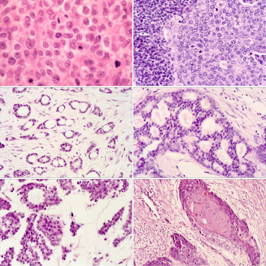
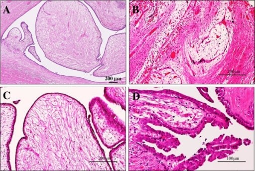
Phyllodes Tumors of the Breast
Phyllodes tumors (or phylloides tumors) are rare breast tumors that start in the connective (stromal) tissue of the breast, not the ducts or glands (which is where most breast cancers start). Most phyllodes tumors are benign and only a small number are malignant (cancer).
Phyllodes tumors are most common in women in their 40s, but women of any age can have them. Women with Li-Fraumeni syndrome (a rare, inherited genetic condition) have an increased risk for phyllodes tumors.
Phyllodes tumors are often divided into 3 groups, based on how they look under a microscope.
0 notes
Text
Invasive or filtering lobular carcinoma
Invasive lobular breast cancer means that the cancer started in the cells that line the lobules and has spread into the surrounding breast tissue. The lobules are the glands that make milk when breastfeeding.
Invasive lobular breast cancer is the second most common type of breast cancer. Around 15 in every 100 breast cancers (around 15%) are invasive lobular carcinoma. This type can develop in women of any age. But it is most common in women between 45 and 55 years old. Breast cancer is very rare in men. It is also very unusual for a man to have an invasive lobular type of breast cancer.
Symptoms
Invasive lobular breast cancer doesn't always form a firm lump. You are more likely to have a thickened area of breast tissue. Possible symptoms include:
- an area of thickening or swelling
- a change in the nipple, for example it might turn inwards (become inverted)
- a change in the skin, such as dimpling or thickening
While invasive lobular breast cancer can cause these particular symptoms, it’s worth being aware of the general symptoms of breast cancer.
0 notes
Text
Triple-negative breast cancer
Triple-negative breast cancer is a kind of breast cancer that does not have any of the receptors that are commonly found in breast cancer.
Think of cancer cells as a house. The front door may have three kinds of locks, called receptors—
- One is for the female hormone estrogen.
- One is for the female hormone progesterone.
- One is a protein called human epidermal growth factor (HER2).
0 notes
Text
Invasive or infiltrating ductal carcinoma
Invasive ductal carcinoma (IDC), also known as infiltrating ductal carcinoma, is a type of breast cancer that starts in the milk ducts of the breast and moves into nearby tissue. In time, IDC may spread (metastasize) through the lymph nodes or bloodstream to other areas of the body. This is known as metastatic breast cancer. Invasive ductal carcinoma is also the most common type of male breast cancer.
Some people may be diagnosed with a subtype of IDC. These diagnoses are rare, with each one making up less than 5 percent of all breast cancers. Less common forms of IDC include:
- Medullary carcinoma
- Metaplastic carcinoma
- Mucinous (or colloid) carcinoma
- Papillary carcinoma
- Tubular carcinoma
IDC may be discovered on a mammogram before presenting any symptoms. However, some individuals may have symptoms including:
- Changes in breast shape
- Breast or nipple pain
- Discharge from the nipple
- Swelling of the breast
- A lump in the breast tissue
- Thickening of the nipple skin
1 note
·
View note
Text
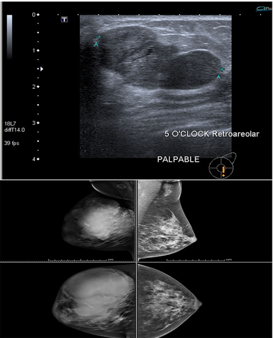
Fibroadenoma
A fibroadenoma is a solid breast lump. This breast lump is not cancer. A fibroadenoma happens most often between ages 15 and 35. But it can be found at any age in anyone who has periods.
A fibroadenoma often causes no pain. It can feel firm, smooth and rubbery. It has a round shape. It might feel like a pea in the breast. Or it may feel flat like a coin. When touched, it moves easily within the breast tissue.
0 notes
Text
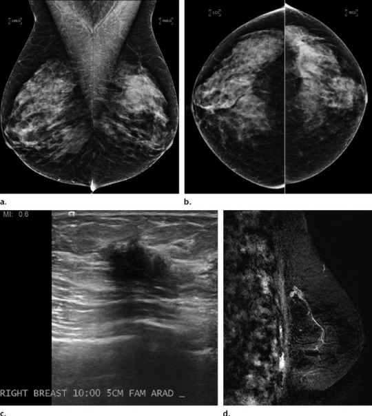
Breast Fat Necrosis
Breast fat necrosis is a non-suppurative inflammation of adipose tissue caused by the disruption of oxygen supply to fat cells, ultimately leading to cell death. It is commonly present in female patients who undergo breast procedures, although it can also be associated with malignancy. In non-surgical patients, the most common etiology is trauma. This activity reviews the etiology, pathophysiology, evaluation, and management of breast fat necrosis in surgical patients.
0 notes
Text
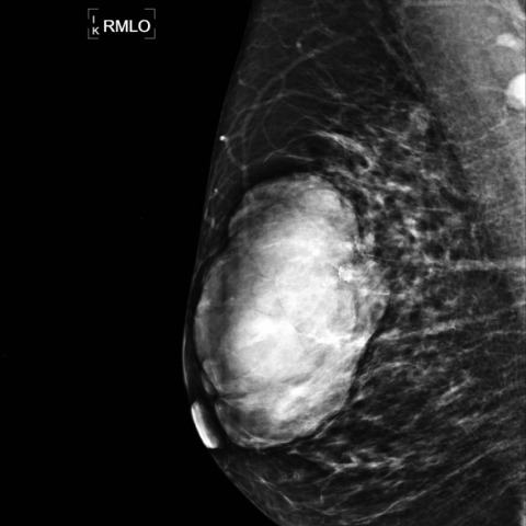
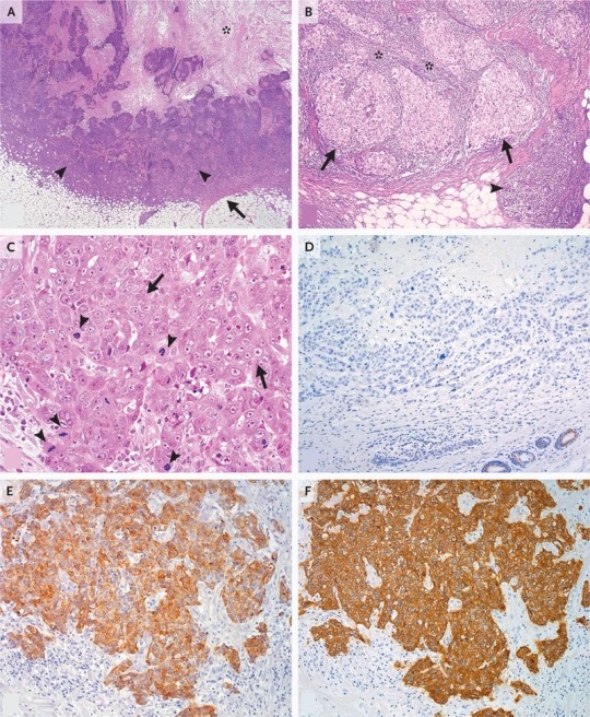
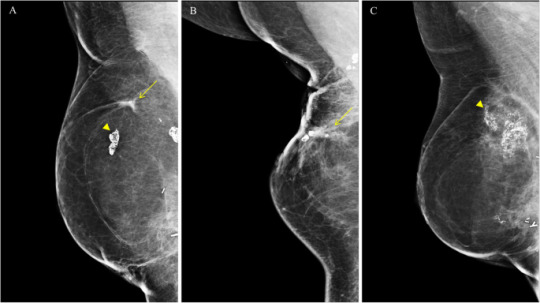
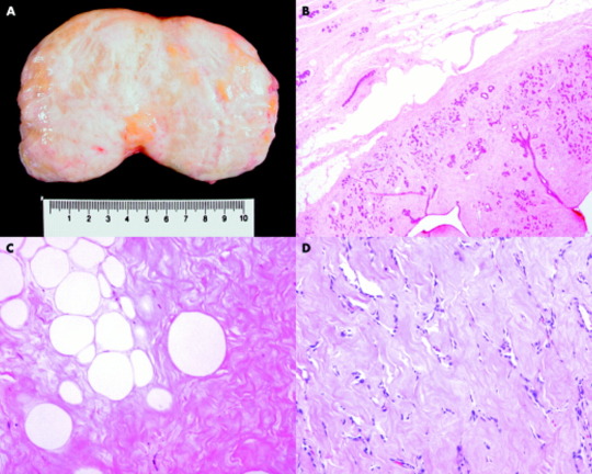
Hamartoma of the breast
Hamartoma is an uncommon breast tumor that histologically contains lobular breast tissue with various admixtures of fibrous, fibrocystic, and adipose tissue. Myoid hamartoma is an extremely rare subtype containing an additional smooth muscle component. Both radiographic correlations and immunohistochemical studies are important to diagnose myoid hamartoma and avoid confusion with other breast diseases. Herein, we report a case of myoid hamartoma of the breast that proved difficult to diagnoses because the findings on morphological evaluation employing various imaging modalities were different from the histological findings of core needle biopsy.
0 notes
Text
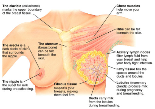
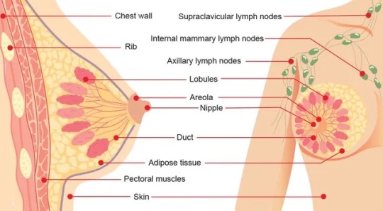
Radiology about breasts: breast embryology, histology and anatomy.
Embryology:
the embryology of the breast shows that the ectoderm and the mesenchyme are responsible for the genesis of the male and female breast, with the ectoderm responsible for the formation of the ducts and alveoli and the mesenchyme responsible for the connective tissue and its vessels. The milk ridge develops in the ventral area of the body, and the pectoral part of the milk ridge produces the right and left mammary. Very little is known about the molecular mechanisms that initiate breast formation. The chronology of breast development is discussed through its developmental and differentiation phases. Developmental anomalies can occur, such as bilateral amastia, unilateral amastia, and bilateral amastia with congenital ectodermal defects, athelia, polythelia, polymastia, and breast asymmetry. The anatomy of the breast is described.
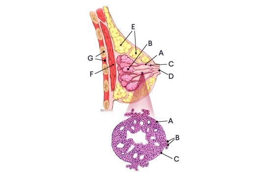
Anatomy:
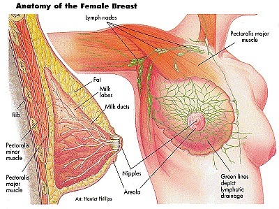
female breasts contain different types of fatty, fibrous, and glandular tissue:
- glandular tissue includes the breast lobes and breast ducts
- fibrous, or supportive or connective, tissue is the same tissue that ligaments and scar tissue are made of
- fatty tissue fills in the spaces between glandular and fibrous tissue and largely determines your breast size
Doctors refer to all non-fatty tissue as fibroglandular tissue. There are also bands of supportive, flexible connective tissue called ligaments, which stretch from the skin to the chest wall to hold the breast tissue in place. Muscle plays an important role too. The pectoral muscle lies against the chest wall underneath both breasts, giving them support. Blood vessels provide oxygen to the breast tissue and carry away waste.
Histology:
Each lobule consists of a large number of alveolar glands, which are the small sac-like structures that produce milk.
The milk produced by the alveolar glands drain into the terminal ducts that join together to form the intralobular ducts.
These ducts converge further and form the lactiferous ducts, which drain the lobes and eventually lead to the nipple.
All of the lobules and ducts are also surrounded by fibro-fatty tissue that provide a supportive and protective function.
Before pregnancy, the mammary glands are considered to be in an inactive state, and they’re structurally different from mammary glands that are in the active state.
The active state occurs during pregnancy and during lactation after childbirth.
During pregnancy, the alveolar glands and the duct system will grow in preparation to produce milk for a newborn baby.
This low power image is an example of lactating mammary glands in the active state.
A portion of a lobe can be seen surrounded by a thick layer of connective tissue that contains a small number of fat cells or adipocytes within the connective tissue as well.
Within the lobe, thinner layers of connective tissue separate the lobe into lobules.
In a neighboring lobe, we can also see a couple of large lactiferous ducts.
These ducts are lined with a double layer of columnar or cuboidal cells with a surrounding layer of connective tissue.
1 note
·
View note