#Fluorescent Antibody Test (FAT) Market
Link
“Rabies Diagnostics Market ” report provides a detailed analysis of global market size, regional and country-level market size, segmentation, market growth, market share, and competitive landscape.
0 notes
Link
Rabies Diagnostics report provides a detailed analysis of global market size, regional and country-level market size, segmentation, market growth, market share, and competitive landscape.
#rabies diagnostics market#Rabies Diagnostics research Rabies Diagnostics report Rabies Diagnostics research report
0 notes
Text
Fluorescent Antibody Test Market Demand To Massive Growth with Leading Players| Thermo Fisher Scientific Inc., Dewinter Optical Inc, Leica Microsystems.
Fluorescent Antibody Test Market is a method used to diagnose rabies in fresh or frozen brain tissues, based on antigen detection which is recommended by world health organisation (WHO) and world organisation for animal health (OIE). Antigens are detected by using reflected light fluorescence microscope.Increasing occurrence of rabies resulted from dog bites, favourable government policies and laws and rising funds for research and development are some of the factors that will accelerate the growth of the fluorescent antibody test (FAT) market in the forecast period of 2020-2027.
Fluorescent antibody test (FAT) market is expected to grow at a CAGR of 6.8% in the forecast period of 2020-2027. The increasing occurrence of rabies in animals and humans has been directly impacting the growth of fluorescent antibody test (FAT) market.
Get Sample Report at :
https://www.databridgemarketresearch.com/request-a-sample/?dbmr=global-fluorescent-antibody-test-fat-market

Competitive Analysis: Global Fluorescent Antibody Test Market
Few of the major competitors currently working in Global Fluorescent Antibody Test Market are Quidel Corporation, BioTek Instruments, Inc., Thermo Fisher Scientific Inc., Dewinter Optical Inc, Leica Microsystems, PicoQuant GmbH, Carl Zeiss AG, Cole-Parmer Instrument Company, LLC., Esco Micro Pte Ltd., BR Technologies, Merck KGaA,, OLYMPUS CORPORATION, labome.com, among other domestic and global players. Market share data is available for Global, North America, Europe, Asia-Pacific (APAC), Middle East and Africa (MEA) and South America separately. DBMR analysts understand competitive strengths and provide competitive analysis for each competitor separately.
Key Pointers Covered in the Global Fluorescent Antibody Test Market Trends and Forecast to 2026
Global Fluorescent Antibody Test Market New Sales Volumes
Global Fluorescent Antibody Test Market Replacement Sales Volumes
Global Fluorescent Antibody Test Market Installed Base
Global Fluorescent Antibody Test Market By Brands
Global Fluorescent Antibody Test Market Size
Global Fluorescent Antibody Test Market Procedure Volumes
Global Fluorescent Antibody Test Market Product Price Analysis
Global Fluorescent Antibody Test Market Healthcare Outcomes
Global Fluorescent Antibody Test Market Cost of Care Analysis
Global Fluorescent Antibody Test Market Regulatory Framework and Changes
Global Fluorescent Antibody Test Market Prices and Reimbursement Analysis
Global Fluorescent Antibody Test Market Shares in Different Regions
Recent Developments for Global Fluorescent Antibody Test Market Competitors
Global Fluorescent Antibody Test Market Upcoming Applications
Global Fluorescent Antibody Test Market Innovators Study
Get Detailed TOC:
https://www.databridgemarketresearch.com/toc/?dbmr=global-fluorescent-antibody-test-fat-market
Global Fluorescent Antibody Test (FAT) Market Scope and Market Size
Fluorescent antibody test (FAT) market is segmented on the basis of technology, product, method, indication, antigen and end-user. The growth amongst these segments will help you analyse meagre growth segments in the industries, and provide the users with valuable market overview and market insights to help them in making strategic decisions for identification of core market applications.
On the basis of technology, fluorescent antibody test (FAT) market is segmented into ELISA/immunohistochemistry, chromatography techniques, PCR and others.
On the basis ofproduct, fluorescent antibody test (FAT) market is segmented into instruments, reagents & kits, accessories. Instruments have been further segmented into fluorescent analyser, fluorescent microscope, flow cytometry, cell sorting, thermal cycler and others. Reagents & kits have been further segmented into c. difficile testing, chlamydia testing, herpes family testing, respiratory family testing and enterovirus testing.
On the basis of method, fluorescent antibody test (FAT) market is segmented into direct and indirect.
On the basis of indication, fluorescent antibody test (FAT) market is segmented into infectious diseases and autoimmune diseases.
Fluorescent antibody test (FAT) market is segmented on the basis of antigen into bacteria and virus.
On the basis of end-user, fluorescent antibody test (FAT) market is segmented into diagnostic centers, hospitals, clinics and others.
Segmentation: Global Fluorescent Antibody Test Market
Global Fluorescent Antibody Test (FAT) Market By Technology (ELISA/Immunohistochemistry, Chromatography techniques, PCR, Others), Products (Instruments, Reagents & Kits, Accessories), Method (Direct, Indirect), Indication (Infectious Diseases, Autoimmune Diseases), Antigen (Bacteria, Virus), End-User (Diagnostic Centers, Hospitals, Clinics, Others), Country (U.S., Canada, Mexico, Germany, Italy, U.K., France, Spain, Netherland, Belgium, Switzerland, Turkey, Russia, Rest of Europe, Japan, China, India, South Korea, Australia, Singapore, Malaysia, Thailand, Indonesia, Philippines, Rest of Asia-Pacific, Brazil, Argentina, Rest of South America, South Africa, Saudi Arabia, UAE, Egypt, Israel, Rest of Middle East & Africa), Market Trends and Forecast to 2027
Inquire Before Buying:
https://www.databridgemarketresearch.com/inquire-before-buying/?dbmr=global-fluorescent-antibody-test-fat-market
Key insights in the report:
Complete and distinct analysis of the market drivers and restraints
Key Market players involved in this industry
Detailed analysis of the Market Segmentation
Competitive analysis of the key players involved
About Us:
Data Bridge Market Research set forth itself as an unconventional and neoteric Market research and consulting firm with unparalleled level of resilience and integrated approaches. We are determined to unearth the best market opportunities and foster efficient information for your business to thrive in the market.
Contact:
Data Bridge Market Research
Tel: +1-888-387-2818
Email: [email protected]
Browse Related Report Here:
Multi Use Bioreactor Market
Europe Ultrasound Imaging Devices Market
#Fluorescent Antibody Test Market#Fluorescent Antibody Test Market share#Fluorescent Antibody Test Market size#Fluorescent Antibody Test Market trends#Fluorescent Antibody Test Market news#Fluorescent Antibody Test Market report
0 notes
Text
Fluorescent Antibody Test Market Astonishing Growth with Top Influencing Key players | Thermo Fisher Scientific Inc., Dewinter Optical Inc, Leica Microsystems.
Fluorescent Antibody Test Market is a method used to diagnose rabies in fresh or frozen brain tissues, based on antigen detection which is recommended by world health organisation (WHO) and world organisation for animal health (OIE). Antigens are detected by using reflected light fluorescence microscope.Increasing occurrence of rabies resulted from dog bites, favourable government policies and laws and rising funds for research and development are some of the factors that will accelerate the growth of the fluorescent antibody test (FAT) market in the forecast period of 2020-2027.
Fluorescent antibody test (FAT) market is expected to grow at a CAGR of 6.8% in the forecast period of 2020-2027. The increasing occurrence of rabies in animals and humans has been directly impacting the growth of fluorescent antibody test (FAT) market.
Get Sample Report at :
https://www.databridgemarketresearch.com/request-a-sample/?dbmr=global-fluorescent-antibody-test-fat-market

Competitive Analysis: Global Fluorescent Antibody Test Market
Few of the major competitors currently working in Global Fluorescent Antibody Test Market are Quidel Corporation, BioTek Instruments, Inc., Thermo Fisher Scientific Inc., Dewinter Optical Inc, Leica Microsystems, PicoQuant GmbH, Carl Zeiss AG, Cole-Parmer Instrument Company, LLC., Esco Micro Pte Ltd., BR Technologies, Merck KGaA,, OLYMPUS CORPORATION, labome.com, among other domestic and global players. Market share data is available for Global, North America, Europe, Asia-Pacific (APAC), Middle East and Africa (MEA) and South America separately. DBMR analysts understand competitive strengths and provide competitive analysis for each competitor separately.
Key Pointers Covered in the Global Fluorescent Antibody Test Market Trends and Forecast to 2026
Global Fluorescent Antibody Test Market New Sales Volumes
Global Fluorescent Antibody Test Market Replacement Sales Volumes
Global Fluorescent Antibody Test Market Installed Base
Global Fluorescent Antibody Test Market By Brands
Global Fluorescent Antibody Test Market Size
Global Fluorescent Antibody Test Market Procedure Volumes
Global Fluorescent Antibody Test Market Product Price Analysis
Global Fluorescent Antibody Test Market Healthcare Outcomes
Global Fluorescent Antibody Test Market Cost of Care Analysis
Global Fluorescent Antibody Test Market Regulatory Framework and Changes
Global Fluorescent Antibody Test Market Prices and Reimbursement Analysis
Global Fluorescent Antibody Test Market Shares in Different Regions
Recent Developments for Global Fluorescent Antibody Test Market Competitors
Global Fluorescent Antibody Test Market Upcoming Applications
Global Fluorescent Antibody Test Market Innovators Study
Get Detailed TOC:
https://www.databridgemarketresearch.com/toc/?dbmr=global-fluorescent-antibody-test-fat-market
Global Fluorescent Antibody Test (FAT) Market Scope and Market Size
Fluorescent antibody test (FAT) market is segmented on the basis of technology, product, method, indication, antigen and end-user. The growth amongst these segments will help you analyse meagre growth segments in the industries, and provide the users with valuable market overview and market insights to help them in making strategic decisions for identification of core market applications.
On the basis of technology, fluorescent antibody test (FAT) market is segmented into ELISA/immunohistochemistry, chromatography techniques, PCR and others.
On the basis ofproduct, fluorescent antibody test (FAT) market is segmented into instruments, reagents & kits, accessories. Instruments have been further segmented into fluorescent analyser, fluorescent microscope, flow cytometry, cell sorting, thermal cycler and others. Reagents & kits have been further segmented into c. difficile testing, chlamydia testing, herpes family testing, respiratory family testing and enterovirus testing.
On the basis of method, fluorescent antibody test (FAT) market is segmented into direct and indirect.
On the basis of indication, fluorescent antibody test (FAT) market is segmented into infectious diseases and autoimmune diseases.
Fluorescent antibody test (FAT) market is segmented on the basis of antigen into bacteria and virus.
On the basis of end-user, fluorescent antibody test (FAT) market is segmented into diagnostic centers, hospitals, clinics and others.
Segmentation: Global Fluorescent Antibody Test Market
Global Fluorescent Antibody Test (FAT) Market By Technology (ELISA/Immunohistochemistry, Chromatography techniques, PCR, Others), Products (Instruments, Reagents & Kits, Accessories), Method (Direct, Indirect), Indication (Infectious Diseases, Autoimmune Diseases), Antigen (Bacteria, Virus), End-User (Diagnostic Centers, Hospitals, Clinics, Others), Country (U.S., Canada, Mexico, Germany, Italy, U.K., France, Spain, Netherland, Belgium, Switzerland, Turkey, Russia, Rest of Europe, Japan, China, India, South Korea, Australia, Singapore, Malaysia, Thailand, Indonesia, Philippines, Rest of Asia-Pacific, Brazil, Argentina, Rest of South America, South Africa, Saudi Arabia, UAE, Egypt, Israel, Rest of Middle East & Africa), Market Trends and Forecast to 2027
Inquire Before Buying:
https://www.databridgemarketresearch.com/inquire-before-buying/?dbmr=global-fluorescent-antibody-test-fat-market
Key insights in the report:
Complete and distinct analysis of the market drivers and restraints
Key Market players involved in this industry
Detailed analysis of the Market Segmentation
Competitive analysis of the key players involved
About Us:
Data Bridge Market Research set forth itself as an unconventional and neoteric Market research and consulting firm with unparalleled level of resilience and integrated approaches. We are determined to unearth the best market opportunities and foster efficient information for your business to thrive in the market.
Contact:
Data Bridge Market Research
Tel: +1-888-387-2818
Email: [email protected]
Browse Related Report Here:
Multi Use Bioreactor Market
Europe Ultrasound Imaging Devices Market
#Fluorescent Antibody Test Market#Fluorescent Antibody Test Market share#Fluorescent Antibody Test Market size#Fluorescent Antibody Test Market trends#Fluorescent Antibody Test Market news#Fluorescent Antibody Test Market report#Fluorescent Antibody Test Market growth
0 notes
Text
Fluorescent Antibody Test Market Shows Strong Growth with Leading Players | Thermo Fisher Scientific Inc., Dewinter Optical Inc, Leica Microsystems.
Fluorescent Antibody Test Market is a method used to diagnose rabies in fresh or frozen brain tissues, based on antigen detection which is recommended by world health organisation (WHO) and world organisation for animal health (OIE). Antigens are detected by using reflected light fluorescence microscope.Increasing occurrence of rabies resulted from dog bites, favourable government policies and laws and rising funds for research and development are some of the factors that will accelerate the growth of the fluorescent antibody test (FAT) market in the forecast period of 2020-2027.
Fluorescent antibody test (FAT) market is expected to grow at a CAGR of 6.8% in the forecast period of 2020-2027. The increasing occurrence of rabies in animals and humans has been directly impacting the growth of fluorescent antibody test (FAT) market.
Get Sample Report at :
https://www.databridgemarketresearch.com/request-a-sample/?dbmr=global-fluorescent-antibody-test-fat-market
Competitive Analysis: Global Fluorescent Antibody Test Market
Few of the major competitors currently working in Global Fluorescent Antibody Test Market are Quidel Corporation, BioTek Instruments, Inc., Thermo Fisher Scientific Inc., Dewinter Optical Inc, Leica Microsystems, PicoQuant GmbH, Carl Zeiss AG, Cole-Parmer Instrument Company, LLC., Esco Micro Pte Ltd., BR Technologies, Merck KGaA,, OLYMPUS CORPORATION, labome.com, among other domestic and global players. Market share data is available for Global, North America, Europe, Asia-Pacific (APAC), Middle East and Africa (MEA) and South America separately. DBMR analysts understand competitive strengths and provide competitive analysis for each competitor separately.
Key Pointers Covered in the Global Fluorescent Antibody Test Market Trends and Forecast to 2026
Global Fluorescent Antibody Test Market New Sales Volumes
Global Fluorescent Antibody Test Market Replacement Sales Volumes
Global Fluorescent Antibody Test Market Installed Base
Global Fluorescent Antibody Test Market By Brands
Global Fluorescent Antibody Test Market Size
Global Fluorescent Antibody Test Market Procedure Volumes
Global Fluorescent Antibody Test Market Product Price Analysis
Global Fluorescent Antibody Test Market Healthcare Outcomes
Global Fluorescent Antibody Test Market Cost of Care Analysis
Global Fluorescent Antibody Test Market Regulatory Framework and Changes
Global Fluorescent Antibody Test Market Prices and Reimbursement Analysis
Global Fluorescent Antibody Test Market Shares in Different Regions
Recent Developments for Global Fluorescent Antibody Test Market Competitors
Global Fluorescent Antibody Test Market Upcoming Applications
Global Fluorescent Antibody Test Market Innovators Study
Get Detailed TOC:
https://www.databridgemarketresearch.com/toc/?dbmr=global-fluorescent-antibody-test-fat-market
Global Fluorescent Antibody Test (FAT) Market Scope and Market Size
Fluorescent antibody test (FAT) market is segmented on the basis of technology, product, method, indication, antigen and end-user. The growth amongst these segments will help you analyse meagre growth segments in the industries, and provide the users with valuable market overview and market insights to help them in making strategic decisions for identification of core market applications.
On the basis of technology, fluorescent antibody test (FAT) market is segmented into ELISA/immunohistochemistry, chromatography techniques, PCR and others.
On the basis ofproduct, fluorescent antibody test (FAT) market is segmented into instruments, reagents & kits, accessories. Instruments have been further segmented into fluorescent analyser, fluorescent microscope, flow cytometry, cell sorting, thermal cycler and others. Reagents & kits have been further segmented into c. difficile testing, chlamydia testing, herpes family testing, respiratory family testing and enterovirus testing.
On the basis of method, fluorescent antibody test (FAT) market is segmented into direct and indirect.
On the basis of indication, fluorescent antibody test (FAT) market is segmented into infectious diseases and autoimmune diseases.
Fluorescent antibody test (FAT) market is segmented on the basis of antigen into bacteria and virus.
On the basis of end-user, fluorescent antibody test (FAT) market is segmented into diagnostic centers, hospitals, clinics and others.
Segmentation: Global Fluorescent Antibody Test Market
Global Fluorescent Antibody Test (FAT) Market By Technology (ELISA/Immunohistochemistry, Chromatography techniques, PCR, Others), Products (Instruments, Reagents & Kits, Accessories), Method (Direct, Indirect), Indication (Infectious Diseases, Autoimmune Diseases), Antigen (Bacteria, Virus), End-User (Diagnostic Centers, Hospitals, Clinics, Others), Country (U.S., Canada, Mexico, Germany, Italy, U.K., France, Spain, Netherland, Belgium, Switzerland, Turkey, Russia, Rest of Europe, Japan, China, India, South Korea, Australia, Singapore, Malaysia, Thailand, Indonesia, Philippines, Rest of Asia-Pacific, Brazil, Argentina, Rest of South America, South Africa, Saudi Arabia, UAE, Egypt, Israel, Rest of Middle East & Africa), Market Trends and Forecast to 2027
Inquire Before Buying:
https://www.databridgemarketresearch.com/inquire-before-buying/?dbmr=global-fluorescent-antibody-test-fat-market
Key insights in the report:
Complete and distinct analysis of the market drivers and restraints
Key Market players involved in this industry
Detailed analysis of the Market Segmentation
Competitive analysis of the key players involved
About Us:
Data Bridge Market Research set forth itself as an unconventional and neoteric Market research and consulting firm with unparalleled level of resilience and integrated approaches. We are determined to unearth the best market opportunities and foster efficient information for your business to thrive in the market.
Contact:
Data Bridge Market Research
Tel: +1-888-387-2818
Email: [email protected]
Browse Related Report Here:
Multi Use Bioreactor Market
Europe Ultrasound Imaging Devices Market
#Fluorescent Antibody Test Market#Fluorescent Antibody Test Market share#Fluorescent Antibody Test Market size#Fluorescent Antibody Test Markettrends#Fluorescent Antibody Test Market news#Fluorescent Antibody Test Market report#Fluorescent Antibody Test Market growth#Fluorescent Antibody Test Market forecast
0 notes
Text
Review on Infectious Bovine Keratoconjunctivitis and its Economic Impacts in Cattle
To know more about journal of veterinary science impact factor: https://juniperpublishers.com/jdvs/index.php To know more about Open Access Publishers: Juniper Publishers

Abstract
Infectious bovine keratoconjunctivitis is one of the most common eye diseases of cattle and is of major eco-nomic importance in the world. It is a bacterial infection of the eye that causes inflammation and in severe cases temporary or permanent blindness. In cattle, the gram-negative bacterium Moraxella bovis is regarded as the main cause of the disease that affects cattle of all ages and occurs worldwide. Also, Moraxella bovoculi and a range of other bacteria, viruses, and environmental conditions seem to be involved. Moraxella bovis has several pathogenic mechanisms; however, only two, pili and the secretion of a β-hemolytic cytotoxin, have been determined to cause clinical disease. The pili allow the bacteria to attach to the dark cells of the corneal epithelium. The hemolysin is a pore-forming toxin that lyses corneal epithelial cells leading to ulceration and causes lysis of bovine leukocytes. The virulence of Moraxella bovis is influenced by both host and environmental factors. It is one of the examples of the diseases that may cause production losses in both dairy and beef farms in many countries.
The economic impact of the disease is significant due to its high contagious nature. Most cattle producers are familiar with this disease but may not know how to best treat it and minimize its spread within the herd. The cost and time used in treating infected cattle adds to the economic losses. The best strategies to prevention and control of an outbreak are maximizing the herd’s immune status, minimizing the concentration of the Moraxella bacteria, and maintaining as irritant-free environment as possible. Treatment decisions are influenced by numerous factors such as effectiveness of the drug selected, cost, labor availability, withholding times, facilities, and availability of veterinary support. Vaccines are partially protective and cannot be completely relied upon to prevent the disease. Coming up with one solution is difficult because of all the contribution factors. Therefore, isolation and a swift reaction are keys in reducing the spread of the disease.
Keywords: Cytotoxin; Economic; Hemolysin; Infectious bovine keratoconjunctivitis
Abbrevations: IBK: Infectious bovine keratoconjunctivitis; BVD: Bovine Virus Diarrhea; IBR: Infectious Bovine Rhinotracheitis; BHV1: Bovine Herpes Virus 1; RTX: Repeats in-Toxin; FAT: Fluorescent Antibody Testing; PCR: Polymerase Chain Reaction
Introduction
Infectious bovine keratoconjunctivitis (IBK), or commonly known as pinkeye is a highly contagious and infectious ocular disease of cattle characterized by conjunctivitis and ulcerative keratitis, which occurs worldwide [1]. The disease also occurs in other livestock [2] and wildlife [3] and is generally regarded as a multifactorial disease. The most common causative agent of IBK is Moraxella bovis (M. bovis) [4]. The pathogenesis of the disease is influenced by many factors, such as season, mechanical irritation, host immune response, eyelid pigmentation, and concurrent presence of pathogenic bacteria, and strain of M. bovis [5]. Also, Moraxella bovoculi and a range of other bacteria, viruses, and environmental conditions seem to be involved [6].
At the present time, it is not known if Moraxella bovoculi (M. bovoculi) plays a primary or secondary role in the pathogene sis of IBK [7]. For a long period of time it had been thought the bacterium M. bovis was the primary cause of IBK. However, M. bovoculi can be isolated with or without M. bovis from eyes of cattle with IBK. Morever, several other infectious agents such as Adenovirus, Mycoplasma, Branhamella (Neisseria), and Listeria have been recovered from the eyes of cattle showing clinical signs like those seen in Moraxella-induced IBK [8].
There are a lot of contributing factors involved with the disease IBK. These include environmental factors like bright UV sunlight, conditions in the paddock like long stalky grass, dust and overhead hay feeders. Nutritional deficiencies also play a role with vitamin A, and the minerals copper and selenium. A high concentration of face flies, breeds of cattle lacking eye pigment and young cattle as well as compromised immunity from other viruses such as Bovine Virus Diarrhea (BVD) [9].
Therefore, the objectives of this paper are
a. To give an overview on infectious bovine keratoconjunctivis cause, occurrence, predisposing factors, method of spread and treatment and as well as its control and prevention.
b. To highlight the economic impacts of the disease (IBK) in cattle producers.
Overview on Infectious Bovine keratoconjunctivitis
Infectious bovine keratoconjunctivitis is a bacterial eye disease of cattle. The disease is perceived to be of economic importance due to poor thrift in affected animals. The financial loss is due to decreased weight gain, increased treatment costs, and market discounts due to eye disfigurement and blindness. Certain strains of M. bovis can produce pit‐like depressions in conjunctival and corneal epithelial cells causing impaired vision in affected animals. This disease is the most common condition affecting beef and dairy heifers, and the second most common disease of nursing calves greater than three weeks old [10].
Etiology
The gram-negative rod bacterium M. bovis is the most primary organism incriminated to cause IBK in cattle and the most frequently isolated. The bacterium adheres to the cells via its fimbriae and pili proteins and produces β–hemolysin toxins which lyse the corneal epithelial cells [11]. Apart from the etiologic agent M. bovis, many factors including exposure to UV light, accumulation of dust and trauma at ocular region etc., predisposes the infection. The ability of M. bovis to cause the disease is influenced by host (the cattle) and environmental factors [10].
There are also several pathogens associated with IBK in cattle, such as Bovine Herpes Virus 1 (BHV 1) which is the causative agent of Infectious Bovine Rhinotracheitis (IBR). However, M. bovis has thus far been the only organism demonstrated to cause IBK in cattle [12]. There are other organisms which can result in severe conjunctivitis and edema of the cornea but they are not known to cause central corneal ulceration [13].
Moraxella bovoculi is a recently described bacterial species that associates with outbreaks of IBK [14]. This new species of Moraxella can be distinguished from two other Moraxella species, M. bovis and M. bovis, since phenylalanine deaminizes activity, as well as divergence at 6 housekeeping genes, and genetic variation within a large ribosomal RNA (rRNA) encoding locus [15]. Moraxella bovoculi has not been reported to cause IBK. However, M. bovoculi isolates do contain known pathogenesis factors including a Repeats in-Toxin (RTX) class operon which encodes a cytotoxin that lyses and kills neutrophils and corneal epithelial cells [16], and a pili (fimbriae) gene which is required for adherence to the corneal epithelium by M. bovis [17]. The extent of host range, niche specialization, and genetic diversity of M. bovoculi is unknown. In addition to IBK cases, M. bovoculi has been detected in ocular secretions from horse and reindeer conjunctivitis cases [18], IBK asymptomatic cattle [19], as well as human respiratory tracts [20] and dog teeth [21]. Other causes like M. bovis, M. catarrhalis, Neisseria ovis, and Aspergillus flavus were also isolated from IBK in cattle and other ruminants [22].
Epidemiology
Infectious bovine keratoconjunctivitis is seen worldwide but mainly in areas with high temperature climates and thus is widespread in Asia, Africa and all-American continents. It is also seen in parts of Europe and UK. In seasonal countries, this disease is most prevalent in the summer months and it usually seen in young animals. During the warmer months, fly numbers are higher and intense sunlight and dust predispose the eye to infection [23].
Occurrence
Infectious bovine keratoconjunctivitis is a highly contagious and infectious bacterial eye disease in cattle which occurs worldwide [4]. It is mainly a disease of young cattle commonly occurring in their first summer. Calves are more susceptible to infection than adults but immunologically naïve cattle can be severely affected when the herd has not been previously exposed [24,25]. Severe outbreaks may occur in older cattle if they have never been exposed to the disease. After infection, cattle develop a temporary immunity which lasts up to a year. Exposure to the causative agents in following years gives further immunity, usually without eye changes being obvious [26]. Natural outbreaks usually peak in the third or fourth week, when as much as 80% of a herd may be infected [27]. Variations, among cattle in breeds, the susceptibility to IBK have been demonstrated Hereford cattle were found to be more susceptible compared with all other purebreds such as Angus and Bos Indicus breeds [5].
Infection can occasionally persist in a few animals and these are a source of infection in the following summer. The infection rate increases to a peak about 3-4 weeks after the first cases appear, and then gradually decreases. The prevalence of IBK in districts and on individual farms varies from year to year, depending on seasons and weather, the fly population and whether cattle are grazing long grass. On some farms there may be only occasional cases while on others 60-80% of cattle may be affected in very severe outbreaks [26].
Predisposing (Risk) factors
The bacteria M. bovis reportedly causes IBK. However, numerous physical factors have been shown to influence the appearance of the ocular disease such as breed and age of the animal, UV light exposure, wind and pollen conditions, and pasture conditions. The presence of other infectious organisms in the tissues surrounding the eye, as well as concurrent upper respiratory infections, can cause the disease problem to be much more severe [28].
Like many diseases, IBK can be considered a complex of organisms and predisposing factors, which result in ocular changes that favor bacterial colonization of the eye. Predisposing factors are a largely variable component in initiation of disease and may be a more important component of the IBK ocular disease complex when dealing with less virulent strains of Moraxella. Other gram-negative bacterial cocci related to M. bovis, Moraxella ovis (formerly Branhamella ovis), and M. bovoculi have been isolated from clinical cases of IBK. A newly isolated strain of bacteria known as M. bovoculi may play an important role in IBK but research has not confirmed this. Other problems such as physical trauma or trauma due to squamous cell carcinoma may also predispose the eye to secondary bacterial infection [29].
And, Mycoplasma, Chlamydia spp., bovine herpes virus-1 and bovine adenovirus, are among the microbial agents suspected to predispose cattle to Moraxella colonization [30] or to add to the severity of IBK [31]. Mycoplasma bovis can cause eye infections resembling those seen with Moraxella bovis as well [32].
Method of Spread (Transmission)
Infectious bovine keratoconjunctivitis is transmitted by direct contact, aerosols and fomites. Flies may serve as mechanical vectors of the bacteria M. bovis [5]. The face fly Musca autumnalis is the important species in transmission of M. bovis. Moreover, the ocular and nasal discharges of infected animals can carry the pathogens, hence direct transmission from animal-to-animal contact, contaminated equipment and animal handlers can also transmit the disease [33]. Transmission occurs when a non- infected animal meets secretions infected with M. bovis. Secretions from the eye, nose, or vagina can be infected. Carrier animals can shed the organism for long periods of time so they are an important factor in the spread of the disease and its survival over winter. When the eyes of a carrier animal are irritated, its tear production increases and promoting the shedding of M. bovis [34]. And, eye irritation from dust, bright sunlight, thistles and long grass can cause lacrimation which attracts flies. The flies feed on the infected secretions and move from animal to animal, this spreading the bacteria within the herd of cattle [9].
Pathogenic Mechanisms of M. bovis
The pathogenic strains of M. bovis are piliated strains that initially bind through their pili to receptors on the surfaces of corneal epithelial cells [35]. The bacterium adheres to the cells via its fimbriae and pili proteins and produces β-haemolysin toxins which lyse the corneal epithelial cells [11]. Moraxella bovis also secretes cytotoxic toxin and pathogenic fibrinolysis, phosphatase, hyaluronidase and aminopeptidases. The bacterial membrane proteins and lipopolysaccharide are also pathogenic [36]. Moraxella bovis invades the lacrimal and tarsal glands of the eye, causing keratitis, opacity, uveitis, aqueous flare and corneal ulcers. Non-pathogenic strains of M. bovis exist, strains that do not produce pili or cytotoxins are much less capable of producing clinical disease [30]. And, the hemolytic and cytolytic activity from culture filtrates of M. bovis isolated from cattle with IBK has been reported recently and this suggests a possible role for gram-negative cocci in the pathogenesis of IBK [37].
Clinical Signs
Infectious bovine keratoconjunctivitis is ocular disease of cattle, which is clinically characterized by corneal ulceration, edema, blepharospasm, photophobia, ocular pain, lacrimation, corneal perforation and permanent blindness in severe cases [38,39]. Blepharospasm and photophobia suggest IBK is painful and pain mitigation therapies may be useful adjuncts to antibiotic therapy by improving animal welfare and reducing weight loss [40]. Since blepharospasm, photophobia and ocular discharge are the earliest indications of IBK, suggesting that detection occurs only once the condition is quite advanced [25]. There are four stages of IBK. The disease may resolve at any of these stages while, without treatment, the most severe cases will progress through all four stages [34].
a. Stage I: This stage is indicated when cattle’s have excessive tearing and increased sensitivity to light. They will blink frequently and there is redness along the eyelids. Cattle will often seek shade, which will decrease their grazing time. Pain associated with IBK also decreases their feed intake. Stage I will progress to a small ulcer in the center of the cornea which appears as a small white spot (Figure 1). The cornea develops a slightly cloudy grey appearance due to inflammation [34].
b. Stage II: The clinical signs described in Stage I continue, but this stage is indicated when the ulcer spreads across the cornea. As more inflammation occurs, the cornea becomes increasingly cloudy. At this point, some of the dark color of the iris can still be seen. Blood vessels from the outside portion of the cornea begin to grow across the cornea to help with healing (Figure 2). These blood vessels make the cornea appear pink, which is how the disease received its name [34].
c. Stage III: This is indicated when the ulcer covers most of the cornea and the inflammation continues to spread into the inner parts of the eye. When this occurs, the inside of the eye fills with fibrin, which is a plus-like substance that gives the eye a yellow appearance versus the typical brown appearance (Figure 3) [34]. The hemolytic M. bovis strains produce a pore forming cytotoxin [41] that promotes the development of corneal ulcers by lysis (death) of corneal epithelial cells [42].
d. Stage IV: Some animals recover spontaneously in three to five weeks, the ulcer heals and reduces, leaving a scar. In some cases, the process becomes chronic, and the opacity takes 1–2 months to resolve. In other cases, depending on the severity of the disease, a white scar may be present even after full resolution of the disease. Occasionally, perforation of the corneal ulcer results in iris prolapse, in which case, blindness may result. This stage becomes obvious when the ulcer extends completely through the cornea, and the iris may protrude through the ulcer (Figure 4). The iris will become stuck in the cornea even after healing [34].
Diagnosis
The clinical examination of IBK revealed mild to severe swelling surrounding affected eyes, and profuse lacrimation. Lesions typically affected either one or both of eyes, and involved the eyelid skin, conjunctiva and corneal opacity [33]. And season and history of infection and presence of flies will raise suspicion of IBK before an animal is examined. Pathology remains confined to the eye and does not reach the bloodstream [23]. On clinical examination, early disease is detectable as a raised area of cloudiness in the cornea indicating keratitis [43].
Ocular secretion specimens were collected by inserting a separate sterile swab into the inferior conjunctival fornix, and then directly inoculating the secretions on blood agar plates. And inoculated plates were subsequently streaked for isolation and incubated aerobically for 24 hrs. at 37°C and then examined for bacterial colonies morphologically characteristic of M. bovis. The colonies typical of M. bovis were subculture and identified, by using described morphologic and biochemical criteria [44].
The causative organism is identified based on cultural, morphological and biochemical characteristics [45]. Characteristic hemolytic colonies are observed on blood agar where it forms small, round, shiny, friable colonies but no colonies were developed on MacConkey agar plate. The pattern of hemolysis was very peculiar 1–2 mm diameter with corrosion of the agar at the edges of colony. Further, some of the colonies were found to be surface spreading. The organism is gram negative diplococci resembling tumbles, non-motile, catalase and oxidase positive. Gelatin agar is liquefied by the organism within in 24hrs of stab culture and able to auto agglutinate normal saline in sugar tubes [23].
Bacteriological examination revealed the production of virulent factors such as hemolysin and proteolytic enzyme production which could have caused opacity or cloudiness of the affected eye [46]. However, fimbriae also help in colonization of the organism in cornea along with capsule, the main virulence factor of M. bovis and the spreading nature of the hemolysis may be due to the presence of fimbriae which is also responsible for the auto agglutination of normal saline [47]. Further the laboratory results are correlated with clinical evidence such as blepharospasm, epiphora, photophobia, chemosis, corneal edema, corneal ulceration and blindness.
Fluorescent antibody testing (FAT) is available for identification and the bacterium may be visible on smears of lacrimal secretions. Polymerase chain reaction (PCR) has become an important tool for research and clinical diagnosis of infectious diseases. Multiplex real-time PCR assay was developed for the detection and differentiation of M. bovis, M. bovoculi and M. bovis [48,49].
Differential Diagnosis
Differential diagnosis includes traumatic conjunctivitis is usually easily differentiated because of the presence of foreign matter (e.g. grass awns) within conjunctival sac of the eye or evidence of a physical injury [50]. Unlike IBK, cases of bovine iritis rarely develop corneal ulceration or prulent ocular discharge, as the pathology is limited to the uveal structures. And, IBR causes conjunctivitis within rare blepharospasm and there is normally no corneal involvement [51]. Mycoplasma bovis has been isolated from the eyes of steers with an outbreak of severe conjunctivitis with corneal opacity, ulceration, and involvement of the eyelids with marked swelling was prominent. Conjunctivitis is prominent in other mycoplasmal infections that produce keratoconjunctivitis [52]. Moreover, chlamydial keratoconjunctivitis presents with identical clinical findings but has a protracted course despite treatment and a higher morbidity [53].
Treatment
Effective treatment of IBK can be done by use of a specific antimicrobial therapy along with proper manage approach. Early treatment of cattle with IBK is important, first for a successful outcome for the affected individual animal and then to stop the shedding of the bacteria, decreasing the risk of transmission to other cattle [54]. Appropriate antimicrobial selection requires knowledge of antimicrobial sensitivities and distribution in ocular tissues and tears. While therapeutic efficacy is affected by the frequency and mode of drug delivery, variations between intensive and extensive enterprises dictate the practical method of antimicrobial delivery. Specific recommendations for antimicrobial therapies targeting Australian IBK outbreaks are dependent upon antimicrobial pharmacokinetics, drug regulations and associated costs [55]. Generally, effective treatment of IBK is very important, as in untreated cases the corneal opacity may lead to corneal ulceration and blindness in turn it finally leads to production loss of animals. Drugs may be delivered to the eye in several ways: subconjunctival injection, topical application and systemic administration and in severe cases surgical treatment options are indicated.
Subconjunctival injection: Subconjunctival administration of antimicrobials [56] aims to reduce treatment costs and total dosages of drug while achieving higher ocular drug concentrations [57]. This probably led to some direct diffusion across the sclera and choroid; alternatively, the drug may gradually leak from the injection site, entering the tear film and eventually the eye via the cornea as if it were applied topically [58]. It also provides pharmacological advantages over deep muscle administration. Most importantly, lower dosages may be used which yield higher ocular concentrations. Difficulty of subconjunctival administration is a drawback which must be considered. Penicillin and aminoglycosides are the most commonly used subconjunctival preparations [59]. Although these drugs result in high ocular concentration, healing rates are not markedly different from deep muscle parenteral oxytetracycline [57].
Topical application: Topical administration of antimicrobial formulations has been recommended as a potentially cost-effective and less labor-intensive method for treatment of IBK [57]. Showing much promise for topical administration is oil- based formulations of benzathine cloxacillin which reduces the shedding of M. bovis and hasten the resolution of corneal ulcers [60]. Topical instillation of silver nitrate (1%) and zinc sulphate (0.4%) eye drops along with oxytetracycline parenterally, twice daily for 7–15 days to all the infected animals, which also exhibited corneal opacity were found to be more effective and led to cure within fortnight. Zinc sulphate is antiseptic, immunostimulant and astringent. It is reported that in catarrhal conditions of conjunctiva, application of zinc sulphate lotion had a proven recovery in later stage of acute infection [61]. It is also reported that zinc sulphate act as integral part of several enzymes important for wound healing and ophthalmic solution is used as mild astringent for relief of eye irritation [62].
Systemic administration: Systemic antimicrobial therapy has been recommended as to target M. bovis located within lacrimal glands and nasal passages. Drugs administered systemically may enter the eye via the tear film or through the perilimbal or intraocular circulation. Generally, lipophilic drugs achieve higher intracorneal and intraocular concentrations and are more effective at penetrating the blood-tear barrier than hydrophilic drugs. Elimination of M. bovis in calves with IBK has been demonstrated following parenteral treatment with oxytetracycline [63] or florfenicol [64].
Surgical treatment options: Surgical treatment options that have been used in treating cattle with IBK include third eyelid flaps and tarsorrhaphy. In cases where globe rupture has occurred or where severe scar formation and globe protrusion represents a potential liability to the animal, exenteration may be indicated.
Controls and Prevention
Management practices that reduce the risk factors associated with IBK are the most effective tools in decreasing the incidence of the disease. Topping pastures can be a good way to reduce seed heads, and thistles which can irritate the eye. Good quality nutrition and minerals available always, will improve the overall condition of the cattle and decrease the incidence of this disease. The pre-corneal tear film is essential in eye defense mechanisms as tears wash away pathogens and tear proteins are an important part of protecting the eye. With a lower incidence rate of the disease, the overall concentration of the bacteria on the farm will be lowered, reducing the risk of a large outbreak. Shaded areas need to be provided to so cattle can get out of bright UV light when it is most intense.
Prevention of IBK is difficult because of the different types of M. bovis, its ability to change from one type to another, and the predisposing environmental conditions. Fly control is one of the most important factors. Insecticide impregnated ear tags in both ears has been shown to decrease the spread of disease. Alternatively, or additionally, insecticide sprays, pour-on, dusters, and back oilers can be used.
Vaccination can be done using bacterin such as pilli from the organism M. bovis. Cellular vaccine comprises of vaccines developed to prevent IBK include live, killed, whole cell or subunit vaccines [65]. Efforts to develop an efficacious vaccine have primarily focused upon the use of surface pili or cytolysin to stimulate host immunity; however, M. bovis possesses other virulence determinants that include proteases, fibrinolysins, phospholipases and other cell surface components such as outer membrane proteins. These potentially conserved antigens provide additional possibilities for vaccine development. Examination of appropriate antigen presentation is necessary to attain an adequate immune response. Further, the potential for antigenic diversity as well as epitope conversion requires continuous epidemiological surveillance of isolates recovered from outbreaks. Current work targeting conserved immunogens provides hope for efficacious vaccines that when used in tandem with proper management may control, if not prevent, IBK.
Most of these vaccines require a booster dose to be effective during the first year of use, then require a yearly booster thereafter. It is important to note that there are several different strains of M. bovis, many of which are not covered by vaccines. The disease symptoms can also be linked to another bacterium known as M. bovoculi, which is related to M. bovis. Incidentally, M. bovoculi is not included in any commercial IBK vaccine. Moraxella bovoculi appears to be associated with more severe IBK symptoms as well as cases that occur sporadically or outside the normal IBK season. In general, vaccination will help limit the number of outbreaks in a herd but may not eliminate the occurrence of disease. However, vaccination combined with careful management for the predisposing factors provides the best chance for preventing disease [66].
The Economic Impacts of the Disease in Cattle
Infectious bovine keratoconjunctivitis cause a significant economic loss throughout the world, due to a very painful condition affecting beef and dairy cattle worldwide. In Ethiopia, the disease causes economic losses arising from decreased weight gain in beef breeds, loss of milk production, short-term disruption of breeding programs, and treatment costs [67]. The bacterium, M. bovis is known to be responsible for this condition. It has been estimated that annual losses associated with only decreased weight gain from infected cattle exceeds 150 million dollars [68]. Major economic losses are the result of in appetence and poor weight gain in affected animals suffering from ocular pain and visual impairment.
Although IBK is rarely fatal, the associated impaired vision results in adverse economic impact of decreased weight gain, low calf growth rate, increased treatment costs, and market discounts due to eye disfigurement and blindness. It has been estimated that IBK costs cattle producers 150 million US$ in the United States and 22million AUD$ in Australia per annum as a result of in appetence and poor weight gain in affected animals suffering from ocular pain and visual impairment [69]. The largest economic loss is incurred through decreased growth as affected calves are on average 35-40 pounds lighter at weaning compared to healthy calves. Lower performance in post-weaning cattle also has also been documented with reduced average daily gain, 365th day weight, and final weight. Additionally, the drug cost for treatment, decreased market value due to corneal scarring, the loss of value of show and breeding stock, and reduced milk production from dairy animals also make this disease a significant economic consideration [29].
Conclusion and Recommendations
Infectious bovine keratoconjunctivitis (IBK) is infectious and a highly contagious eye disease of cattle, causes a great economic impact in both beef and dairy cattle farms worldwide. In cattle, the gram-negative bacterium Moraxella bovis is regarded as the main cause of the disease. This bacterium has several pathogenic mechanisms; however, only two, pili and the secretion of a β-hemolytic cytotoxin, have been determined to cause clinical disease. Environmental factors include UV light exposure, face fly populations, climate and pasture conditions and host factors include genetics, breed, age, nutrition, immune status and current infections influence the virulence of M. bovis. Carrier animals are asymptomatic but they shed the organism. M. bovis may be harbored in the nasal, ocular, and vaginal secretions; and it may be transmitted by direct contact, aerosol, or fomites. Cattles are the primary natural reservoir for M. bovis and there is a high nasal carrier state. The face fly, Musca autumnal is, is a primary mechanical vector for IBK and serves as an irritant. Though IBK is rarely fatal, it causes considerable economic losses to the cattle and dairy industries because of decreased weight gain, decreased milk production, devaluation because of eye disfigurement, and because of the high cost of treatment.
Based on the above conclusions, the following recommendations are forwarded:
a. Any cattle herd producer who has experienced IBK outbreak aware of the discomfort and loss of performance that can occur.
b. Early detection, segregation and treatment of infected stock.
c. Reduce the incidence of flies and subsequent spreading of bacteria with the application of pesticide self-application devices or ear tags and pour-on treatments.
d. Development of a breeding program that selects for pigmented eyelids and hair surrounding the eye.
To know more about journal of veterinary science impact factor: https://juniperpublishers.com/jdvs/index.php
To know more about Open Access Publishers: Juniper Publishers
0 notes
Text
Rabies Diagnostics Market Expected To Witness A Sustainable Growth Over 2023
MRFR is the Leading Brand in The Research Company Who Recently Published Global Rabies Diagnostics Market Research Reports which includes Market Size, growth, Regional Analysis, Top Industry Players Formation, Major Drivers, Trends and Forecast to 2023
Rabies Diagnostics Market Overview:
The global report, with substantial studies of the rabies diagnosis market, reveals that the market is showing signs of expansion with 4.36% CAGR during the forecast period (2018-2023). Market Research Future (MRFR) made predictions related to this growth, and it would rise considerably from its USD 1964 million valuation of 2017. Rabies is a virus often found in animals, and if they bite humans, they transmit the virus to the human body. The disease is deadly, as a result is fatal. Only a few have survived the disease. That is why the preventive measure to stop the rabies virus from taking hold of the body is important.
Read more news on https://www.medgadget.com/2020/03/rabies-diagnostics-market-analysis-2020-growing-awareness-among-pet-handlers-to-ensure-proper-growth-for-rabies-diagnostics-market.html
This has made the diagnosis all the more necessary. Growing number of pet owners and awareness among them are expected to boost the market. The global rabies diagnosis market is witnessing a strong surge in research and development plans, which can boost the market prospect. Various non-profit organizations are also taking proper steps using collaboration as an effective measure to inspire market growth.
However, the rabies diagnosis market may find its growth hampered by the rising number of vaccination as a preventive measure and high cost that comes with the diagnostic procedure.
Rabies Diagnostics Market Segmentation:
MRFR’s take on the global rabies diagnosis market can be segmented into several segments like method, technology, and end user. These segments reveal inputs and insights that can be later used for a better understanding of the market and devising strategies.
By method, the global report on the rabies diagnosis market can be segmented into an immunohistochemical test, histologic examination, amplification methods, fluorescent antibody test (FAT), and serology tests.
By technology, the global report on the rabies diagnosis market can be segmented into chromatography techniques, PCR, ELISA/immunohistochemistry, and others. The ELISA/immunohistochemistry is quite popular among physicians who look for such changes.
By end user, the report on the rabies diagnosis would encompass hospitals, diagnostic centers, cancer palliative care clinics, and others. The hospitals segment would lead the market and has the potential to register the highest CAGR.
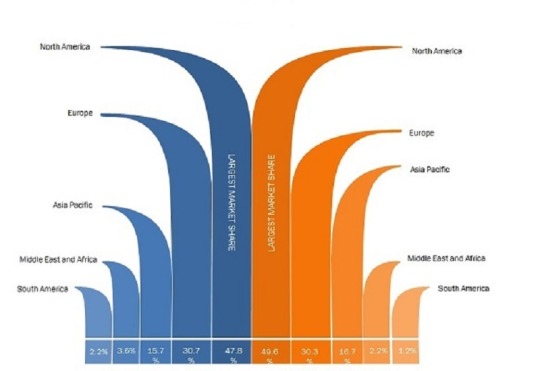
Rabies Diagnostics Market Regional Analysis:
The Asia Pacific region has a chance to dominate the global market as the market is witnessing growth for the high number of population. Improving the healthcare sector is expected to boost regional growth and ensure the sector gets proper funding. India and China are expected to play a crucial role in ensuring substantial revenue for the market.
The Americas would be the second-largest region with the US and Canada playing prominent roles. High level of awareness, better healthcare involvement, the inclusion of non-profit organizations to boost the market prospect, and others could help the market grow. Also, the market has a better chance of growth with the hike in awareness among pet owners.
Obtain Premium Research Report Details, Considering the impact of COVID-19 @ https://www.marketresearchfuture.com/reports/rabies-diagnostics-market-7796
Rabies Diagnostics Market Competitive Landscape:
Several top-ranked companies are taking notable initiatives to improve their market stance. The global market would get substantial support from companies like Merck KGaA, Bio-Rad Laboratories, Inc., Creative Diagnostics, Aviva Systems Biology Corporation, Abbexa Ltd., Demeditec Diagnostics GmbH, BioNote, Inc., Norgen Biotek Corp., MyBioSource.com., Express Biotech International Inc., and BioGen Technologies. These companies would explore the market possibilities using tactical moves like a merger, acquisition, collaboration, innovation, hike in research related funding, better awareness creation, and other moves. MRFR’s report on the market includes mentions of the latest moves of these companies. This is to facilitate an easy understanding of the market and ensure the chance of developing better strategies.
#Rabies Diagnostics Market#Rabies Diagnostics Market Size#Rabies Diagnostics Market Share#Rabies Diagnostics Market Growth#Rabies Diagnostics Market Analysis
0 notes
Text
Rabies Diagnostics Market 2023 Competitive Landscape, Opportunities, Challenges, Key Updates by Creative Diagnostics, Demeditec Diagnostics GmbH, Abbexa Ltd., Norgen Biotek Corp., BioNote, Inc., etc
Rabies Diagnostics: Information by Method (Fluorescent Antibody Test (FAT), Serology Tests, and Others), by Technology (ELISA/Immunohistochemistry, and Others), by End User (Hospitals Diagnostic centers, others)—Global Forecast till 2023
The Global Rabies Diagnostics Market is growing pervasively, mainly due to the increasing prevalence of rabies. Besides, spreading awareness of diagnostic procedures is driving the growth of the market. Also, rising research and development activities by non-profit organizations alongside, the aggressive strategies by top players such as mergers & acquisition, expansion, and collaboration for the development of new products and launches are accelerating the growth of the market on a global level.
According to a leading research firm Market Research Future (MRFR), the Global Rabies Diagnostics Market Size had created a valuation of USD 1964 MN in 2017 which is expected to further appreciate at a CAGR of 4.36% throughout the forecast period (2018–2023). In its analysis, MRFR asserts that the market will perceive phenomenal accruals by 2023. The major outburst of the rabies virus, mainly in various regions across the globe is a significant driving force for the market.
Governments, across the world, have taken up various initiatives to spread awareness about rabies viruses. They are introducing multiple diagnostic procedures in the hospitals, allotting significant budgetary programs. These factors, as a result, substantiate the growth of the market, commutatively. Simultaneously, diligent efforts put in by vendors and pharma companies to develop novel diagnostic devices foster the growth of the market.
Conversely, high costs associated with the diagnostic and vaccination is acting as a significant headwind to the growth of the market. Also, the growing number of new entrants with absolutely limited medical equipment business history, and a considerable price gap between the top players and small unorganized players are impeding the market growth.
Nevertheless, continuous technological advancements, rising demand for quick test results would support the growth of the market. Furthermore, increasing governmental endeavors to spread awareness towards the viruses are estimated to propel the market from 2018 to 2023.
Global Rabies Diagnostics Market – Competitive Analysis
Highly competitive, the Rabies diagnostics market appears to be widely expanded with the presence of many players. Expected extensions in products & services would drive the competition in the market, intensifying it further. This market demonstrates pretty good growth prospects that are expected to attract new entrant, also introducing the market with fierce competition.
Rabies Diagnostics Market Major Players:
Players leading the global rabies diagnostics market are Merck KGaA, Bio-Rad Laboratories, Inc., Aviva Systems Biology Corporation, Demeditec Diagnostics GmbH, Creative Diagnostics, Abbexa Ltd., BioNote, Inc., Norgen Biotek Corp., Express Biotech International Inc., BioGen Technologies, and MyBioSource.com, among others.
Global Rabies Diagnostics Market – Segments
The report is segmented into four dynamics to widen the scope of understanding,
By Method: Fluorescent Antibody Test (FAT), Immunohistochemical Test, Amplification Methods, Histologic Examination, and Serology Tests, among others.
By Technology: ELISA/Immunohistochemistry, Chromatography Techniques, and PCR, among others.
By End-user: Hospitals, Diagnostic Centers, and Cancer Palliative Care Clinics, among others.
By Regions: Europe, North America, Asia Pacific, and the Rest-of-the-World.
Browse More Information @ https://www.marketresearchfuture.com/reports/rabies-diagnostics-market-7796
Global Rabies Diagnostics Market – Geographical Analysis
The Asia Pacific has the maximum market coverage in the global rabies diagnostics market. Despite the several adversaries, the APAC market is surging rapidly due to the ongoing programs to increase awareness among people, developments in healthcare, and the growing per capita healthcare expenditure in the region.
With the rising investments in healthcare and expansions by market players, the region is expected to continue its dominance over the global rabies diagnostics market throughout the forecast period. Also, Government support for R&D focused on the development and adoption of advanced technology alongside, the availability of best treatment options in India and China are substantiating the regional market growth.
North America is the second-largest market for rabies diagnostics accounting for a substantial market share. Rising strategic alliances between significant players and well-spread awareness about rabies drive the growth of the market in the region. Additionally, the involvement of non-profit organization to control the disease and well-established healthcare infrastructure would speed up the growth of the regional market.
Also, the awareness towards the advantages of the test methods and the augmented governmental spending for R&D activities to develop novel diagnostic methods propel the growth of the regional market. Furthermore, technological advancements transpired into the field of rabies diagnostics driving forces behind the growth of the market in the region.
The rabies diagnostics market in Europe is multiplying. The market growth in this region attributes to the government funding, well-established healthcare sector, and support from the government for research and development. European governments spend substantially on diagnostic tests, innovations, and treatments for the virus.
0 notes
Text
Rabies Diagnostics Market – Global Industry Analysis, Size, Share, Growth, Trends, and Forecast 2019 - 2023
The Global Rabies Diagnostics Market is growing pervasively, mainly due to the increasing prevalence of rabies. Besides, spreading awareness of diagnostic procedures is driving the growth of the market. Also, rising research and development activities by non-profit organizations alongside, the aggressive strategies by top players such as mergers & acquisition, expansion, and collaboration for the development of new products and launches are accelerating the growth of the market on a global level.
According to a leading research firm Market Research Future (MRFR), the Global Rabies Diagnostics Market Share had created a valuation of USD 1964 MN in 2017 which is expected to further appreciate at a CAGR of 4.36% throughout the forecast period (2018–2023). In its analysis, MRFR asserts that the market will perceive phenomenal accruals by 2023. The major outburst of the rabies virus, mainly in various regions across the globe is a significant driving force for the market.
Governments, across the world, have taken up various initiatives to spread awareness about rabies viruses. They are introducing multiple diagnostic procedures in the hospitals, allotting significant budgetary programs. These factors, as a result, substantiate the growth of the market, commutatively. Simultaneously, diligent efforts put in by vendors and pharma companies to develop novel diagnostic devices foster the growth of the market.
Conversely, high costs associated with the diagnostic and vaccination is acting as a significant headwind to the growth of the market. Also, the growing number of new entrants with absolutely limited medical equipment business history, and a considerable price gap between the top players and small unorganized players are impeding the market growth.
Nevertheless, continuous technological advancements, rising demand for quick test results would support the growth of the market. Furthermore, increasing governmental endeavors to spread awareness towards the viruses are estimated to propel the market from 2018 to 2023.
Global Rabies Diagnostics Market – Competitive Analysis
Highly competitive, the Rabies diagnostics market appears to be widely expanded with the presence of many players. Expected extensions in products & services would drive the competition in the market, intensifying it further. This market demonstrates pretty good growth prospects that are expected to attract new entrant, also introducing the market with fierce competition.
Major Players:
Players leading the global rabies diagnostics market are Merck KGaA, Bio-Rad Laboratories, Inc., Aviva Systems Biology Corporation, Demeditec Diagnostics GmbH, Creative Diagnostics, Abbexa Ltd., BioNote, Inc., Norgen Biotek Corp., Express Biotech International Inc., BioGen Technologies, and MyBioSource.com, among others.
Global Rabies Diagnostics Market – Segments
The report is segmented into four dynamics to widen the scope of understanding,
By Method : Fluorescent Antibody Test (FAT), Immunohistochemical Test, Amplification Methods, Histologic Examination, and Serology Tests, among others.
By Technology : ELISA/Immunohistochemistry, Chromatography Techniques, and PCR, among others.
By End-user : Hospitals, Diagnostic Centers, and Cancer Palliative Care Clinics, among others.
By Regions : Europe, North America, Asia Pacific, and the Rest-of-the-World.
Browse More Information:- https://www.marketresearchfuture.com/reports/rabies-diagnostics-market-7796
Global Rabies Diagnostics Market – Geographical Analysis
The Asia Pacific has the maximum market coverage in the global rabies diagnostics market. Despite the several adversaries, the APAC market is surging rapidly due to the ongoing programs to increase awareness among people, developments in healthcare, and the growing per capita healthcare expenditure in the region.
With the rising investments in healthcare and expansions by market players, the region is expected to continue its dominance over the global rabies diagnostics market throughout the forecast period. Also, Government support for R&D focused on the development and adoption of advanced technology alongside, the availability of best treatment options in India and China are substantiating the regional market growth.
North America is the second-largest market for rabies diagnostics accounting for a substantial market share. Rising strategic alliances between significant players and well-spread awareness about rabies drive the growth of the market in the region. Additionally, the involvement of non-profit organization to control the disease and well-established healthcare infrastructure would speed up the growth of the regional market.
Also, the awareness towards the advantages of the test methods and the augmented governmental spending for R&D activities to develop novel diagnostic methods propel the growth of the regional market. Furthermore, technological advancements transpired into the field of rabies diagnostics driving forces behind the growth of the market in the region.
The rabies diagnostics market in Europe is multiplying. The market growth in this region attributes to the government funding, well-established healthcare sector, and support from the government for research and development. European governments spend substantially on diagnostic tests, innovations, and treatments for the virus.
#Rabies Diagnostics Market#Rabies Diagnostics Market Size#Rabies Diagnostics Market Share#Rabies Diagnostics Market Trends#Rabies Diagnostics Market Growth#Merck KGaA#Bio-Rad Laboratories#Aviva Systems Biology Corporation#Demeditec Diagnostics GmbH
0 notes
Text
Rabies Diagnostics Market Industry Booming Demand by Top Regions and Major Segments Till 2023
Rabies is a life treating a viral disease which is caused by the biting of an infected rabid animal. It is fever, headache, muscle aches, loss of appetite, nausea, and tiredness to the infected animal or human. The surge in the prevalence of rabies, increasing awareness about rabies, and a rising number of pet animals are expected to drive the growth of the global rabies diagnostics market during the forecast period. Moreover, ongoing research and development activities by non-profit organizations and aggressive strategies by top players such as mergers, expansion, and collaboration for the development of new products and new product launches by major players are also boosting the growth of the market. According to the report published by World Organization for Animal Health, Asia-Pacific region is hugely affected by rabies owing to the increasing cases of rabies, in Asia approximately 31000 deaths were caused by rabies which is 56% of the total cases.
The high costs associated with the diagnostic, absence of awareness about rabies and scarcity of skilled workforce may hamper the growth of the market during the assessment period.
Global Rabies Diagnostics Market: Competitive Landscape
Some of the key players in the global Rabies Diagnostics Market are
Bio-Rad Laboratories, Inc.
Merck KGaA
Aviva Systems Biology Corporation
Creative Diagnostics
Demeditec Diagnostics GmbH
Abbexa Ltd.
Norgen Biotek Corp.
BioNote, Inc.
Express Biotech International Inc.
com.
BioGen Technologies
Rabies Diagnostics Market Segmentation
The global rabies diagnostics market has been segmented on the basis of method, technology, end-user, and region.
Based on method, the market has been classified as a fluorescent antibody test (FAT), immunohistochemical test, amplification methods, histologic examination, and serology tests. Global rabies diagnostics market on the basis of technology has been segmented into ELISA/immunohistochemistry, chromatography techniques, PCR, and others.
Based on market has been segmented into hospitals, diagnostic centers, cancer palliative care clinics, and others.
Rabies Diagnostics Market Regional Analysis
The market is likely to dominate by Asia-Pacific during the forecast period owing to the improving healthcare infrastructure, increasing investments in the healthcare sector and expansions by market players in the region. Also, a country such as India and China are considering the fastest growing region due to a government focused toward adopting new technology, and the best treatment option from a developed country.
The Americas are expected to be the second-largest market in the global rabies diagnostics market due to the rising collaborative activities by major players, raising awareness about rabies, the involvement of non-profit organization to control rabies, and well-established healthcare infrastructure.
The European market is likely to hold a significant market share in the rabies diagnostics market. The government funding, well-established healthcare sector, and support of the healthcare sector coupled with increasing research and development are responsible for the growth of this market in the region.
The market in the Middle East & Africa is expected to account for the smallest share of the global rabies diagnostics market due to an underdeveloped healthcare sector, lack of technical knowledge, and poor medical facilities.
Table Of Content
1. Report Prologue
2. Market Introduction
2.1. Definition
2.2. Scope Of The Study
2.2.1. Research Objective
2.2.2. Assumptions
2.2.3. Limitations
3. Research Methodology
3.1. Introduction
3.2. Primary Research
3.3. Secondary Research
3.4. Market Size Estimation
4. Market Dynamics
4.1. Drivers
4.2. Restraints
4.3. Opportunities
4.4. Challenges
4.5. Macroeconomic Indicators
TOC Continued….!
Browse Complete Report Details @
https://www.marketresearchfuture.com/reports/rabies-diagnostics-market-7796
About US:
Market Research Future (MRFR), enable customers to unravel the complexity of various industries through Cooked Research Report (CRR), Half-Cooked Research Reports (HCRR), Raw Research Reports (3R), Continuous-Feed Research (CFR), and Market Research & Consulting Services.
Contact Us:
Market Research Future
Office No. 528, Amanora Chambers
Magarpatta Road, Hadapsar,
Pune - 411028
Maharashtra, India
Phone: +1 646 845 9312
Email: [email protected]
0 notes
Photo

Fluorescent Antibody Test (FAT) Market – Industry Trends and Forecast to 2027
Fluorescent antibody test (FAT) market is expected to grow at a CAGR of 6.8% in the forecast period of 2020-2027. The increasing occurrence of rabies in animals and humans has been directly impacting the growth of fluorescent antibody test (FAT) market.
Get Sample Report at :
https://bit.ly/2YbAtZL
#Fluorescent Antibody Test Market#Fluorescent Antibody Test Market share#Fluorescent Antibody Test Market size#Fluorescent Antibody Test Market trends#Fluorescent Antibody Test Market news#Fluorescent Antibody Test Market report#Fluorescent Antibody Test Market groiwth#Fluorescent Antibody Test Market forecast
0 notes
Link
Fluorescent antibody test (FAT) market is expected to grow at a CAGR of 6.8% in the forecast period of 2020-2027. The increasing occurrence of rabies in animals and humans has been directly impacting the growth of fluorescent antibody test (FAT) market.
#Fluorescent Antibody Test (FAT) Market#Fluorescent Antibody Test (FAT) Market sharee#Fluorescent Antibody Test (FAT) Market size#Fluorescent Antibody Test (FAT) Market trends#Fluorescent Antibody Test (FAT) Market growth#Fluorescent Antibody Test (FAT) Market report
0 notes
Text
Covid 19 Impact on Rabies Diagnostics Market Leading Players, Current Trends, Market Challenges, Growth Drivers And Business Opportunities
Rabies Diagnostics Market Overview
The global rabies diagnostics market is growing pervasively, mainly due to the increasing prevalence of rabies. Besides, spreading awareness of diagnostic procedures is driving the growth of the market. Also, rising research and development activities by non-profit organizations alongside, the aggressive strategies by top players such as mergers & acquisition, expansion, and collaboration for the development of new products and launches are accelerating the growth of the market on a global level.
According to a leading research firm Market Research Future (MRFR), the global rabies diagnostics market had created a valuation of USD 1964 MN in 2017 which is expected to further appreciate at a CAGR of 4.36% throughout the forecast period (2018–2023). In its analysis, MRFR asserts that the market will perceive phenomenal accruals by 2023. The major outburst of the rabies virus, mainly in various regions across the globe is a significant driving force for the market.
Governments, across the world, have taken up various initiatives to spread awareness about rabies viruses. They are introducing multiple diagnostic procedures in the hospitals, allotting significant budgetary programs. These factors, as a result, substantiate the growth of the market, commutatively. Simultaneously, diligent efforts put in by vendors and pharma companies to develop novel diagnostic devices foster the growth of the market.
Conversely, high costs associated with the diagnostic and vaccination is acting as a significant headwind to the growth of the market. Also, the growing number of new entrants with absolutely limited medical equipment business history, and a considerable price gap between the top players and small unorganized players are impeding the market growth.
Nevertheless, continuous technological advancements, rising demand for quick test results would support the growth of the market. Furthermore, increasing governmental endeavors to spread awareness towards the viruses are estimated to propel the market from 2018 to 2023.
Global Rabies Diagnostics Market – Segments
The report is segmented into four dynamics to widen the scope of understanding,
By Method: Fluorescent Antibody Test (FAT), Immunohistochemical Test, Amplification Methods, Histologic Examination, and Serology Tests, among others.
By Technology: ELISA/Immunohistochemistry, Chromatography Techniques, and PCR, among others.
By End-user: Hospitals, Diagnostic Centers, and Cancer Palliative Care Clinics, among others.
By Regions: Europe, North America, Asia Pacific, and the Rest-of-the-World.
Global Rabies Diagnostics Market – Geographical Analysis
The Asia Pacific has the maximum market coverage in the global rabies diagnostics market. Despite the several adversaries, the APAC market is surging rapidly due to the ongoing programs to increase awareness among people, developments in healthcare, and the growing per capita healthcare expenditure in the region.
With the rising investments in healthcare and expansions by market players, the region is expected to continue its dominance over the global rabies diagnostics market throughout the forecast period. Also, Government support for R&D focused on the development and adoption of advanced technology alongside, the availability of best treatment options in India and China are substantiating the regional market growth.
North America is the second-largest market for rabies diagnostics accounting for a substantial market share. Rising strategic alliances between significant players and well-spread awareness about rabies drive the growth of the market in the region. Additionally, the involvement of non-profit organization to control the disease and well-established healthcare infrastructure would speed up the growth of the regional market.
Also, the awareness towards the advantages of the test methods and the augmented governmental spending for R&D activities to develop novel diagnostic methods propel the growth of the regional market. Furthermore, technological advancements transpired into the field of rabies diagnostics driving forces behind the growth of the market in the region.
The rabies diagnostics market in Europe is multiplying. The market growth in this region attributes to the government funding, well-established healthcare sector, and support from the government for research and development. European governments spend substantially on diagnostic tests, innovations, and treatments for the virus.
Global Rabies Diagnostics Market – Competitive Analysis
Highly competitive, the Rabies diagnostics market appears to be widely expanded with the presence of many players. Expected extensions in products & services would drive the competition in the market, intensifying it further. This market demonstrates pretty good growth prospects that are expected to attract new entrant, also introducing the market with fierce competition.
Obtain Premium Research Report Details, Considering the impact of COVID-19 @ https://www.marketresearchfuture.com/reports/rabies-diagnostics-market-7796
Rabies Diagnostics Market Major Players:
Players leading the global rabies diagnostics market are Merck KGaA, Bio-Rad Laboratories, Inc., Aviva Systems Biology Corporation, Demeditec Diagnostics GmbH, Creative Diagnostics, Abbexa Ltd., BioNote, Inc., Norgen Biotek Corp., Express Biotech International Inc., BioGen Technologies, and MyBioSource.com, among others.
Rabies Diagnostics Industry/ Innovations/ Related News
July 03, 2019 ----- Ceva Sante Animale S.A. (France), announced the successful completion of the acquisition of veterinary biopharmaceutical and research and development activities of IDT Biologika GmbH (Germany). The acquisition would position Ceva closer to its 2020 growth objective of a 50:50 bio-pharma split. The product portfolio would be added to Ceva's portfolio soon. A new innovation center focused on swine vaccines would be created within IDT's existing Dessau site.
IDT's strong range of swine vaccines would strengthen Ceva's presence in this segment. The deal would boost Ceva's Veterinary Public Health offer in rabies control, with Rabitec, a rabies vaccine that can be used as oral immunization of foxes and other species. This would play a crucial role in campaigns focused on the successful eradication of rabies worldwide.
#Covid 19 Impact on Rabies Diagnostics Market#Rabies Diagnostics Market#Rabies Diagnostics Market Size#Rabies Diagnostics Market Share
0 notes
Text
Rabies Diagnostics Market Analysis By Development Status, Business Opportunities, Trends And Competitive Landscape
Rabies Diagnostics Market Overview
Global rabies diagnostics market is growing pervasively, mainly due to the increasing prevalence of rabies. Besides, spreading awareness of diagnostic procedures is driving the growth of the market. Also, rising research and development activities by non-profit organizations alongside, the aggressive strategies by top players such as mergers & acquisition, expansion, and collaboration for the development of new products and launches are accelerating the growth of the market on a global level.
According to a leading research firm Market Research Future (MRFR), the global rabies diagnostics market had created a valuation of USD 1964 MN in 2017 which is expected to further appreciate at a CAGR of 4.36% throughout the forecast period (2018–2023). In its analysis, MRFR asserts that the market will perceive phenomenal accruals by 2023. The major outburst of the rabies virus, mainly in various regions across the globe is a significant driving force for the market.
Governments, across the world, have taken up various initiatives to spread awareness about rabies viruses. They are introducing multiple diagnostic procedures in the hospitals, allotting significant budgetary programs. These factors, as a result, substantiate the growth of the market, commutatively. Simultaneously, diligent efforts put in by vendors and pharma companies to develop novel diagnostic devices foster the growth of the market.
Conversely, high costs associated with the diagnostic and vaccination is acting as a significant headwind to the growth of the market. Also, the growing number of new entrants with absolutely limited medical equipment business history, and a considerable price gap between the top players and small unorganized players are impeding the market growth.
Nevertheless, continuous technological advancements, rising demand for quick test results would support the growth of the market. Furthermore, increasing governmental endeavors to spread awareness towards the viruses are estimated to propel the market from 2018 to 2023.
Global Rabies Diagnostics Market – Segments
The report is segmented into four dynamics to widen the scope of understanding,
By Method: Fluorescent Antibody Test (FAT), Immunohistochemical Test, Amplification Methods, Histologic Examination, and Serology Tests, among others.
By Technology: ELISA/Immunohistochemistry, Chromatography Techniques, and PCR, among others.
By End-user: Hospitals, Diagnostic Centers, and Cancer Palliative Care Clinics, among others.
By Regions: Europe, North America, Asia Pacific, and the Rest-of-the-World.
Global Rabies Diagnostics Market – Geographical Analysis
The Asia Pacific has the maximum market coverage in the global rabies diagnostics market. Despite the several adversaries, the APAC market is surging rapidly due to the ongoing programs to increase awareness among people, developments in healthcare, and the growing per capita healthcare expenditure in the region.
With the rising investments in healthcare and expansions by market players, the region is expected to continue its dominance over the global rabies diagnostics market throughout the forecast period. Also, Government support for R&D focused on the development and adoption of advanced technology alongside, the availability of best treatment options in India and China are substantiating the regional market growth.
North America is the second-largest market for rabies diagnostics accounting for a substantial market share. Rising strategic alliances between significant players and well-spread awareness about rabies drive the growth of the market in the region. Additionally, the involvement of non-profit organization to control the disease and well-established healthcare infrastructure would speed up the growth of the regional market.
Also, the awareness towards the advantages of the test methods and the augmented governmental spending for R&D activities to develop novel diagnostic methods propel the growth of the regional market. Furthermore, technological advancements transpired into the field of rabies diagnostics driving forces behind the growth of the market in the region.
The rabies diagnostics market in Europe is multiplying. The market growth in this region attributes to the government funding, well-established healthcare sector, and support from the government for research and development. European governments spend substantially on diagnostic tests, innovations, and treatments for the virus.
Global Rabies Diagnostics Market – Competitive Analysis
Highly competitive, the Rabies diagnostics market appears to be widely expanded with the presence of many players. Expected extensions in products & services would drive the competition in the market, intensifying it further. This market demonstrates pretty good growth prospects that are expected to attract new entrant, also introducing the market with fierce competition.
OBTAIN PREMIUM RESEARCH REPORT DETAILS @ https://www.marketresearchfuture.com/reports/rabies-diagnostics-market-7796
Rabies Diagnostics Market Major Players:
Players leading the global rabies diagnostics market are Merck KGaA, Bio-Rad Laboratories, Inc., Aviva Systems Biology Corporation, Demeditec Diagnostics GmbH, Creative Diagnostics, Abbexa Ltd., BioNote, Inc., Norgen Biotek Corp., Express Biotech International Inc., BioGen Technologies, and MyBioSource.com, among others.
Rabies Diagnostics Industry/ Innovations/ Related News
July 03, 2019 ----- Ceva Sante Animale S.A. (France), announced the successful completion of the acquisition of veterinary biopharmaceutical and research and development activities of IDT Biologika GmbH (Germany). The acquisition would position Ceva closer to its 2020 growth objective of a 50:50 bio-pharma split. The product portfolio would be added to Ceva's portfolio soon. A new innovation center focused on swine vaccines would be created within IDT's existing Dessau site.
IDT's strong range of swine vaccines would strengthen Ceva's presence in this segment. The deal would boost Ceva's Veterinary Public Health offer in rabies control, with Rabitec, a rabies vaccine that can be used as oral immunization of foxes and other species. This would play a crucial role in campaigns focused on the successful eradication of rabies worldwide.
#Rabies Diagnostics Market#Rabies Diagnostics Market Size#Rabies Diagnostics Market Growth#Rabies Diagnostics Market Analysis
0 notes
Last Seen Blogs

comraderaccoon
🌹🍉Spooky Scary Socialist🍉🌹

gregparkersru
Greg Parker: Flashpoint RP Character

fnaf-movie-countdown
We’re Waiting Every Night~🎶

lovelyluna-glow
Moonlover

loverjpg
111 UNREAD 💌