#microradiographic
Text
Raychiel twerk to chunk by Starleon frm Starmacxmedia
My girlfriend sucking my hard cock in the parking lot
Plumber lays some pipe in bitchy blonde
Fatass Latina
Restrained Lydia Black receives merciless pussy hammering
Skinny Blonde Teen Jerking Cock
Aged man had a crazy sex session with a hot legal age teenager slut
Sweet schoolgirl banged in realsex action
CHUPADA DE PIJA Y GARCHE INTENSO BAREBACK EN MASAJES
Nina Kayy with a big anal ass
#acad#carpetbagged#carpogenic#do.#Messere#girtline#pilchard#dipteron#predeceaser#cavus#mateo-#jaspagate#runkling#pampas#prefiction#Orangemen#quadroons#unfimbriated#microradiographic#primevarous
2 notes
·
View notes
Photo
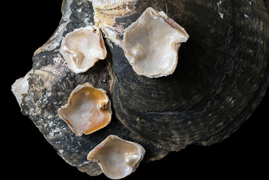
Figure 1: Four Pinctada maculata bead-cultured blister pearls attached to their hosts. Photo by Nuttapol Kitdee/GIA.
Pinctada maculata is the smallest mollusk of the Pinctada genus, averaging 40 mm at its widest point. The mollusk is found in the Pacific Ocean, including the waters off Penrhyn Island in the northern Cook Islands (Strack, 2001). P. maculata produces small natural pearls, also referred to as “pipi pearls,” ranging from yellow to orange (“golden”), pink, green, white, black, and gray, and commonly ranging from 4 to 5 mm in diameter (Buscher, 1999). Natural blister pearls* are sometimes encountered and are often referred to as “Puku” (Strack, 2001).
Figure 2: Map showing the location of Penrhyn Island (inset), approximately 3,430 miles (5,520 kilometers) from Brisbane, Australia.
This work describes the characteristics of four Pinctada maculata cultured blister pearls (figure 1) originating from Penrhyn Island, the northernmost atoll of the Cook Islands (figure 2). The cultured blister pearls were produced by Mr. Taruia Matara, who also experimented in culturing pearls using Pinctada magaritifera in the waters off Penrhyn Island in the early 1990s. His Pinctada maculata experiments took place in the late 1990s. However, he was unsuccessful with the latter and only a few examples were produced, four of which were donated to GIA by Mr. Umit Koruturk of Australian Pure Pearls in Sharjah, United Arab Emirates.
The cultured blister pearls supplied incorporated a freshwater bead as the nucleus. In keeping with the usual cultured blister pearl production procedures, the bead was placed within the shell between the inner shell surface and overlying mantle tissue (figure 3). This allows the epithelium cells in the mantle tissue to secrete nacre over the bead (Strack, 2001; figure 4).
Figure 3: Diagram of a bead nucleus (green sphere) glued to the shell beneath the mantle tissue.
Figure 4: The bead nucleus (green sphere) is overgrown with newly deposited nacre layers secreted by the mantle.
MATERIALS AND METHODS
We studied four P. maculata shells, with attached bead-cultured blister pearls. The shells measured approximately 49.03 mm x 48.94 mm, 41.51 mm x 40.45 mm, 38.43 mm x 38.53 mm, and 39.05 mm x 35.50 mm, and weighed 12.92, 7.97, 6.74 and 5.42 grams, respectively. We used a number of methods commonly employed in the analysis of pearls to examine the samples. The techniques applied helped determine their identity, the environment in which they formed, possible treatments used, and the possible mollusk origin. A Matrix-FocalSpot XT-3 series real-time X-ray (RTX) machine (voltage 90 kV and current 0.18 mA) fitted with a Toshiba image intensifier was used to identify the nature of the nuclei. A FocalSpot Verifier PF-100 model FSX-PF100 X-ray fluorescence unit (voltage 100 kV and current 3.2 mA ) was then used to provide information about the environment in which the shells and nuclei formed since marine and freshwater materials react differently when exposed to X-rays (Hänni et al., 2005). Finally, a Nikon SMZ18 microscope incorporating NIS-Elements photomicrography software and a Canon G16 camera were used to record the appearance of the samples. The former, together with a GIA binocular gemological microscope, were used to observe any evidence of possible treatments applied, and support some of the conclusions resulting from the other tests.
RESULTS
The shells and their blister pearls were photographed with other shells from the Pinctada genus in order to show the size of the shells in relation to the other species and capture the more obvious external features (figure 5). In theory, cultured pearls have a “newer” appearance than natural pearls, although newly found natural pearls do still appear in the market from time-to-time. Hence the surfaces of most cultured pearls are less likely to be as worn as those of some natural pearls. A pearl’s appearance is also usually related to the appearance of its shell (Southgate and Lucas, 2008). For instance, “golden pearls” mostly originate from mollusks that exhibit a degree of yellow on the shell (Strack, 2001).
Figure 5: Pinctada species shells showing the range of sizes in the genus. From left to right, P. maxima, P. margaritifera, P. mazatlanica, and P. maculata. The latter is the smallest species in the Pinctada genus. Photo by Nuttapol Kitdee/GIA.
This study focused on bead-cultured blister pearls still attached to their hosts. The majority of bead-cultured pearls are round to near-round in shape (Strack, 2001). The samples examined were more baroque to semi-baroque owing to their uneven outlines where they met the shell, but their main bodies were more toward near-round (figure 6).
Figure 6: A Pinctada maculata bead-cultured blister pearl attached to its host shell. The pearl exhibits a near-round form, as would be expected for a bead-cultured pearl. Photo by Promlikit Kessrapong/GIA.
To see the detail more clearly, we employed photomicrography to capture the fine features of the samples. One specimen in particular was of interest: a blister pearl that possessed a large opening between the nacre overgrowth and the host shell, permitting clear observation of the bead nucleus (figure 7 and figure 8).
Figure 7: This photomicrograph of the exposed area clearly shows there is a near-round bead nucleus within, surrounded by nacre that enfolds it and meets the shell. Photo by Kwanreun Lawanwong/GIA.
Figure 8: Magnified view of the bead nucleus revealing an abraded area in more detail. The multiple layers of nacre are easy to see, as are the gaps between the nacre, bead, and shell. Photo by Kwanreun Lawanwong/GIA.
While the external appearance of pearls can be very useful in pearl testing, it is the internal structure that gemologists ultimately need to consider when reaching a conclusion (Sturman, 2009). Various RTX and X-ray computed tomography units exist for this purpose, and when it comes to identifying most bead-cultured pearls the structures are quite obvious. Normally, bead-cultured pearls exhibit a clear demarcation between the bead nucleus and surrounding nacre, while the radio-opacity of the bead nucleus and nacre overgrowth differ (Wehrmeister et al., 2008). In some cases, obvious banding within the shell bead nucleus (figure 9) may be seen when the nucleus is correctly oriented in relation to the detector or, in rare cases these days, the film.
Figure 9: Characteristic bead-cultured pearl structures observed in samples examined in GIA’s Bangkok laboratory. A drilled bead-cultured pearl showing clear banding within the shell bead nucleus (A). A drilled bead-cultured pearl showing a very clear demarcation and organic-rich/void feature between the bead and nacre in one area (B). A bead nucleus with a thinner nacre coating and less-defined boundary (C).
The four samples examined in this study revealed clear demarcations, while the degree of radio-opacity of their beads and that of the surrounding nacre were easy to see. The fact that they are naturally attached to their shells was clearly evident on the RTX images as well. X-ray images captured from side-on (figure 10) proved the nacre covers the bead nucleus of each and blends in with the surface of their shell host without any joints or boundaries that would imply the shell was manipulated. All were clearly naturally attached cultured blister pearls.
Figure 10: Microradiograph of one of the bead-cultured blister pearls, showing the clear demarcation between the relatively large bead positioned against the host’s shell and the overlying nacre.
When X-rayed in the direction seen in figure 11, a very clear round white shape was visible. This relates to the much greater density (volume of material) the X-rays have to pass through in comparison to the much thinner section of shell. The bead and nacre overgrowth thus show as one form; higher magnification (zoom work) and refocusing, as well as the adjustment of the contrast, would be required to see the boundary between the bead and nacre clearly again.
Figure 11: The shell nucleus of this bead-cultured blister pearl, attached to its host shell and covered by nacre (left), is clearly revealed as a white radio-opaque sphere (right) on the microradiograph. Photo by Promlikit Kessrapong/GIA.
We determined the environment in which the shells and their blister pearls formed using an X-ray fluorescence unit where the results were captured via a camera within the unit. The X-ray fluorescence reactions of two of the samples studied are shown in figure 12 and figure 13, and they reveal a definite variation between the different components. A strong greenish yellow fluorescence was observed emanating from the bead nucleus. The strongest fluorescence comes from the bead nucleus because the vast majority of the cultured pearl industry uses freshwater shell (mostly sourced from the United States) which contains Mn in sufficient quantities to react with the X-rays (Hänni et al., 2005). In the saltwater areas possessing little or no Mn, the lack of a reaction is notable, and this is especially evident in the three images in figure 12, where the exposed bead reacts with an intense greenish yellow glow and the surrounding nacre is mostly inert. The weaker yellowish green fluorescence generally shown by areas of the nacre is due to a partial masking of the bead’s fluorescence by the overlying non-reactive SW nacre.
Figure 12: X-ray fluorescence images of a bead-cultured blister pearl on its host showing variations in fluorescence. A: Overall image of the piece. B: Reaction of the bead nucleus to the X-rays taken at an ISO setting of 6400. C: Reaction of the bead nucleus to the X-rays taken at a higher ISO setting (12800). Photos by Promlikit Kessrapong/GIA.
Figure 13: X-ray fluorescence image of one of the bead-cultured blister pearls studied exhibiting an X-ray fluorescence reaction. The image on the left shows the nacre overgrowth with no open or blemished areas. The image on the right shows the X-ray fluorescence reaction of the freshwater bead nucleus emanating from beneath the nacre (ISO setting 12800). Photos by Promlikit Kessrapong/GIA.
X-ray fluorescence is a useful test in the arsenal of techniques available to gemological laboratories. The reactions of beads and their pearls can vary according to the thickness of their nacre for two main reasons. The first is the thickness of the nacre overgrowth, and the second is the degree to which the freshwater bead itself fluoresces, since not all beads fluoresce with the same intensity. When considering both points, it should be easy to comprehend that beads with thinner nacre tend to produce a stronger reaction than those with a thicker nacre overgrowth, while a freshwater bead that shows weak fluorescence may show no fluorescence at all if it is surrounded by saltwater nacreous layers that are able to grow thick enough to completely subdue the fluorescence.
CONCLUSIONS
The existence of Pinctada maculata (pipi) bead-cultured blister pearls proves that limited success has been achieved in producing them. Nevertheless, it is also very apparent that we should not expect to see this product in the trade anytime soon. While bead-cultured blister pearls provide an alternative product in the market, they need to be of high quality to be marketable. Unfortunately, the experiments carried out on Pinctada maculata shells in Penrhyn Island did not result in suitably high quality end-products, and so they apparently were discontinued for a variety of reasons. It is very interesting to note that work was carried out to develop such bead-cultured blister pearls, and the results obtained from the scientific analysis of the four samples donated to GIA proved that their identification is straightforward, as is the case with almost all bead-cultured blister pearls. Their distinctive external appearance was the first clue to their identity, as the size of the blister pearls in relation to their small hosts was something perhaps a bit unusual, and of course what appeared to be a bead nucleus on one example visually was later confirmed by RTX, as were the three remaining samples. The RTX work clearly revealed the characteristic round outlines of shell beads used in the culturing process. Finally, the freshwater origin of the bead nuclei, in keeping with the majority of saltwater cultured pearls, was confirmed by observing their luminescence reactions after exposure to X-rays. The information from this study shows that Pinctada maculata bead-cultured blister pearls do exist, but only on an experimental basis. No commercial bead-cultured Pinctada maculata pearls of any kind are known to the authors, even after asking a few individuals involved principally in the trade of natural pearls produced by this, the smallest of all the Pinctada species.
DISCLAIMER
The term “blister pearl” has been applied throughout this article, since such products are often referred to this way in the trade. However, the correct nomenclature should not include the word "pearl" since the objects that formed on these shells are not “pearls” in the strict sense of the word. Blister pearls technically form within a pearl sac in the soft body of the mollusk and then move to the shell, where they are covered by additional nacreous layers.
https://www.gia.edu/gia-news-research/pinctada-maculata-bead-cultured-blister-pearls-shells
0 notes
Text
Microradiographic evaluation of bone from cows with experimental hypervitaminosis D, diet-induced hypocalcemia
http://dlvr.it/Nnh90K
0 notes
Last Seen Blogs

nocturnalfandomartist
Nocturnal Artist

bangla24net
Untitled

luxuryvision
VisionBoard

lightningelite
THOSE WHO RIDE THE LIGHTNING

albertfrazierdetroit
AlbertFrazierDetroit