#aminotransferase
Text
Case of Necrotizing Pancreatitis following COVID-19 Infection by Faezeh Sehatpour in Journal of Clinical Case Reports Medical Images and Health Sciences

ABSTRACT
New aspects of COVID-19 are increasingly being recognized. Although the virus is mainly known to affect the lungs, involvement of other organs including the heart, liver, gastrointestinal, renal and pancreas is also detected. Acute pancreatitis is detected as one of both the early and late presentations of COVID -19. Cytokine storm or the presence of angiotensin-converting enzyme 2 (ACE2) receptor in pancreatic cells, are both two causes of pancreatic injury in COVID-19 infection. In this study, we reported a 25-year-old man admitted to our department with the impression of necrotizing pancreatitis concomitant with COVID-19 infection. Patient's lab data, imaging and outcomes were documented in full detail.
Abbreviations:
WBC, white blood cell;HB, hemoglobin; MCV, mean corpuscular volume; PLT, platelet; BUN, blood urea nitrogen; Na, sodium; K, potassium; ; AST, aspartate aminotransferase; ALT, alanine aminotransferase; ALK.P, alkaline phosphatase; ALB, albumin; LDH, Lactate dehydrogenase ; CPK, creatine phosphokinase; CRP,c-reactive protein; AFP,alpha-fetoprotein; CEA,carcinoembryonic antigen; CA19-9,cancer antigen 19-9; Immunoglobulin G4.
INTRODUCTION
The Covid-19 pandemic is an ongoing pandemic that started in December 2019 and spread rapidly around the word. COVID-19 was caused by severe acute respiratory syndrome corona virus 2 (SARS-CoV-2), first identified in Wuhan, China. So far, more than 200 countries have been affected by the pandemic. (1)
New aspects of COVID-19 are increasingly being recognized. Although the virus is mainly known to affect the lungs, involvement of other organs including the heart, liver, gastrointestinal, renal and pancreas is increasingly being reported. (2)
The involvement of the gastrointestinal system is maybe due to the expression of the angiotensin-converting enzyme2 (ACE2) on the hepatocyte, cholangiocyte and other parts of the GI tract. (3) In a recent survey, acute pancreatitis was detected as one of both early and late presentations of COVID -19. (4-6) However, it is still unclear whether SARS-COV-2 directly affects pancreatic cells because of ACE2, if it is a cytokine storm which causes pancreatic injury. (7)
We reported a case of COVID-19 with subsequent acute necrotizing pancreatitis.
CASE REPORT
A 25-year-old man without any known medical disease presented to our emergency department with progressive epigastric pain, nausea and vomiting and anorexia one week prior to admission. He has no history of alcohol consumption. He also had a history of admission to another hospital about two weeks ago with a diagnosis of COVID-19 pneumonia. On admission, he has a blood pressure of 115/75 mm HG, a heart rate of 100 beats per minute, a temperature of 37.1 ⁰C and oxygen saturation of 95% while the patient is breathing in the room air. Primary investigations summarized in Table-1. Amylase and lipase were 146 IU/L and 82 IU/L respectively. Nasal swab test for COVID-19 (RT-PCR for SARS-CoV-2) was positive. Abdominal sonography showed markedly prominent pancreas with in homogeneous parenchymal echogenicity and large cystic lesion arising from the pancreas, in favor of acute complicated pancreatitis with pseudo cyst. The gall bladder has a normal size and wall thickness without any gall stones. The pancreatic duct was not dilated. Due to the finding of abdominal ultra sound, CT scan of abdomen was done on him which revealed an enlarged pancreas with necrosis of the main portion of pancreatic parenchyma. Large cystic lesion measuring 15×7×11 cm in size arising from the pancreatic neck with extension to the right and left side of the abdomen suggestive of large pancreatic pseudo cyst (figure1). Lung HRCT (low dose) also showed bilateral peripheral ground glass opacities in favor of COVID-19 pneumonia (figure2). According to the findings of a physical exam, laboratory data and clues in imaging immediate management of acute necrotizing pancreatitis (invasive intravenous hydration and pain control) was started for him. He was finally discharged from the hospital with a full recovery.
Table 1: laboratory data
Figure 1: Abdominal CT scan: large loculated pseudo cystic structure measuring about 158mm*100mm in lesser sac due to post pancreatitis pseudo cyst formation.
Figure 2: lung HRCT: multiple ground glass and bilateral pleural effusion
DISCUSSION
Acute pancreatitis is an acute inflammation of the pancreas characterized by abdominal pain, nausea, vomiting and elevated exocrine pancreatic enzymes; amylase and lipase. Gallstones and chronic alcohol abuse are the most common causes of acute pancreatitis. Viruses are uncommon causes of acute pancreatitis. Pancreatitis has been reported with several viruses, including mumps,
coxsackievirus, hepatitis A and B virus, cytomegalovirus, varicella-zoster, herpes simplex and human immunodeficiency virus. (8)
Although we have not conclusively proven the presence of the virus in the pancreas, the causes of COVID-19 and acute pancreatitis and the lack of other clear causes for pancreatitis strengthen the relationship between the two diseases. In this study, the patient presented with necrotizing COVID-19in 19 in the early post period of COVID-19 infection.
In Fan Wang and colleagues' survey, 52 COVID-19 cases followed and showed that 17% of COVID-19 patients developed pancreatic injury and presented with mild elevated pancreatic enzymes; serum amylase and lipase without clinically severe pancreatitis. The possibility of drug induced acute pancreatitis in patients who have received medication due to COVID-19 is also expressed as one of the reasons for acute pancreatitis in COVID-for19 infection. (9) Saffa Saeed Al Mazrouei and his teammates reported a 24-year-old patient with acute non-necrotizing pancreatitis with concurrent COVID-19. No evidence of pseudo cyst or abscess was detected in his imaging. (10)
Pancreatic damage can be due to the direct effect of the virus on pancreatic cells or indirectly secondary to the immune system. In another study in Wuhan, it showed that ACE2 was expressed in the pancreas higher than the lung in the normal population, indicating that SARS-CoV-2 can bind to ACE2 in the pancreas and cause pancreatic cell damage. (7, 11)
Acute pancreatitis is one of the presentations or complications of COVID-19 infection. Further investigation with samples is needed to reveal the pathophysiology, presentation, treatment and prognosis of acute pancreatitis in COVID-19 infection.
For more information: https://jmedcasereportsimages.org/about-us/
For more submission : https://jmedcasereportsimages.org/
#COVID-19#Cytokine storm#blood cell;HB#aminotransferase#CRP#c-reactive protein#carcinoembryonic antigen#alpha-fetoprotein#anorexia#RT-PCR for SARS-CoV-2#HRCT#Faezeh Sehatpour#jcrmhs
0 notes
Text
The aminotransferases in chloroplasts may have a significant role in amino acid biosynthesis, because plant leaves or isolated chloroplasts exposed to radioactively labeled carbon dioxide rapidly incorporate the label into glutamate, aspartate, alanine, serine, and glycine.
"Plant Physiology and Development" int'l 6e - Taiz, L., Zeiger, E., Møller, I.M., Murphy, A.
#book quote#plant physiology and development#nonfiction#textbook#aminotransferase#chloroplast#plant cells#organelles#amino acids#biosynthesis#biochemistry#glutamate#aspartate#alanine#serine#glycine#radioactive carbon
0 notes
Text


he ain't wrong
46 notes
·
View notes
Text
Learn to interpret your liver function test results with our comprehensive guide. Understand commonly used liver tests and their implications for your health.
Do Visit: https://www.healixhospitals.com/blogs/reading-and-interpreting-your-liver-function-test-a-guide-to-commonly-used-liver-tests
#Liver Function Test#Liver Health#Diagnostic Tests#Liver Enzymes#Blood Tests#Hepatic Function#Liver Panel#Bilirubin Levels#Liver Disease#Alanine Aminotransferase (ALT)#Aspartate Aminotransferase (AST)#Alkaline Phosphatase (ALP)#Gamma-Glutamyl Transferase (GGT)#Liver Health Markers#Hepatobiliary Disorders#Serum Biomarkers#Hepatic Enzymes#Liver Damage#Medical Laboratory Tests#Hepatology
1 note
·
View note
Text
Understanding Liver Function Test
What is a liver function test?
A liver function test is a blood test that measures the levels of various enzymes and proteins in your blood. These substances are produced by the liver, and they can be a sign of liver damage or disease.
#liver function test#Bilirubin#Alkaline Phosphatase (ALP)#Aspartate Aminotransferase (AST)#and Alanine Transaminase (ALT)
0 notes
Text
Role of Alpha Fetoprotein in hepatocellular carcinoma by MuhammadWaqar Mazhar in Journal of Clinical and Medical Images, Case Reports

Abstract
Hepatocellular carcinoma prevelance rate is higher in Pakistan due to HCV mortality rate, consumption of Alchol, and regular smoking, higher level of AFP progression normal liver cells into fatty liver cells, after inflammation it convert into HCC.In this study, we find the correlation between AFP and hepatocellular carcinoma. AFP involve in development of liver cancer, LFT’s test elevation and HCV also cause of cancer.
Keywords: Hepatocellular Carcinoma; Alpha Fetoprotein; alanine amino transferases; aspartate aminotransferases.
Introduction
Hepatocellular carcinoma is the 4th most common malignancy in worldwide and it is leading cause of cancer like disease in liver, and it exceed more than 1 million deaths per year by 2030 [1]. Acute hepatitis and acute liver failure are the most serious medical condition that require early diagnosis by release of IL-6, TNF-α and elevated alanine amino transferases, aspartate aminotransferases, alkaline phosphatase and α -Fetoprotein that progress healthy liver in to fatty liver known as steatosis and then inflammation occur in this and leads to hepatocellular carcinoma [2]. Most cases of HCC due to the virus like HCV and HBV, Diabetic and obesity, alcohol related diseases, non- alcohol related diseases, carcinogens like aflatoxins compounds [3]. HCC is the most common cancer that have high mortality rate in cancers due to mortality of HCV and NLFD. In Pakistan HCC ratio high due to prevalence and mortality rate of HCV [4]. The major treatment of HCC are chemotherapy, radiotherapy, transplantation and surgery. Because the most cases diagnose at the late stage, surgery cannot be performed and drugs are the only treatment of HCC [5]. Most patients in HCC become more drug resistance drug resistance. Drug treatment is the best choice of patients who are not edible for surgery. HCC is usually resistance to chemotherapeutic drugs. Because it hinders liver cancer treatment. In recent years targeted drugs use as medication and immune checkpoint inhibitors are introduce for treatment [6].
In the previous research evidence indicates that alpha-fetoprotein has high false-positive rate in diagnosis of early stage of HCC. The EASL clinic practices shows that AFP as a biomarker for liver transplantation and drug indicator [7]. The AFP level increased in many patients’ ad its risk for progression of HCC. AFP, currently the only biomarker available for HCC drug treatment, function as immune suppressor and promote malignancy transformation in HCC [8]. HCC is resistant to traditional chemotherapeutic agents such as doxorubicin, tetrahydrofolate, oxaliplatin, cisplatin, and gemcitabine. currently the recommended drugs include such as targeted therapeutics and immune checkpoint inhibitors [9].
AFP is a glycoprotein that secreted by endoderm embryonic tissue. The lower level of AFP in blood due to AFP is decrease in mature hepatocytes and that AFP gene expression is blocked. It is possible that AFP involved in HCC development and progression become an important factor affecting HCC diagnosis and treatment. AFP plays an important role in promoting cancer cell proliferation and, inhibition cancer cell apoptosis.
LFT’s test performed for liver injury, alanine aminotransferases, aspartate aminotransferases and alkaline phosphatase. These enzymes are commonly elevated in liver disease patients. Alkaline phosphatase and AFP play important role in the diagnosis of cancer.
Case Study
The patient name was sikandar, age 56 patient feel pain in their abdomen and sudden loss of weight. The patient has already hepatitis C infection and their PCR results were positive with high viral load. Due to serious illness it admitted in emergency ward 12, Nishter Hospital Multan. The doctors panel referred some test and kept in observations for better health condition.
The total bilirubin level was 2.05mg/dl in their blood and their normal values 0.6 - 1.2. The serum glutamate-pyruvate transaminase level is 43U/L and normal values up to 40. Aspartate amino transferases and alkaline phosphatase level were high in blood respectively 151 U/L and 493 U/l show in (Figure 1). Its indicate liver injury and cirrhosis. The AFP test indicates correlation with Hepatocellular carcinoma. The AFP level in patient was 6101ng/ml and normal values were 0.1 – 10. Higher level of AFP indicates that HCC have positive relation with AFP to proliferate cancer. The test formed by fully automated state of the Art analyzer Beckman Coulter 700 AIJ.

Figure 1: Liver function and Alpha Feto Protein test in patient.
After blood reports, doctor suggest ultarosund Computrised Tomography whole abdominal view. In view, spleen size becomes enlarged 6cm, calculi in gall bladder, heterogeneous patchy atrial enhancement of right lobe, and some nodules seen in both lobes of liver. The doctor findings the AFP correlation with HCC, splenomegaly, ascites, cholelithiasis and protosystematic collaterals.

Figure 2: Ultrasound Computrised Tomography whole abdomen.
The patient diagnosed with hepatocellular carcinoma at last stage, and doctor reffered to liver transplantation in india. But after 4 weeks he cannot survive.
Conclusion
Hepatitis C was the major risk of hepatocellular carcinoma in Pakistan. Smoking and alcohol have big problem to influence HCC in humans. The case study show that alpha fetoprotein has correlation with HCC. Higher Alkaline phosphatase and serum Bilirubin level enhance the liver carcinoma. AFP play role in cell proliferation, cancer cell differentiation and cell cycle arrest.
For more details : https://jcmimagescasereports.org/author-guidelines/
#Hepatocellular Carcinoma#Alpha Fetoprotein#alanine amino transferases#aspartate aminotransferases#malignancy#HCV#HCC#doxorubicin#tetrahydrofolate#oxaliplatin#Tomography#MuhammadWaqar Mazhar#JCMICR
0 notes
Link
The aspartate aminotransferase test (AST) is a liver function test. Your liver is responsible for making a fluid called bile, which helps in the digestion of food. It also eliminates waste products and other toxins from your blood.
0 notes
Text
Non-Operatively Managed Primary Small Bowel Volvulus: A Case Report by Ewnte B*

Abstract
Background: Primary small intestinal volvulus is one of the common causes of intestinal obstruction in various localities of the developing world. Although operative intervention has been the usual mode of treatment; this case report depicts meticulous follow-up & care, there is a possibility for relief of obstruction with non-operative management.
Case presentation: this is a case report of a 20-year-old male patient presented with crampy abdominal pain and frequent bilious vomiting. Plain abdominal film showed multiple distended small bowel loops with air fluid level, consistent with small bowel obstruction. Ruling out other etiologies primary small bowel volvulus was entertained and naso-gastric tube inserted, patient catheterized and kept nil per oral. After 48 hours of admission all symptoms resolved the patient resumed feeding and was discharged home.
Conclusions: The reported case shows evidence in which the patient’s primary small bowel volvulus was relieved non-operatively with insertion of naso gastric tube keeping nil per oral.
Key words: Small bowel volvulus; Primary volvulus; Non-operative management
Abbreviations: BPM: Beats Per Minute; WBC: White Blood Cells; RBC: Red Blood Cells; HGB: Hemoglobin; HCT: Hematocrit; MCV: Mean Corpuscular Volume; BUN: Blood Urea Nitrogen; ALT: Alanine Aminotransferase; AST: Aspartate Aminotransferase; ALP: Alkaline Phosphatase
Introduction
Small bowel volvulus is a condition in which there is a torsion of all or a segment of the small bowel and its mesentery: this can lead to bowel obstruction, ischemia, infarction, or perforation. The typical patient with the primary volvulus of the small intestine was found to be a young adult, male, muscular, farmer, from a rural area whose diet was bulky and mainly made of cereals [1,2].
Case Presentation
A 20-year-old male patient presented to Nefas Mewcha primary hospital Emergency department in January 2020 with the main complaint of crampy abdominal pain and distention of 14 hours duration. Associated with this, he also had nausea and frequent bilious vomiting eight times. He had passed feces 24 hours ago. He had no fever, cough, chest pain or night sweating. He had no history of similar illness before, no history of previous abdominal surgery.
He is not married and claims to be not sexually active. Lives with his parents and has three sisters and two brothers. He makes a living as a farmer. There are no medical illnesses that run in the family. There was no history of tobacco smoking or substance abuse. He consumes a local alcohol made of sorghum occasionally.
At presentation, his blood pressure was 105/60 mm Hg, pulse rate was 68 Beats Per Minute (BPM), respiratory rate was 18 per minute and temperature was 36.2 oC axillary. Physical examination of the patient at presentation, the patient was acutely sick looking in pain; not in cardio respiratory distress. He had a dry tongue and buccal mucosa. No palpable lymph adenopathy in all accessible areas. Chest was clear and resonant. S1 and S2 cardiac sounds were well heard and there were no added cardiac sounds. Abdomen was slightly distended, moves with respiration, flanks were full, there were no scars, no distended veins and hernia sites were free. Palpation revealed a tense abdomen with no area of tenderness, no shifting dullness, hyperactive tympanic percussion note, bowel sounds were 35 per minute. There is scanty stool on the examining finger, with no blood on it from digital rectal examination, no palpable mass was detected. The patient was conscious and neurological examination was intact.
A complete blood count of our patient showed: White Blood Cells (WBC) 12500 mcL, Red Blood Cells (RBC) 4.6 mcL, Hemoglobin (HGB) 16 gm/dL, Hematocrit (HCT) 48%, Mean Corpuscular Volume (MCV) 89.1fL, platelets 470×103, creatinine 0.6, Blood Urea Nitrogen (BUN) 30, Alanine Aminotransferase (ALT) 28, Aspartate Aminotransferase (AST) 24, Alkaline Phosphatase (ALP) 48, albumin 4.3, total bilirubin 1.1 and direct bilirubin 0.4.
Plain abdominal X-Ray showed centrally distributed, distended small bowel loops and rectal gas shadow (Figure 1). CT scan is not available at this setup so it was not possible to do one.
Management and Outcomes
The diagnosis of acute abdomen secondary to small bowel obstruction secondary to primary small bowel volvulus plus stage I shock was entertained, Double intravenous line was inserted and Trans-urethral catheter inserted, Naso-gastric tube was inserted. Three Liters of normal saline was given over a course of 2 hours, at emergency department. The patient was admitted to the ward and was advised on the possible options of management, consented on conservative management, associated risks and the possibility of surgical intervention at any time in the course of the management. The patient was kept Nil per oral, put on maintenance fluid and replacement of ongoing losses. Nasogastric tubes produced 600 ml of bilious content during the first 6 hours; which was replaced with an equal amount of ringer lactate. The abdominal cramp subsided after 4 hours of inpatient admission. After 12 hours of admission, the Blood pressure was 100/70 mmHg, pulse rate 68 per minute and the abdominal distension decreased significantly and the bowel sounds were 26 per minute, there was no area of tenderness and the patient passed flatus.
Following 24 hours of admission, the patient passed feces and vital signs were within normal range. Naso-gastric was removed and the patient was initiated with sips. The patient tolerated sips very well and was observed for 24 more hours and discharged on the next day. He was appointed to the surgical referral clinic after a week.
In subsequent weeks, the patient was seen at a referral clinic; he had no change in bowel habit or any other complaint. His vital signs were stable and physical examination was detected with no abnormality. He has been followed every month for 3 consecutive months and has reported no recurrence of symptoms.
Discussion
Volvulus is the Latin word for rolled up or twisted and is derived from the verb ‘volvere’, meaning to roll or turnabout. By definition, volvulus is an abnormal twisting of the intestine, which can impair the blood flow to the intestine. Volvulus can lead to gangrene and death of that segment of the gastrointestinal tract, intestinal obstruction, perforation of the intestine and peritonitis [3]. Small intestinal volvulus in adults can be classified as primary or secondary. In the former there is no obvious anatomical cause involving the mesentery or the small bowel, whereas in the latter there is an abnormal fixation due to adhesions or bands leading to the twisting of the mesentery. The primary type is often seen in Africa and Asia [4]. It is a significant cause of primary bowel obstruction in sub-Saharan Africa [5]. It is the leading cause of small bowel obstruction in Ethiopia [6].It is a rare entity in Western adults [7].
Clinical signs & symptoms were unspecific & resembled intestinal obstruction [8]. The most frequent symptom was observed to be sudden abdominal pain [9]. Vomiting was also observed in most of the patients while abdominal distention and constipation were reported less frequently [10]. Clinical examination reveals abdominal distension and/or diffuse tenderness with or without signs of peritonitis [8]. Small bowel volvulus is a rare but life-threatening surgical emergency. Owing to its rarity, it is seldom entertained as a differential for small bowel obstruction [11].
One of the challenges in managing primary small bowel volvulus operatively has been the risk of subsequent adhesion obstructions. The risk of occurrence of adhesion obstructions among patients that underwent laparotomy in general was reported to be 4.6% [12]. This gives rise to the endless circle of obstruction and subsequent operation, which further increase the risk more.
This case report presents a case of primary small bowel volvulus causing small bowel obstruction; which was managed non-operatively. Treatment of primary volvulus has mostly been via surgical intervention. This report depicts with close follow-up and Naso-gastric tube decompression, primary small bowel volvulus can also be treated without surgical intervention.
In the course of managing patients with primary small bowel volvulus, spontaneous resolutions has been observed [3]. This is because of natural de-rotation of the volvulus segment and relief of the obstruction. The case reported presented with symptoms and signs of small bowel obstruction. The patient has frequent vomiting with severe abdominal cramp associated with mild abdominal distension. The vital signs were within normal range supporting the diagnosis of non-ischemic obstruction. Abdominal x-ray showed multiple air fluid levels, which confirmed the diagnosis of small bowel obstruction. Ruling out other causes and considering the epidemiological prevalence, primary small bowel volvulus was entertained as a cause of obstruction.
As per the request of the patient to be followed conservatively, the patient was managed non-operatively with insertion of naso gastric tube and keeping nil per oral. The patient responded well for the management and were discharged subsequently. Showed no recurrence during the follow-up period.
Conclusion
Primary Small bowel volvulus is a rare cause of small bowel obstruction. The reported case shows evidence in which the patient’s primary small bowel volvulus was relieved non-operatively with insertion of naso gastric tube keeping nil per oral.
For more information about Article : https://ijclinmedcasereports.com/
https://ijclinmedcasereports.com/ijcmcr-cr-id-00131/
https://ijclinmedcasereports.com/pdf/IJCMCR-CR-00131.pdf
#Beats Per Minute#White Blood Cells#Red Blood Cells#Hemoglobin#Hematocrit#Mean Corpuscular Volume#Blood Urea Nitrogen#Alanine Aminotransferase#Aspartate Aminotransferase#Alkaline Phosphatase#Ewnte B*#IJCMCR#clinical studies
0 notes
Text
Toko yang Jual alat pengukuran aktivitas ALT dari Prima Medikatama Untuk Pusat Dialisis

Toko yang Jual alat pengukuran aktivitas ALT KLIK https://primamedikatama.com/, Reagen Kimia Darah AST/GOT EVOGEN, Reagen Pemeriksaan Golongan Darah, Probe Cleanser 50ml, Reagen Widal Test, Blood Control Hematology.
Apakah Anda mencari reagen yang dapat memberikan hasil pengukuran aktivitas ALT (Alanin Aminotransferase) dengan akurat?
Prima Medikatama mempersembahkan Reagen ALAT (GPT) FS (IFCC mod.) yang dapat memenuhi kebutuhan pusat dialisis Anda.
Spesifikasi Produk
Volume: 2 x 50 ml
Metode: IFCC (International Federation of Clinical Chemistry)
Jenis Metode: UV Enzymatic, KINETIC
Berat setelah dipacking: 500 gram
Dimensi: Panjang: 10 cm, Lebar: 10 cm, Tinggi: 12 cm
Harga: Rp350.000 per box (2 x 50 ml)
Fungsi Produk
Reagen ini dirancang khusus untuk pengukuran kuantitatif aktivitas ALT dalam serum manusia.
ALT, atau dikenal sebagai SGPT, adalah salah satu indikator penting dalam tes fungsi hati.
Peningkatan kadar ALT dapat mengindikasikan adanya kerusakan atau peradangan pada jaringan hati.
Keunggulan Produk
Reagen ALAT (GPT) FS (IFCC mod.) adalah reagen diagnostik yang diandalkan.
Pengukuran dilakukan dengan menggunakan metode spektrofotometri UV pada panjang gelombang 340 nm sesuai dengan standar IFCC.
Produk ini terdiri dari dua reagen, yaitu R1 dan R2, yang berbentuk cairan siap pakai.
Keunggulan lainnya adalah stabilitasnya hingga masa kedaluwarsa, sehingga Anda dapat mempercayai hasil yang konsisten.
Dengan menggunakan reagen ini, pusat dialisis Anda dapat memastikan hasil pengukuran ALT yang andal dan akurat.
Membantu dalam diagnosis penyakit hati, dan mengawasi perkembangan pasien dengan lebih baik.
Percayakan kualitas pengukuran ALT pada Prima Medikatama.
Dapatkan reagen ALAT (GPT) FS (IFCC mod.) sekarang juga, dan pastikan bahwa pasien Anda menerima perawatan yang terbaik.
Untuk informasi lebih lanjut atau untuk memesan alat pengukuran aktivitas ALT, jangan ragu untuk menghubungi kami melalui https://wa.me/6282311150090 atau dengan mengklik tombol "alat pengukuran aktivitas ALT" Untuk Pemesanan.
#Diluent Mindray 20L#Probe Cleanser 50ml#Alat Kesehatan Habis Pakai#Probe Cleanser Mindray#Lyse Mindray#Reagen Widal Salmonella Typhi O#Reagen Pemeriksaan Golongan Darah#Reagen Widal
77 notes
·
View notes
Text
Rheumatoid Arthritis:
Refer to rheumatologist.
●Nonpharmacologic measures – Nonpharmacologic measures, such as patient education, psychosocial interventions, and physical and occupational therapy, should be used in addition to drug therapy. Other medical interventions that are important in the comprehensive management of RA in all stages of disease include cardiovascular risk reduction and immunizations to decrease the risk of complications of drug therapies.
●Initiation of DMARD therapy soon after RA diagnosis – We suggest that all patients diagnosed with RA be started on disease-modifying antirheumatic drug (DMARD) therapy as soon as possible following diagnosis, rather than using antiinflammatory drugs alone, such as nonsteroidal antiinflammatory drugs (NSAIDs) and glucocorticoids (Grade 2C). Better outcomes are achieved by early compared with delayed intervention with DMARDs.
●Tight control of disease activity – Tight control treatment strategies to "treat to target" are associated with improved radiographic and functional outcomes compared with less aggressive approaches. Such strategies involve reassessment of disease activity on a regularly planned basis with the use of quantitative composite measures and adjustment of treatment regimens to quickly achieve and maintain control of disease activity if targeted treatment goals (remission or low disease activity) have not been achieved. (
●Pretreatment evaluation – Laboratory testing prior to therapy should include a complete blood count, erythrocyte sedimentation rate (ESR), C-reactive protein (CRP), aminotransferases, blood urea nitrogen, and creatinine. Patients receiving hydroxychloroquine (HCQ) should have a baseline ophthalmologic examination, and most patients who will receive a biologic agent or Janus kinase (JAK) inhibitor should be tested for latent tuberculosis (TB) infection. Screening for hepatitis B and C should be performed in all patients. Some patients may require antiviral treatment prior to initiating DMARD or immunosuppressive therapy, depending upon their level of risk for hepatitis B virus (HBV) reactivation.
●Adjunctive use of antiinflammatory agents – We use antiinflammatory drugs, including NSAIDs and glucocorticoids, as bridging therapies to rapidly achieve control of inflammation until DMARDs are sufficiently effective. Some patients may benefit from longer-term therapy with low doses of glucocorticoids.
●Drug therapy for flares – RA has natural exacerbations (also known as flares) and reductions of continuing disease activity. The severity of the flare and background drug therapy influence the choice of therapies. Patients who require multiple treatment courses with glucocorticoids for recurrent disease flares and whose medication doses have been increased to the maximally tolerated or acceptable level should be treated as patients with sustained disease activity. Such patients require modifications of their baseline drug therapies.
●Monitoring – The monitoring that we perform on a regular basis includes testing that is specific to evaluation of the safety of the drugs being; periodic assessments of disease activity with composite measures; monitoring for extraarticular manifestations of RA, other disease complications, and joint injury; and functional assessment.
●Other factors affecting target and choice of therapy – Other factors in RA management that may influence the target or choice of therapy include the disabilities or functional limitations important to a given patient, progressive joint injury, comorbidities, and the presence of adverse prognostic factors.
Osteoarthritis
General principles – General principles of osteoarthritis (OA) management include providing continuous care that is tailored to the patient according to individual needs, goals, and values and should be patient-centered. Treatment can be optimized by OA and self-management education, establishing treatment goals, and periodic monitoring.
●Monitoring and assessment – The management of OA should include a holistic assessment which considers the global needs of the patient. Patient preferences for certain types of therapies should also be assessed, as compliance and outcomes can be compromised if the care plan does not meet the patient's preferences and beliefs.
●Overview of management – The goals of OA management are to minimize pain, optimize function, and beneficially modify the process of joint damage. The primary aim of clinicians should include targeting modifiable risk factors. Due to the modest effects of the individual treatment options, a combination of therapeutic approaches is commonly used in practice and should prioritize therapies that are safer.
●Nonpharmacologic therapy – Nonpharmacologic interventions are the mainstay of OA management and should be tried first, followed by or in concert with medications to relieve pain when necessary. Nonpharmacologic therapies including weight management and exercises, braces and foot orthoses for patients suitable to these interventions, education, and use of assistive devices when required.
●Pharmacologic therapy – The main medications used in the pharmacologic management of OA include oral and topical nonsteroidal antiinflammatory drugs (NSAIDs). Other options include topical capsaicin, duloxetine, and intraarticular glucocorticoids. Our general approach to pharmacotherapy is described below.
•In patients with one or a few joints affected, especially knee and/or hand OA, we initiate pharmacotherapy with topical NSAIDs due to their similar efficacy compared with oral NSAIDs and their better safety profile.
•We use oral NSAIDs in patients with inadequate symptom relief with topical NSAIDs, patients with symptomatic OA in multiple joints, and/or patients with hip OA. We use the lowest dose required to control the patient's symptoms on an as-needed basis.
•We use duloxetine for patients with OA in multiple joints and concomitant comorbidities that may contraindicate oral NSAIDs and for patients with knee OA who have not responded satisfactorily to other interventions.
•Topical capsaicin is an option when one or a few joints are involved and other interventions are ineffective or contraindicated; however, its use may be limited by common local side effects.
•We do not routinely use intraarticular glucocorticoid injections due to the short duration of its effects (ie, approximately four weeks).
•We avoid prescribing opioids due to their overall small effects on pain over placebo and potential side effects (eg, nausea, dizziness, drowsiness), especially for long-term use and in the older adult population.
•We do not routinely recommend nutritional supplements such as glucosamine, chondroitin, vitamin D, diacerein, avocado soybean unsaponifiables (ASU), and fish oil due to a lack of clear evidence demonstrating a clinically important benefit from these supplements. Other nutritional supplements of interest that may have small effects on symptoms include curcumin (active ingredient of turmeric) and/or Boswellia serrata, but the data are limited.
●Role of surgery – Surgical treatment is dominated by total joint replacement, which is highly effective in patients with advanced knee and hip OA when conservative therapies have failed to provide adequate pain relief.
●Factors affecting response to therapy – The discordance of radiographic findings to pain supports the notion that the mechanisms of pain are complex and likely multifactorial. The placebo effect is also known to impact response to therapy.
●Prognosis – Although there is great variability among individuals and among different phenotypes of OA, courses of pain and physical functioning have been found to be predominantly stable, without substantial improvement or deterioration of symptoms over time.
2 notes
·
View notes
Text
Unitron stick with natural ways. LAb pastry dish growing method
frequency in autoimmune hepatitis sera [96–98]. In one study on 30 cases, there was reactivity to the 70-kDa U1-RNP in 8, and to the Ro/La (SS-A/SS-B) antigens in 3 (97), and in another report on cases from North America and Asia, 95 and 47, respectively, the frequencies for the North American cases were 43% for snRNPs and 38% for Ro/La, and frequencies for the Asian cases were generally higher [98]. One reactivity described was to an unidentified 52-kDa RNP, in 23% of 65 cases of AIH [82], but there has been no follow-up on this.
Delivery method
Chronic active hepatitis (CAH) is characterized by persisting high aminotransferase activities in blood and inflammation in the liver. An immunologic basis for CAH was proposed, based on antinuclear antibodies (ANA) first detected by the test for lupus erythematosus (LE) cells, and a complement fixation (CF) reaction for anticytoplasmic antibodies. Autoimmune hepatitis is a female-predominant disease. Autoimmune hepatitis (AIH) belongs to that puzzling group of autoimmune disorders in which there is a disease-specific target organ yet non-organ-specific serologic markers, and it remains one of the less well understood in regard to both inductive and effector processes. The chapter provides an analysis of the range of antigenic reactants in the context of the defined serologic types, 1 and 2. Furthermore, contemporary protocols for diagnosis and therapy are so well accepted and effective that research into the basic nature of AIH lacks the profile that pertains for the more prevalent and less successfully treatable autoimmune diseases.
Absorption without outside body and fuctions.
13 notes
·
View notes
Text
Cholestasis during pregnancy
Cholestasis is a condition that affects the liver, gallbladder, and bile ducts. It can occur in two ways. Extrahepatic cholestasis occurs outside the liver (also known as non-obstetric cholestasis). Intrahepatic cholestasis, also called obstetric cholestasis, happens inside the liver. In pregnant women, hormones change the way bile flows through the gallbladder and the bile ducts. This causes bile to build up in the liver and spill into the bloodstream.
Obstetric cholestasis is a pregnancy-related liver disorder that occurs in approximately 1 in 200 women during their third trimester. It is marked by itching, high levels of serum aminotransferases and bile acids, and signs and symptoms that go away on their own two to three weeks after birth.
In severe obstetric cholestasis, the condition can be very serious and lead to premature birth, fetal distress, and stillbirth. Surgical treatment is usually recommended to prevent these complications and lessen the risk for you and your baby.
Fortunately, there is an effective treatment for obstetric cholestasis called ursodeoxycholic acid. It protects interstitial Cajal-like cells in the gallbladder from undergoing apoptosis by inhibiting TNF-a expression, thus preventing oxidative stress and thrombotic complications.
Cholestasis of pregnancy (also called intrahepatic cholestasis) is a liver disorder that can develop during your pregnancy. Your doctor will diagnose it by doing a physical exam and a blood test that shows how well your liver is functioning.
The tests will also measure how much bile acid is in your bloodstream. The more bile acids you have, the more likely you are to have cholestasis during pregnancy.
Intrahepatic cholestasis of pregnancy is a rare, reversible disease that typically occurs in the second half of pregnancy. It causes severe pruritus and an elevation of total serum bile acids, which may lead to jaundice in 10% of patients.
A pregnant woman with obstetric cholestasis (also called intrahepatic cholestasis of pregnancy) may experience itching without a rash. It typically starts in the third trimester and goes away after delivery.
Cholestasis occurs when bile, the digestive fluid that helps break down fats, does not leave the liver for the small intestine. This causes bile acids to build up in the bloodstream and cause itching.
The bile acids can also cause problems with a pregnant woman’s ability to absorb fat, and this can affect her blood clotting. This can also affect her baby, increasing the risk of stillbirth and premature birth.
Cholestasis occurs because of an impairment in the normal flow of bile. This leads to the accumulation of bile acids, bilirubin, and cholesterol.
The main cause of cholestasis is obstruction of the bile flow in the hepatocytes or cholangiocytes. This can happen for a number of different reasons.
One of the most common causes is a blockage in your gallbladder (bile ducts). Another cause is an obstruction in the liver or the tubes that carry bile from your stomach to your intestine.
You may need to take medication to help your bile move through your body more easily. This can reduce your itching and help your bile levels return to normal.
Cholestasis is a condition in which the flow of bile from your liver to your small intestine slows down or stops. This can happen due to problems with your liver, bile duct, or pancreas.
If you have obstetric cholestasis, your doctor will check for other health issues to help determine the cause. You'll have a physical exam and get blood tests to measure the level of bile acid in your blood as well as your liver function.
In most cases, obstetric cholestasis resolves after you deliver your baby. But you'll need to keep up with monitoring for a while after you give birth. You may have ultrasounds and fetal heart monitors to make sure your baby is okay.
3 notes
·
View notes
Text
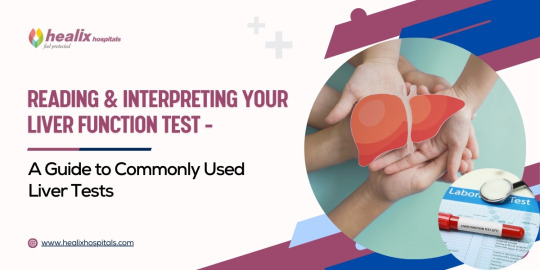
Reading And Interpreting Your Liver Function Test - A Guide To Commonly Used Liver Tests

The liver is a vital organ responsible for numerous metabolic functions in the body, including detoxification, protein synthesis, and bile production. Monitoring liver health is crucial for early detection and management of liver diseases. One of the primary tools for assessing liver function is the Liver Function Test (LFT). In this guide, we will delve into the commonly used liver tests, how to interpret the results, and what they indicate about your liver health.
Understanding Liver Function Tests
Liver Function Tests (LFTs) are a group of blood tests that provide valuable insights into the health and function of the liver. These tests measure various enzymes, proteins, and substances in the blood that are indicative of liver health.
Key components of Liver Function Tests
Alanine Aminotransferase (ALT): Elevated levels suggest liver damage, commonly caused by conditions like hepatitis or fatty liver disease.
Aspartate Aminotransferase (AST): Similar to ALT, elevated AST levels indicate liver damage but may also be elevated in conditions affecting the heart or muscles.
Alkaline Phosphatase (ALP): Elevated ALP levels may suggest liver or bone disease.
Total Bilirubin: Increased levels may indicate liver dysfunction or obstruction of bile ducts.
Albumin and Total Protein: These are measures of liver synthetic function; decreased levels may suggest liver disease.
What are the causes of abnormal liver function test results?
Causes of abnormal liver function test results can vary and may indicate different underlying conditions. Some common causes include:
1. Build-up of Fat in the Liver:
* Non-alcoholic fatty liver disease (NAFLD) can lead to abnormal liver function tests, especially in overweight or obese individuals.
2. Liver Inflammation and Damage:
* Infections, toxic substances like alcohol or certain medications, and immune conditions can cause liver inflammation and subsequent abnormal test results.
3. Liver Overworking:
* When the liver is under stress from processing medicines or toxic substances like alcohol or paracetamol, it can result in abnormal liver function tests.
4. Bile Duct Blockage:
* Blockages in the bile ducts, such as by gallstones, can lead to abnormal liver function test results.
5. Liver Conditions and Diseases:
* Underlying conditions like Wilson's disease, haemochromatosis, or Gilbert's syndrome can affect liver function and result in abnormal test values.
6. Liver Injury:
* Physical injury to the liver, trauma, or presence of abscesses or tumors within the liver can cause abnormal liver function tests.
7. Medications and Supplements:
* Certain medications, over-the-counter drugs, herbal remedies, and traditional medicines can also impact liver function test results.
8. Other Factors:
* Factors like high alcohol intake, viral infections, autoimmune conditions, metabolic liver diseases, heart problems, and tumors in the liver can contribute to abnormal liver function tests.
Continue Reading: https://www.healixhospitals.com/blogs/reading-and-interpreting-your-liver-function-test-a-guide-to-commonly-used-liver-tests
#Liver Function Test#Liver Health#Diagnostic Tests#Liver Enzymes#Blood Tests#Hepatic Function#Liver Panel#Bilirubin Levels#Liver Disease#Alanine Aminotransferase (ALT)#Aspartate Aminotransferase (AST)#Alkaline Phosphatase (ALP)#Gamma-Glutamyl Transferase (GGT)#Liver Health Markers#Hepatobiliary Disorders#Serum Biomarkers#Hepatic Enzymes#Liver Damage#Medical Laboratory Tests#Hepatology
1 note
·
View note
Text
Get Precise Insights into Your Liver Health with PACE Hospitals' SGPT Test in Hyderabad
Your liver plays a vital role in your overall health, performing numerous functions like detoxification, protein synthesis, and blood sugar regulation. Monitoring its health is crucial for early detection of potential problems. The SGPT test in Hyderabad, also known as the ALT (alanine aminotransferase) test, is a simple blood test offered by PACE Hospitals that measures the level of an enzyme present in the liver. Elevated SGPT levels can indicate liver damage or disease.
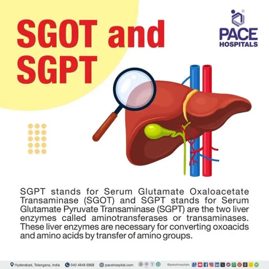
What is SGOT and SGPT ?
SGPT stands for Serum Glutamate Oxaloacetate Transaminase (SGOT) and SGPT stands for Serum Glutamate Pyruvate Transaminase (SGPT) are the two liver enzymes called aminotransferases or transaminases. These liver enzymes are necessary for converting oxoacids and amino acids by transfer of amino groups.
SGPT and SGOT are obsolete (old) names. While SGPT is now currently termed as Alanine Aminotransferase (ALT), SGOT is termed as Aspartate Aminotransferase (AST).
The SGOT and SGPT normal range is.
Alanine transaminase (SGPT or ALT): 4 to 36 IU/L
Aspartate transaminase (SGOT or AST): 5 to 30 IU/L
Benefits of Getting an SGPT Test at PACE Hospitals:
Accurate and Reliable Results: PACE Hospitals utilizes state-of-the-art equipment and adheres to stringent quality control measures to ensure the accuracy of your test results.
Expert Guidance: Our team of experienced healthcare professionals can interpret your test results and provide personalized recommendations based on your specific needs.
Proactive Management: Early detection of liver issues allows for timely intervention and treatment, potentially preventing complications.
Convenience and Comfort: Schedule your SGPT test appointment online or by calling our dedicated team. Our patient-centric approach ensures a smooth and comfortable experience.
Who Should Consider an SGPT Test?
An SGPT test may be recommended for individuals with:
A history of liver disease, such as hepatitis
Symptoms of liver problems, including fatigue, nausea, abdominal pain, or jaundice
Certain medical conditions like diabetes or high cholesterol
Individuals who consume excess alcohol
Take Charge of Your Liver Health Today
PACE Hospitals is committed to providing comprehensive and advanced diagnostic services to empower you to make informed decisions about your well-being.
Don't wait to prioritize your liver health. Take the first step towards optimal well-being with PACE Hospitals' SGPT test.
0 notes
Text
Role of Alpha Fetoprotein in hepatocellular carcinoma by MuhammadWaqar Mazhar in Journal of Clinical and Medical Images, Case Reports

Abstract
Hepatocellular carcinoma prevelance rate is higher in Pakistan due to HCV mortality rate, consumption of Alchol, and regular smoking, higher level of AFP progression normal liver cells into fatty liver cells, after inflammation it convert into HCC.In this study, we find the correlation between AFP and hepatocellular carcinoma. AFP involve in development of liver cancer, LFT’s test elevation and HCV also cause of cancer.
Keywords: Hepatocellular Carcinoma; Alpha Fetoprotein; alanine amino transferases; aspartate aminotransferases.
Introduction
Hepatocellular carcinoma is the 4th most common malignancy in worldwide and it is leading cause of cancer like disease in liver, and it exceed more than 1 million deaths per year by 2030 [1]. Acute hepatitis and acute liver failure are the most serious medical condition that require early diagnosis by release of IL-6, TNF-α and elevated alanine amino transferases, aspartate aminotransferases, alkaline phosphatase and α -Fetoprotein that progress healthy liver in to fatty liver known as steatosis and then inflammation occur in this and leads to hepatocellular carcinoma [2]. Most cases of HCC due to the virus like HCV and HBV, Diabetic and obesity, alcohol related diseases, non- alcohol related diseases, carcinogens like aflatoxins compounds [3]. HCC is the most common cancer that have high mortality rate in cancers due to mortality of HCV and NLFD. In Pakistan HCC ratio high due to prevalence and mortality rate of HCV [4]. The major treatment of HCC are chemotherapy, radiotherapy, transplantation and surgery. Because the most cases diagnose at the late stage, surgery cannot be performed and drugs are the only treatment of HCC [5]. Most patients in HCC become more drug resistance drug resistance. Drug treatment is the best choice of patients who are not edible for surgery. HCC is usually resistance to chemotherapeutic drugs. Because it hinders liver cancer treatment. In recent years targeted drugs use as medication and immune checkpoint inhibitors are introduce for treatment [6].
In the previous research evidence indicates that alpha-fetoprotein has high false-positive rate in diagnosis of early stage of HCC. The EASL clinic practices shows that AFP as a biomarker for liver transplantation and drug indicator [7]. The AFP level increased in many patients’ ad its risk for progression of HCC. AFP, currently the only biomarker available for HCC drug treatment, function as immune suppressor and promote malignancy transformation in HCC [8]. HCC is resistant to traditional chemotherapeutic agents such as doxorubicin, tetrahydrofolate, oxaliplatin, cisplatin, and gemcitabine. currently the recommended drugs include such as targeted therapeutics and immune checkpoint inhibitors [9].
AFP is a glycoprotein that secreted by endoderm embryonic tissue. The lower level of AFP in blood due to AFP is decrease in mature hepatocytes and that AFP gene expression is blocked. It is possible that AFP involved in HCC development and progression become an important factor affecting HCC diagnosis and treatment. AFP plays an important role in promoting cancer cell proliferation and, inhibition cancer cell apoptosis.
LFT’s test performed for liver injury, alanine aminotransferases, aspartate aminotransferases and alkaline phosphatase. These enzymes are commonly elevated in liver disease patients. Alkaline phosphatase and AFP play important role in the diagnosis of cancer.
Case Study
The patient name was sikandar, age 56 patient feel pain in their abdomen and sudden loss of weight. The patient has already hepatitis C infection and their PCR results were positive with high viral load. Due to serious illness it admitted in emergency ward 12, Nishter Hospital Multan. The doctors panel referred some test and kept in observations for better health condition.
The total bilirubin level was 2.05mg/dl in their blood and their normal values 0.6 - 1.2. The serum glutamate-pyruvate transaminase level is 43U/L and normal values up to 40. Aspartate amino transferases and alkaline phosphatase level were high in blood respectively 151 U/L and 493 U/l show in (Figure 1). Its indicate liver injury and cirrhosis. The AFP test indicates correlation with Hepatocellular carcinoma. The AFP level in patient was 6101ng/ml and normal values were 0.1 – 10. Higher level of AFP indicates that HCC have positive relation with AFP to proliferate cancer. The test formed by fully automated state of the Art analyzer Beckman Coulter 700 AIJ.
https://jcmimagescasereports.org/wp-content/uploads/2022/10/fig-1-10.jpg
Figure 1: Liver function and Alpha Feto Protein test in patient.
After blood reports, doctor suggest ultarosund Computrised Tomography whole abdominal view. In view, spleen size becomes enlarged 6cm, calculi in gall bladder, heterogeneous patchy atrial enhancement of right lobe, and some nodules seen in both lobes of liver. The doctor findings the AFP correlation with HCC, splenomegaly, ascites, cholelithiasis and protosystematic collaterals.
https://jcmimagescasereports.org/wp-content/uploads/2022/10/fig-2-10.jpg
Figure 2: Ultrasound Computrised Tomography whole abdomen.
The patient diagnosed with hepatocellular carcinoma at last stage, and doctor reffered to liver transplantation in india. But after 4 weeks he cannot survive.
Conclusion
Hepatitis C was the major risk of hepatocellular carcinoma in Pakistan. Smoking and alcohol have big problem to influence HCC in humans. The case study show that alpha fetoprotein has correlation with HCC. Higher Alkaline phosphatase and serum Bilirubin level enhance the liver carcinoma. AFP play role in cell proliferation, cancer cell differentiation and cell cycle arrest.
For more details : https://jcmimagescasereports.org/author-guidelines/
#Hepatocellular Carcinoma#Alpha Fetoprotein#alanine amino transferases#aspartate aminotransferases#AFP#HCC#HCV#aminotransferases#enzymes#MuhammadWaqar Mazhar#JCMICR
0 notes
Text

Welcome to see more details about Mazdutide in Phcoker!!
About GLORY-1 (NCT05607680), this Phase 3 clinical trial is evaluating the efficacy and safety of mazdutide in Chinese adults with overweight or obesity. The trial is multi-center, randomized, double-blind, and placebo-controlled, ensuring the highest standards of research. During the 48-week double-blind treatment period, 610 participants were randomized to receive mazdutide 4 mg, 6 mg, or placebo. We are excited to see the results of this groundbreaking study!
The GLORY-1 study was a resounding success! Both mazdutide 4 mg and 6 mg outperformed the placebo in two key areas: the percentage change in body weight from baseline to week 32 and the proportion of participants with a weight loss of ≥5% at week 32. And the best part? The weight-loss efficacy continued to improve from week 32 to week 48!
Furthermore, the study achieved all key secondary endpoints with flying colors! This includes the proportion of participants who experienced significant weight loss of 10% or 15%, as well as improvements in waist circumference, systolic blood pressure, triglycerides (TG), low-density lipoprotein cholesterol (LDL-C), total cholesterol, serum uric acid, and alanine aminotransferase (ALT). It's worth noting that mazdutide outperformed the placebo in all of these weight-loss and cardiometabolic endpoints.
During the double-blind treatment period, the safety profile of mazdutide was similar to that observed in previous clinical studies, and no new safety signals were detected. Additionally, its friendly safety profile and lack of new safety signals make it a promising candidate for future treatments. We are confident in the potential of Mazdutide and its ability to positively impact patients' lives.
Excitingly, Mazdutide has become the first GLP-1R/GCGR dual agonist to succeed in Phase 3 trials. As the first registration trial for weight management using mazdutide, the results of GLORY-1 not only confirm the efficacy and safety of mazdutide in a large population but also provide high-quality clinical evidence of long-term pharmacotherapy for weight management, specifically for the Chinese population with overweight or obesity.
#double-blind#Mazdutide#MazdutidePhcoker#MazdutideResearch#Mazdutidepeptide#Mazdutidepowder#Mazdutidesupply#Mazdutidemanufacture#Mazdutidesale#MazdutideWeightloss#MazdutideForobesity#MazdutideVSsemaglutide#MazdutideVStirzepatide#MazdutideVSretatrutide#MazdutideVScagrilintide#polypeptide#weight loss#tirzepatide#semaglutide
1 note
·
View note
Last Seen Blogs

car98765
Untitled

kmultiverse-archive
the dimension of imagination

brotherstakeon
Brothers Take On

poly111
Sin título

i-amsoup
I Am Soup