#darkfield microscopy
Photo
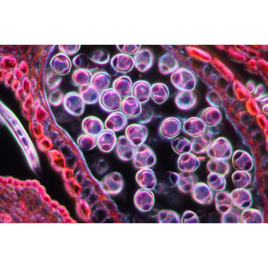
This is a darkfield microscopy image of pollen grains captured using a ZEISS Axiolab microscope.
29 notes
·
View notes
Text
A good video if you want to learn more about the self-assembling nanobots. 🤔
#pay attention#educate yourselves#educate yourself#knowledge is power#reeducate yourself#reeducate yourselves#think about it#think for yourselves#think for yourself#do your homework#do some research#do your own research#ask yourself questions#question everything#nano technology#micro bots#science#you decide#news
20 notes
·
View notes
Text
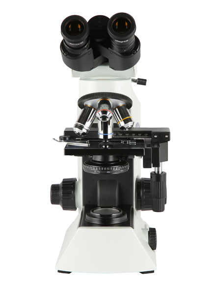
Michael scope
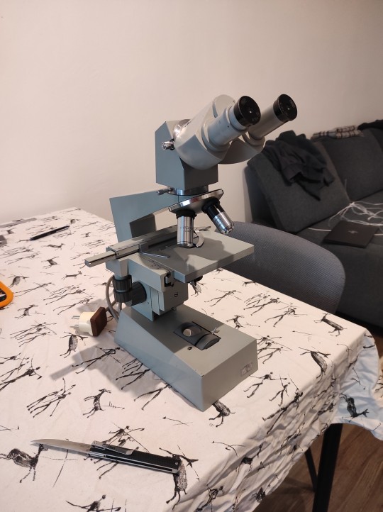

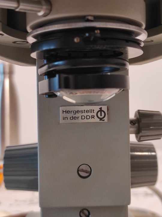
Zeiss Jena Laboval ?3? I think, documentation for the Laboval series online starts and ends with the Laboval 4. By all accounts this is their upper mid range lab scope, with Amplival being the top of the line before you get to big scopes that have to be installed by a technician.
There's a large variety of Zeiss scopes that were manufacturered on this side of the iron curtain, as you can see this is an East German piece. Pretty good condition, scratchy condenser optics but the objectives and eyepiece seem to be in great nick, from a quick inspection, and that's what matters. Eyepieces are 10×'s, Objectives are a 3.2 semiplanar, a 10, a 40 planar (which I think will get a lot of use), and a 100 oil immersion. Pretty normal setup.
I don't have slides and slips yet so I can't do a proper mount, but this is pretty promising for just sliding a sheet of paper into the slide holder. Proper mounting will improve the focus plane immensely. 32× and 100× so far.


The illumination is off-centre and uneven but that's resolvable and anyway it might be fun to move to LED. I also want to build some top illumination brackets for opaque subjects.
I had like. A kiddies toy microscope growing up and I got as far as trying to make it do darkfield with pieces of cardboard, but never something this professional, binocular optics is such a big step up on its own.
The only real issue I've seen so far is that the stage Z axis is very sloppy, huge backlash. Everything has been stuffed with new grease recently so at least it moves smoothly, old scopes and old typewriters both have a tendency to seize if they're forgotten for more than a few months at a time.
I'll swing by the lake and pick up some algae and protist samples later, and I need to order slides and slips. Also I can print some darkfield and oblique illumination filters.
Probably not going to fuck too much with oil immersion, but who knows, also I'll keep an eye out for water immersion objectives.
I think that with an appropriate head replacement and some filter hacking I could get phase contrast microscopy up and running, probably scavenging some Amplival parts. I'd need to see. I can definitely get fluorescence microscopy working with an illumination upgrade. It would also be nice to gut the electronics and put in a simple battery powered illumination system I can charge over USB so I don't have to rely on wall power. Even the stock tungsten lighting is only 5W at 6V so that's easy to swing.
Objectives are DIN 45mm I think, and it sounds like many Eastern Bloc microscopes use standardized head mounts. I'll also be able to print a lot of parts for this, but I'll probably want to get some black filament for optical reasons.
First I want to do some protist sketches, I picked up a protists book at a used bookstore a while ago and it got me really hyped to do protist watching. This is definitely at some level me trying to replace macroscopic wildlife spotting in my life.
19 notes
·
View notes
Text

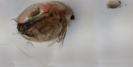
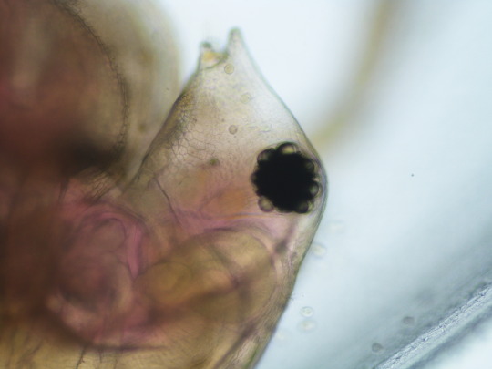

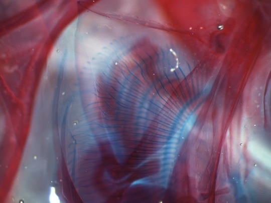
Photos i submitted to a microscopy contest.... first two are Daphnia magna (1st photo is one of the babies i accidentally flushed out of the mama (2nd photo)). third photo is a close up on a different daphnia
3rd photo is a composite darkfield of a stained and cleared black-banded sunfish's teeth, 4th is its gill filaments
#my creations#I checked the winners from last year and i doubt ill place#some of those photos are high quality like youd see in a textbook#but i figured id share some since its free#microscopy
4 notes
·
View notes
Text
C19 Unvaccinated Turned Magnetic From Shedding - Correlation With Darkfield Microscopy Live Blood Analysis
Image: C19 unvaccinated blood with many construction sites and clot formation seen This is an interesting case report of a C19 unvaccinated healthcare worker who works in a hospital, who came to see me for C19 vax shedding symptoms. The patient did not have any vaccination since a influenza shot in 2013. In 2021 she had 3 days of suspected Covid symptoms. Interestingly during that time she had…
View On WordPress
0 notes
Text
Magnus Magic: Microscope Hacks for Unique Observations
For a very long time, microscopes have been the key to the invisible, opening doors to an amazing world of minute details and hidden structures. Nevertheless, traditional observations are only the beginning of the wonders of microscopy. In this blog, we delve into the fascinating world of "Magnus Magic"—hacks for microscopes and other non-traditional methods that enhance the microscope experience, providing one with fresh insights and a more profound comprehension of the microscopic marvels all around us.

Polarized Light: Revealing Latent Patterns
Make use of polarized light's ability to highlight structures and patterns in your microscopic specimens. With Magnus Microscopes, you can really see birefringent things like biological tissues and crystals in a whole new light by inserting a polarizing filter in the light path. This method makes visible complex patterns and vivid colors that are frequently hidden by ambient light.

Iluminating the Unseen with Darkfield Illumination
By carefully illuminating specimens against a black backdrop, darkfield illumination modifies your observations. By increasing the contrast of transparent or semi-transparent materials, this method is especially good at bringing features that could be lost in brightfield illumination to light. Discover the hidden beauty of diatoms, microbes, and fragile structures using Magnus Microscopes with darkfield lighting.
Do It Yourself Microscope Photography: Seeing What's Not Seen
Investigate do-it-yourself (DIY) microscope photography to expand your Magnus Microscope's possibilities. High-resolution pictures and films of your microscopic observations can be taken using a dedicated microscope camera or an adapter for smartphones. Make a visual record of your investigations, share your findings with others, or even use eye-catching microscope pictures to further scientific outreach.

Microbial Life as Seen in Wet Mounts and Microaquariums
Use wet mounts and micro aquariums to create your own little aquatic ecosystem. Magnus Microscopes make it simple to prepare wet mounts, which are slides that include a droplet of liquid containing your specimen in them. Examine living microorganisms in their native environment, such as aquatic plants or pond water creatures. Under a microscope, the dynamic motions and behaviors of these small organisms present a captivating spectacle.

Turn Your Observations Into Custom Microscope Slides
Try making your own microscope slides to personalize your findings. To examine different samples side by side, make double-layer slides. You can also use colored filters to highlight particular characteristics of your specimens. You can make your observations more visually striking by including some imagination into your slide presentation so that you can concentrate on particular aspects or highlight specific structures.

Time-Lapse Microscopy: Seizing Changing Events
Time-lapse microscopy, which provides a dynamic picture of biological processes and changes over time, is made possible by Magnus Microscopes with a motorized stage. Magnus Microscopes' time-lapse microscopy offers insightful observations into the dynamic realm of the microscopic, whether examining chemical processes, microbial growth, or cell division.
Reflective Lighting: Highlighting Surface Features
Try reflecting illumination for specimens with detailed surface features, such textured materials or complex biological systems. Magnus Microscopes can accentuate surface details and offer a distinctive viewpoint on the sample's topography by placing a mirror or other reflecting surface underneath the specimen. Studies on biological surfaces and materials science benefit greatly from this technique.
Cross-Polarization: Improving Contrast and Diminishing Glare
In microscopy, cross-polarization is a technique that improves contrast and lowers glare. Magnus Microscopes increase the visibility of details in translucent or reflective specimens and efficiently limit undesired reflections by combining a second polarizer on the observation side with a polarizer in the illumination path.
The Invisible Spotted by Fluorescence Microscopy
With Magnus Microscopes, explore the fluorescent universe beyond sight. Fluorescence dyes or proteins that change color when exposed to particular wavelengths are used in fluorescence microscopy. Magnus Microscopes allow the viewing of fluorescently labeled structures, offering insights into molecular interactions and cellular processes when used with the appropriate filters and light.
Microscopic Artistry: Creating Art from Observations
Investigate the creative aspect of microscopy by combining unusual materials into your findings. Think of transforming your specimens into works of "microscopic art" by including colored gels, liquid crystals, or even commonplace items like paper or cloth. Magnus Microscopes become canvases for creative expression as well as instruments for observation.

Conclusion: Enchanting Microscopic Explorations
To sum up, Magnus Microscopes go beyond conventional observations with a dash of magic—microscope tricks that reveal obscure information and revolutionize your investigation of the microscopic world. Magnus Microscopes facilitate the creation of microscopic art, as well as the enhancement of contrast and capturing dynamic processes, for educators, researchers, and enthusiasts. As you set out on your voyage of original observations with Magnus Magic, keep in mind that the invisible is a vast field of opportunity just waiting to be unlocked with ingenuity and inventiveness.
#magnusopto#teachingmicroscopes#microscopy#microscopysolutions#scientificresearch#educationalmicroscopes#compoundmicroscopes#optics
1 note
·
View note
Text
Darkfield Microscopy And A Dark Agenda | MSOM Ep. 851
https://rumble.com/v3wkkly-darkfield-microscopy-and-a-dark-agenda-msom-ep.-851.html
View On WordPress
0 notes
Text
Live Blood Analysis in Surrey: What You Need to Know

Live blood analysis, also known as dark field microscopy, is a non-invasive health assessment technique examining a tiny drop of fresh blood from the fingertip under a high-powered microscope. This method allows practitioners to assess the quality of an individual’s blood, providing insights into their overall health and well-being. Now, if you are looking for a well-esteemed live blood analysis in Surrey, read along to exercise complete knowledge about it.
What to Expect During Live Blood Analysis
Live blood analysis is a quick and painless procedure that typically takes 15-30 minutes. During the procedure, a practitioner will collect a small drop of blood from your fingertip and place it on a glass slide. The slide will then be placed under a darkfield microscope, allowing the practitioner to examine the blood cells in their live state.
What Can Live Blood Analysis Reveal?
Through your live blood analysis in Surrey, you can get information about a variety of health factors, including:
Red blood cell morphology:
The shape and structure of RBCs can indicate oxygen-carrying capacity and overall blood health.
White blood cell count and morphology:
White blood cells are essential for immune function. Irregularities in their numbers or shapes may suggest underlying infections or immune system imbalances.
Platelet count and morphology:
Platelets play a very important role in blood clotting. Abnormalities in platelet count or morphology may indicate clotting disorders.
Presence of parasites or fungi:
Live blood analysis can detect the presence of certain parasites or fungi that may not be detectable through conventional blood tests.
Overall blood quality:
The blood’s general appearance, including debris or toxins, can provide insights into overall blood health and circulation.
Is Live Blood Analysis Considered a Diagnostic Tool?
Live blood analysis is not considered a diagnostic tool and should not be used to replace conventional medical tests. However, through a live blood analysis in Surrey, you can get valuable information that may complement traditional diagnostic methods and help guide further investigations or treatment decisions.
Who Should Consider Live Blood Analysis?
Go for live blood analysis in Surrey if you need a comprehensive assessment of their overall health and well-being.
It may be beneficial for those with:
Chronic health conditions:
Live blood analysis can provide insights into the progression of chronic conditions and help monitor the effectiveness of treatment.
Nutritional deficiencies: Live blood analysis can detect signs of nutritional deficiencies, such as anemia or vitamin D deficiency.
Underlying toxicities:
Live blood analysis can identify the presence of heavy metals or other environmental toxins in the bloodstream.
Finding a Qualified Practitioner in Surrey
Choosing a qualified practitioner adequately trained in the technique is essential if you are considering live blood analysis. In Surrey, several practitioners offer live blood analysis services. You can find a list of practitioners on the Canadian Association of Naturopathic Doctors (CAND) website. Live blood analysis can be valuable for assessing overall health and well-being. While it is not a diagnostic tool, it can provide insights that may complement traditional diagnostic methods and guide further investigations or treatment decisions. If you are considering live blood analysis in Surrey, be sure to choose a qualified practitioner who has been adequately trained in the technique.
0 notes
Text
Dental Anesthetic Septocaine Mixed With Live Blood Shows Same Nano Sensors As In Budesonide - Attack Red Blood Cells And Cause Clot Formation. Darkfield Microscopy Up To 4000x Magnification
0 notes
Text

This is salt captured under the microscope using darkfield microscopy.
11 notes
·
View notes
Text
Analysis of Scientific Imagery Animations
In order to gather inspiration for my storyboard I’ve been looking at a variety of scientific animations that depict cellular structures and and scientific concepts.
Some of the things that I’m focusing on include:
The storytelling technique used
The techniques used to simplify and explain concepts
Visual styles
Animation style
Voice overs, music and sound effects
The Whole Brain Catalog- Animated by Drew Berry
youtube
I like the way that this animation starts with the subject; a mouse and takes your through his skull into his brain and into the different sections of the brain and cellular structures.
Many of the scientific imagery and animations I’ve been looking at feel so foreign and abstract that setting the scene in this way makes it more comprehensive.
Sound: Contemporary classical music

Drew Berry- Organelles of a human cell
youtube
His animations include interesting sounds that at times feel exaggerated or mechanical. Especially when you consider that all these minute entities are inside our bodies. Questions that I’m thinking about…If you could record the sound of Dinatoms swirling around in the ocean what sound would they make?

California Academy of Sciences- Travel Deep inside a Leaf
youtube
This animation takes an approach similar to what I’ve been sketching out. The animation begins by depicting a tree and zooming into the leaves, displaying tiny ants crawling on leaves and zooms into the leaf and inside a microscopic perspective within the structure of the leave.
It’s effective because it provides the viewer with context rather than just showcasing microscopic images that can sometimes feel quite abstract.
Seeing this animation and comparing it to others, has provided me with affirmation that I’m heading in the right direction. Instead of a voiceover it uses text.
The screenshots below demonstrate the intro sequence that starts off with imagery of a tree to set the context, then zooms through the leaves to focus on a single leaf with ants crawling over it before entering into the cellular structures…

The Journey to the Microcosmos
This channel explores the tiny, unseen world that surrounds us. The quality of the videos is pretty amazing, they use powerful microscopes to capture microorganisms in action with colourful vibrancy. I was curious about how they capture the images and whether they are modified with dyes or animation effects. So I did some investigation and found a video in which they go through the process.
I learnt that they use a variety of lenses and light to shine through the microscopes. Light field, darkfield, brightfield, and phase contrast microscopy techniques are all used on the mostly translucent microorganisms which are referred to as ‘amplitude objects, because the different types of light including polarized, UV light and other lights that reflect on them and through them, so the shifting movements inside them become visible to us. I’ll need to work out how to play with translucent effects and light to capture similar appearances in the models I create.
Here is the video that outlines the process they take to capture the imagery
youtube
I did verify that they don't use coloured dyes, unlike other scientific imagery-capturing techniques.
This image shows the same microorganism displayed using different types of light as mentioned above.

The Journey to the Microcosmos -The tiny worlds of single-celled organisms
youtube
Soundtrack is ambient and their video features an engaging voiceover that takes the viewer through a variety of organisms.
Colours- The images are captured through microscopes and I’m surprised by the vibrancy of the colours. They do seem real and not recreated with effects because you can actually see the cells moving around. Phase contrast microscopy. Because many of the microorganisms are transparent the light.

Harvard Online- Electron Transport Chain
youtube
Explainer animation that combines animation, text and a voice-over. The colours are overly saturated, I personally don’t like this visual approach as it distances the viewer from the reality of what they are seeing. These are cellular structures are within us, the animation style is very synthetic looking.
This style of animation and approach is an example of what I’ll try to avoid.
Sound: Voice-over and ambient electronic music in the background.

Nucleus Medical Media- Biology: Cell Structure
youtube
This was an award-winning animation back in 2012. The imagery is crisp and designed in a way that takes complex biological concepts into simple easy to easy-to-understand explanations. The voice-over and the overlaid text as well as the ambient electronic soundtrack all work really well with the lively and vibrant animation style.

0 notes
Text
Microstructural analysis empowers researchers to design advanced materials, enhance quality control
Metallurgy is a fascinating field that deals with the study of metals and their alloys, focusing on their structure, properties, and performance. One of the fundamental aspects of metallurgical research is microstructural analysis. This technique involves the examination of small-scale structures within metals and alloys, providing valuable insights into their behavior and characteristics. In this article, we will delve into various techniques used for microstructural analysis in metallurgy and explore their significant applications in the industry.

Metnmat Research and Innovation
Table of Contents
Introduction
Understanding Microstructural Analysis
Sample Preparation Techniques
Optical Microscopy
Scanning Electron Microscopy (SEM)
X-ray Diffraction (XRD)
Transmission Electron Microscopy (TEM)
Ultrasonic Testing
Image Analysis and Digital Microscopy
Applications of Microstructural Analysis in Metallurgy Future Trends in Microstructural Analysis
Conclusion
FAQs
Introduction
Metals and alloys are integral to various industries, from aerospace and automotive to electronics and construction. To ensure their optimal performance and reliability, it is essential to understand their microstructure, which directly influences their mechanical, thermal, and electrical properties.
Understanding Microstructural Analysis
Microstructural analysis is a vital tool in the field of metallurgy, as it allows researchers to observe and understand the internal structure of metals and alloys at a microscopic level. By examining the arrangement of grains, phases, inclusions, and defects within the material, metallurgists can gain valuable information about its properties and performance. This knowledge is crucial for various applications, from material design and manufacturing to failure analysis and quality control.
The first step in microstructural analysis is sample preparation. Proper sample preparation is essential to obtain accurate and reliable results. It involves cutting the metal sample into manageable sections and mounting it on a substrate to facilitate handling during subsequent preparation steps. Grinding and polishing are then performed to achieve a smooth and flat surface, ready for microscopic examination. Finally, etching is conducted to reveal the microstructure's features, making them more visible under a microscope.
Sample Preparation Techniques
Before conducting microstructural analysis, proper sample preparation is crucial to obtain accurate and reliable results. The following techniques are commonly used:
Cutting and Mounting
Samples are carefully sectioned and mounted on a substrate to facilitate handling during subsequent preparation steps.
Grinding and Polishing
Grinding and polishing are performed to achieve a smooth and flat surface, ready for microscopic examination.
Etching
Etching is a chemical process used to reveal the microstructure's features, making them more visible under a microscope.
Optical Microscopy
Optical microscopy is one of the most widely used techniques for microstructural analysis in metallurgy. It involves the use of visible light to examine the metal sample's microstructure. With a wide range of magnifications available, optical microscopy is suitable for routine examination of metal structures.
Stereomicroscopy is a variation of optical microscopy that provides a 3D view of the sample's surface. This is particularly useful for studying surface defects and coatings. Polarized light microscopy (PLM) is employed to study birefringent materials, providing information about crystallographic orientations. Darkfield microscopy, on the other hand, enhances the contrast of transparent phases, helping to identify small particles or precipitates.
Light Microscopy
Light microscopy is employed for routine examination of metal structures with a wide range of magnifications.
Stereomicroscopy
Stereomicroscopy provides a 3D view of the sample's surface, aiding in the study of surface defects and coatings.
Polarized Light Microscopy (PLM)
PLM is utilized to study birefringent materials, revealing information about crystallographic orientations.
Darkfield Microscopy
Darkfield microscopy enhances the contrast of transparent phases, useful for identifying small particles or precipitates.
Scanning Electron Microscopy (SEM)
SEM allows for high-resolution imaging and 3D visualization of a sample's surface using electrons.
Scanning Electron Microscopy (SEM) is an advanced technique used for high-resolution imaging and 3D visualization of a metal sample's surface. Instead of using light, SEM utilizes a focused beam of electrons to interact with the sample. As the electrons interact with the surface, various signals, such as secondary electrons and backscattered electrons, are generated, which are then detected to form an image.
SEM is particularly valuable for investigating surface topography and identifying surface features at a nanoscale level. The high magnification and resolution capabilities of SEM make it an essential tool for researchers and engineers in metallurgy.
"Microstructural analysis empowers researchers to design advanced materials, enhance quality control, and drive innovation across diverse industries."
X-ray Diffraction (XRD)
X-ray Diffraction (XRD) is another powerful technique used for microstructural analysis in metallurgy. XRD is based on the principle of X-ray scattering by the crystal lattice of a material. When X-rays interact with the crystal lattice, they are diffracted in specific directions, producing a diffraction pattern. This pattern is characteristic of the crystal structure and phases present in the material.
By analyzing the diffraction pattern, researchers can identify the crystal structures and phases in the metal sample, providing crucial information about its composition and microstructure. XRD is especially useful for analyzing powdered samples and identifying unknown materials.
Transmission Electron Microscopy (TEM)
TEM is an advanced technique that enables the examination of the internal microstructure at the nanoscale.
Transmission Electron Microscopy (TEM) is an advanced technique that enables researchers to examine the internal microstructure of metals and alloys at an incredibly high resolution. TEM works by transmitting a focused beam of electrons through a thin sample, which is then magnified to form an image on a fluorescent screen or a digital detector.
TEM allows for the visualization of individual grains, dislocations, and other fine microstructural features at the nanoscale level. This level of detail is essential for understanding the behavior of materials, especially in high-tech applications and research.
Ultrasonic Testing
Ultrasonic testing measures the ultrasonic waves' velocity through a material, helping to detect defects and assess material integrity.
Ultrasonic Testing (UT) is a non-destructive testing technique used to assess the integrity of a material or detect defects within it. In ultrasonic testing, high-frequency sound waves, usually in the range of 0.5 to 10 MHz, are introduced into the material. These sound waves travel through the material until they encounter a boundary or defect, where they are reflected back to a receiver.
By analyzing the time it takes for the sound waves to return and the amplitude of the returning waves, researchers can determine the presence and location of defects, such as cracks or voids. UT is commonly used for quality control in manufacturing and inspection of welded joints.
Image Analysis and Digital Microscopy
Image analysis software is used to quantify and characterize microstructural features, providing valuable data for research and development.
Digital microscopy, in combination with image analysis, enables researchers to perform complex measurements and calculations on the microstructural features of metals and alloys. This data is used to evaluate the effects of different processing techniques, assess the material's properties, and optimize the performance of metal components.
"From optical microscopy to transmission electron microscopy, explore the cutting-edge techniques that reveal the intricate world of metal microstructures."
Applications of Microstructural Analysis in Metallurgy
Microstructural analysis plays a pivotal role in various applications within the field of metallurgy:
microstructural analysis plays a pivotal role in various applications within the field of metallurgy. The knowledge gained from these analyses informs critical decisions in material design, manufacturing processes, and performance evaluation.
One of the primary applications of microstructural analysis is quality control in manufacturing. By examining the microstructure of metal samples, manufacturers can ensure the consistency and quality of their products. Any variations or defects in the microstructure can be detected and addressed early in the production process, preventing potential issues down the line.
microstructural analysis is essential in evaluating heat treatment processes. Heat treatment is commonly used to alter the microstructure of metals, improving their mechanical properties. By analyzing the microstructure after heat treatment, researchers can optimize the process to achieve the desired material properties.
Quality Control in Manufacturing
By analyzing the microstructure, manufacturers can ensure the consistency and quality of their products.
Failure Analysis
In cases of component failure, microstructural analysis helps identify the root cause and prevent future occurrences.
Material Characterization
The microstructural analysis aids in understanding the material's properties and behavior under different conditions.
Welding and Joining
Evaluation of microstructures in welded or joined materials ensures structural integrity and performance.
Corrosion Studies
Studying the microstructure helps in understanding corrosion mechanisms and selecting corrosion-resistant materials.
Heat Treatment Evaluation
The microstructural analysis is vital for optimizing heat treatment processes and achieving desired material properties.
Future Trends in Microstructural Analysis
The field of microstructural analysis continues to evolve, with ongoing advancements in imaging techniques and data analysis methods.
Another exciting trend is the development of in-situ microstructural analysis methods. In-situ techniques allow researchers to study the evolution of microstructures under specific conditions, such as temperature or mechanical stress. These methods provide valuable insights into how microstructures change over time and can help optimize processes or predict material behavior under different environmental conditions.
Advancements in data analysis and machine learning also hold great promise for microstructural analysis. With the increasing volume of data generated by various microscopy techniques, sophisticated data analysis tools are essential for extracting meaningful information. Machine learning algorithms can identify patterns and correlations within large datasets, facilitating more accurate material property predictions and improved material design.
"Unlocking the hidden secrets within metals and alloys through microstructural analysis is the key to optimizing their performance and reliability."
Conclusion
Microstructural analysis in metallurgy is a critical discipline that provides valuable insights into the properties and behavior of metals and alloys. By employing various advanced techniques, researchers and engineers can make informed decisions in material design, manufacturing, and performance evaluation.
As technology continues to advance, we can expect further innovations in microstructural analysis methods, enabling more detailed and precise examination of materials. These developments will undoubtedly lead to the creation of novel materials with enhanced properties, contributing to the improvement of various industries and applications.
The future of microstructural analysis is promising, and ongoing research in this field will undoubtedly shape the way we understand and utilize metals and alloys in the years to come.
FAQs
Q. What is the significance of microstructural analysis in metallurgy?
Microstructural analysis helps in understanding the relationships between a material's structure and its properties, enabling better material design and selection for specific applications.
Q. Which technique is best suited for studying grain boundaries in metals?
Transmission Electron Microscopy (TEM) is exceptionally well-suited for studying grain boundaries at the nanoscale.
Q. Can microstructural analysis identify the presence of impurities?
Yes, microstructural analysis can reveal the presence of impurities and their distribution within the material.
Q. How does microstructural analysis contribute to alloy development?
The microstructural analysis allows researchers to assess the microstructure-property relationships, aiding in the development of new and improved alloys.
Q. Is microstructural analysis applicable to non-metallic materials?
Yes, microstructural analysis techniques can be adapted to study the microstructures of non-metallic materials, including ceramics and polymers.
Reach Us Now.
#Metallurgy#Microstructural Analysis#Techniques#Materials#Metals#Alloys#Material Characterization#Manufacturing#Research
0 notes
Text
My Interview With SGT Report On Darkfield Microscopy Findings Of Nanotechnology In Unvaccinated Blood And Rubbery Clot Investigation
0 notes
Text
Rubbery Clot Formation Shown In Living C19 Unvaccinated Person Via Darkfield Microscopy - Hydrogel Replacing Blood
View On WordPress
0 notes
Text
https://instagr.am/reel/CuVnWw_p6sa/
🪱WormS🪱 Microscopic nematodes have a unique way of moving despite lacking limbs or appendages. The movement of these long slender worms (typically .1 - 2.5mm long) is achieved through the coordinated contraction and relaxation of their body muscles, combined with the hydrostatic pressure within their fluid-filled body cavity.
By contracting the longitudinal muscles that run along the length of their body, nematodes can create a bending motion that propels them forward in a wave-like manner. Their forward progress is still not very fast and IMO they look rather frantic achieving it.
Nematodes possess sensory organs on both ends of their body, which help them detect environmental cues such as light, chemicals, movement, oxygen levels, and temperature. Using these sensory inputs, they can navigate and adjust their movement direction accordingly.
Filmed with @jaredlead Jared Leadbetter at @caltech using Canon Mark III and Zeiss Axioplan II. The looks are achieved with various types of #microscopy including #brightfield #darkfield and #phasecontrast . Song by Füxa. #nematode #worms #worm #microscopic #microscopicworld #motility #motion #hydrostatic
July 05, 2023 at 06:48PM
via Instagram https://instagr.am/reel/CuVnWw_p6sa/
0 notes
Text
yes the main town in the globeland project is called brightfield. yes the alternate dimension version of it is called darkfield. yes the name is significant in that they refer to microscopy techniques and it was a coincidence i decided to roll with.
#auri rambles#globeland in my oc stuff basically refers to the universe-wide simulation where all the stories take place#perpetua is the vaguely space opera hermitcraft one#brightfield is a post-post apocalyptic fantasy a la adventure time#undecided if 'the apotheosis of spica' is set in there because it could stand on its own#thematically apotheosis and perpetua are similar in that theyre both man vs god conflicts (kinda)#but apotheosis doesn't have the meta narrative aspect to it unlike brightfield#brightfield as a story is really old and i kinda dropped it a while ago to focus on hc stuff#its just something i keep around lmao#apotheosis is about a girl who starts spiraling after her crush/best friend gets married to a god#apart from that i dont have much since it more like a backstory for a dnd character HAHAHA#but i think about luciana and spica a lot
0 notes
Last Seen Blogs

crzass-blog
일단 우리 재밌게 살까?(이브에로스.com)

puntofinalizando123
Un usuario más.

bjjjjjeoeooeoe
Hazbin Hotel Stuff

heyyapollo
Apollo

izzyjames-trainer
Innocence, you lost your queen