#Gamma Glutamyl Cysteine
Text
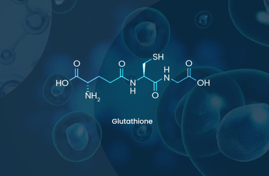
What is Glutathione
Glutathione (GSH) is often termed the “master antioxidant”. This tripeptide is ubiquitous in nature and is produced by every organism from bacteria, to plants to animals that derives energy from oxidative phosphorylation and respiration. Glutathione is synthesized in the cytosol of each cell by the action of two enzymes in an elegantly regulated system that allows it to be maintained at different homeostatic levels in different tissue types, with the liver, given its detoxification role, having the highest amounts. Glutathione plays a pivotal role in most key physiological functions including but not limited to maintenance of cellular redox, neutralising free radicals, cell cycle regulation, proliferation, apoptosis, xenobiotic metabolism, and the recycling of other cellular antioxidants such as Vitamins C and E. Glutathione depletion and a corresponding increase in reactive oxygen species (ROS) during microbial infection is a key driver of the immune response and inflammation. Most chronic diseases are related to oxidative stress arising from the affected tissue losing the capacity to maintain glutathione at adequate healthy levels. The severity of many poisonings from drugs, alcohol, heavy metals and environmental toxins are related to an acute depletion of cellular glutathione. Progressive depletion of cellular glutathione is also used as a mechanism by many viruses to control their replication cycle during infection.
https://www.glutathionereporter.com/what-is-glutathione/
#Glutathione Reporter#Glutathione and it’s importance to Life#The importance of glutathione#importance of glutathione in the body#How do cells produce Glutathione?#gamma-glutamylcysteine#Measuring cellular Glutathione concentration#What is Gamma-Glutamylcysteine#Gamma-Glutamylcysteine (GGC)#GGC supplement#gamma glutamyl cysteine supplement#gamma glutamylcysteine#Gamma Glutamyl Cysteine#glutathione boosting supplements#what is acute inflammation and chronic inflammation#what is acute and chronic inflammation#what is the difference between acute and chronic inflammation
0 notes
Link
#Measuring cellular Glutathione concentration#What is Gamma-Glutamylcysteine#Gamma-Glutamylcysteine (GGC)#gamma glutamylcysteine#Gamma Glutamyl Cysteine#glutathione boosting supplements#what is acute inflammation and chronic inflammation#what is acute and chronic inflammation#what is the difference between acute and chronic inflammation#glutathione and chronic inflammation#chronic glutathione depletion#glutathione & inflammation
0 notes
Text
NMK-BH2, a manuscript Microtubule-Depolymerising Bis (Indolyl)-Hydrazide-Hydrazone, Induces Apoptotic and Autophagic Mobile or portable Loss of life inside Cervical Cancers Cellular material simply by Presenting in order to Tubulin from Dabrafenib -- Internet site
splendens sensu lato chaos in to nine clades understanding that European along with western United states representatives of the taxon usually are not conspecific. Part Splendentes and also supersection "Marginatae" associated with Inocybe, smooth-spored taxa seen as a any symptoms involving special developmental characters (totally or perhaps mainly pruinose stipe, shortage of cortina, frequent presence of marginate basal bulb) aren't monophyletic. The particular varieties principle for We. splendens is actually talked about, and a lectotype with regard to My spouse and i. splendens sensu Heim will be designated. 2 brand-new varieties, We. monticola and that i. praecox, are generally illustrated along with described. The badly recognized varieties We. bakeri can be redescribed via variety material, plus a important for Twenty two types is supplied for detection of those along with other non-reddening kinds along with easy spores and a caulocystidiate stipe documented from The united states along with European countries.Carbonic anhydrase self-consciousness lowers apnoeic events within slumber unhealthy inhaling and exhaling. Zonisamide suppresses carbonic anhydrase, and causes weight reduction within fat patients. This study investigated your comparable impact present in components, which can both relieve obstructive slumber apnoea (OSA). Ongoing good respiratory tract pressure #Link# (CPAP) was adopted like a normal care comparator. 48 patients using moderate-to-severe OSA along with a bmi involving 27-35 kilograms.michael(-2) ended up randomised to obtain either zonisamide, placebo or CPAP with regard to 30 days. The off shoot stage (30 weeks) compared CPAP along with zonisamide. Polysomnography, biochemistry and biology as well as signs or symptoms were assessed. In Four weeks, zonisamide reduced apnoea/hypopnoea catalog (AHI) by way of a indicate +/- SD 33 +/- 39% and also oxygen desaturation index by simply 31 +/- 31% (p=0.02 and also 2.014, respectively; placebo adjusted). The suggest conformity adjusted decrease in AHI following zonisamide and CPAP ended up being 13 and 61%, correspondingly, (p=0.001) at Twenty-four several weeks. Weight was somewhat modified with A month, nevertheless diminished right after zonisamide as well as improved soon after CPAP at Twenty four several weeks (-2.Several +/- 3.0 kilogram as opposed to Two.Three or more +/- Two.3 kilogram, g small compared to 0.001). Zonisamide reduced bicarbonate from Several along with Twenty-four weeks. Side-effects have been more common following zonisamide. Zonisamide diminished OSA outside of bodyweight potentially by simply components in connection with carbonic anhydrase self-consciousness. The consequence had been less evident than that attained through CPAP.gamma-Glutamyltransferase (gamma GT) can be a mobile or portable area enzyme in which catalyzes hydrolysis of the connection #Link# linking your glutamate along with cysteine remains of glutathione along with glutathione-S-conjugates. We have witnessed in which human pancreatic tumor cells and tumor-associated stellate cellular material convey large degrees of this kind of molecule when compared to normal pancreatic epithelial along with stellate cells. Recognition with the proteins within tumor areas correlated along with gamma GT task on the outside of the classy tumor and #Link# stellate tissues. We all analyzed whether or not the growth gamma GT might be used to deliver a beneficial on the cancer endothelial cellular material. GSAO is often a glutathione-S-conjugate of your trivalent arsenical which is stimulated to penetrate endothelial cellular material through gamma GT cleavage with the gamma-glutamyl remains.
0 notes
Text
Thuốc Acezym 600 tác dụng, liều dùng, giá bao nhiêu? | Tracuuthuoctay
TraCuuThuocTay.com chia sẻ: Thuốc Acezym 600 điều trị bệnh gì?. Acezym 600 công dụng, tác dụng phụ, liều lượng.
BÌNH LUẬN cuối bài để biết: Thuốc Acezym 600 giá bao nhiêu? mua ở đâu? Tp HCM, Hà Nội, Cần Thơ, Bình Dương, Đồng Nai, Đà Nẵng. Vui lòng tham khảo các chi tiết dưới đây.
Acezym 600
Tác giả: Dược sĩ Hạnh Nguyễn
Tham vấn y khoa nhóm biên tập.
ngày cập nhật: 16/1/2011

Nhóm thuốc: Thuốc cấp cứu và giải độc
Dạng bào chế:Hộp 1 lọ bột pha tiêm và 1 ống dung môi 10ml
Đóng gói:Hộp 1 lọ bột pha tiêm và 1 ống dung môi 10ml
Thành phần:
Glutathion 600mg
SĐK:VD-14244-11
Nhà sản xuất: Công ty Dược & trang thiết bị Y tế Bình Định (BIDIPHAR) – VIỆT NAM Nhà đăng ký: Nhà phân phối:
Chỉ định:
Hỗ trợ làm giảm độc tính trên thần kinh của xạ trị và của các hóa chất điều trị ung thư bao gồm cisplatin, cyclophosphamid, oxaplatin, 5-fìuorouracil, carboplatin: Tiêm tĩnh mạch glutathion ngay trước khi tiến hành xạ trị và trước phác đò hóa trị liệu của các hóa chất trên.
Hỗ trợ điều trị ngộ độc thủy ngân: Phối hợp các thuốc điều trị ngộ độc thủy ngân đặc hiệu như 2,3- dimercaptoprnpan-l-suỉ/onat và meso-2,3- dimercaptosuccinic acid vói tiêm truyền glutathion và vitamin C liều cao làm giảm nồng độ thủy ngân trong máu.
Hỗ trợ trong điều trị xơ gan do rượu, xơ gan, viêm gan do virus B, c, D và gan nhiễm mỡ: Giúp cải thiện thế trạng của bệnh nhân và các chỉ số sinh hóa như bilirubin, GOT, GPT, GT cũng như giảm MDA và tổn thương tế bào gan rõ rệt.
Hỗ trợ trong điều trị các bệnh lý liên quan đến rổi loạn mạch ngoại vi, mạch vành và các rối loạn huyết học: Cải thiện các thông số huyết động của hệ tuần hoàn lớn và nhỏ, giúp kéo dài khoảng cách đi bộ không cảm thấy đau ở các bệnh nhân bị tắc động mạch chi dưới; Cải thiện đáp ứng vận mạch với các thuốc giãn mạch vành như acetylcholin, nitroglycerin ở những bệnh nhân có các yếu tố nguy cơ bệnh mạch vành; Cải thiện tình trạng thiếu máu ở các bệnh nhân lọc máu do suy thận mãn: Tiêm truyền glutathion cuối mỗi chu kỳ lọc máu giúp làm giảm liều erythropoietin đến 50%.
Hỗ trợ điều trị chảy máu dưới nhện: Giúp cải thiện triệu chứng chảy máu dưới nhện.
Hỗ trợ trong điều trị đái tháo đường không phụ thuộc insulin: Giúp làm tăng nhạy cảm với insulin ở các bệnh nhân này.
Hỗ trợ trong điều trị viêm tụy cấp: glutathion có thể có hiệu quả trong việc bảo tồn các chức năng của các cơ quan khỏi sự tấn công của chất trung gian hóa học của phản ứng viêm.
DƯỢC LỰC HỌC
Glutathion là một tripeptid nội sinh và có mặt trong các tế bào của tất cả các cơ quan và bộ máy của cơ thể. Sự có mặt rộng rãi này có liên quan đến sự đa dạng trong chức năng sinh học của glutathion; bao gồm cả các vai trò quan trọng của nó trong nhiều quá trình sinh hóa và trao đổi chất.
Nhóm sulfridilic của cystein trong glutathion rất ái nhân và do đó nó dễ dàng phản ứng với các chất hóa học hoặc của các chất chuyển hóa khác theo cơ chế ái điện tử, kết quả là làm bất hoạt các chất ngoại sinh có thể gây độc. Hơn nữa,glutathiondạng khử, khi phản ứng với một lượng lớn các chất chuyển hóa thông qua phản ứng oxy hóa sẽ tạo ra các phức hợp kém độc hơn và có thế dễ dàng bị chuyến hóa và bài tiết ra dưới dạng acid mercaptan.
Nhờ vậy, glutathion có thể được ứng dụng trong điều trị nhiễm độc có liên quan đến các cơ chế trên, ví dụ như nhiễm độc gan do rượu ethylic hoặc do thuốc, hoặc do các tác nhân hóa trị liệu chuyên biệt…
DƯỢC ĐỘNG HỌC
Sau khi được đưa vào tĩnh mạch, glutathion nằm phần lớn trong hồng cầu, trong khi ở huyết tương nó bị phân hủy nhanh chóng bởi gamma-glutamyl- transpeptidase và gamma-glutamyl-cyclotransferase. Do đó, nồng độ đỉnh của glutathion dạng khử trong huyết tương là rất nhỏ, mặc dù dùng ở liều cao (nồng độ đỉnh trong huyết tương chỉ đạt khoảng 1nmol/ml sau khi 600mg được đưa vào tĩnh mạch); trong khi các mức độ trao đổi chất cystein là lớn hơn nhiều (nồng độ đỉnh trong huyết tương là 17 nmol/ml). Nồng độ trong máu thì lại ngược lại, sau 5-10 phút đưa 600mg glutathion vào qua đường tĩnh mạch, nồng độ đã đạt khoảng 100nmol/ml. Sau đó nồng độ trong máu giảm dần và đạt nồng độ ổn định sau 60 phút thuốc được đưa vào cơ thể.
Liều lượng – Cách dùng
– Truyền tĩnh mạch: 600 mg/ngày.
– Các tình trạng nghiêm trọng hơn: 600-1200 mg/ngày.
1. Dùng theo đường tiêm truyền tĩnh mạch:
1.1. Hỗ trợ làm giảm độc tính trên thần kinh của xạ trị và của các hoá chất điều trị ung thư:
+ Tiêm truyền tĩnh mạch chậm gluthation ngay trước khi tiến hành xạ trị 15 phút: Liều dùng 1200 mg.
+ Tiêm truyền tĩnh mạch chậm gluthation trong 15 phút trước phác đồ hóa trị liệu của các hóa chất (cisplatin, cyclophosphamid, oxaplatin, 5 fluorouracil, carboplatin): Liều dùng 1500 mg – 2400 mg. Lặp lại liều 900 mg – 1200 mg sau ngày thứ 2 và thứ 5 của đợt điều trị. Có thể lặp lại hàng tuần liều 1200 mg.
1.2. Hỗ trợ trong điều trị ngộ độc thuỷ ngân: Phối hợp các thuốc điều trị ngộ độc thủy ngân đặc hiệu như 2,3 – dimercaptopropan – 1- sulfonat và meso – 1,3 – dimercaptosuccinic acid với tiêm truyền gluthation và vitamin C liều cao làm giảm nồng độ thủy ngân trong máu. Liều dùng trong đợp cấp 1200 – 1800 mg/ngày. Liều duy trì 600 mg/ngày cho đến khi hồi phục.
1.3. Hỗ trợ trong điều trị xơ gan do rượu, xơ gan, viêm gan do vi rút B,C,D và gan nhiễm mỡ:
+ Hỗ trợ điều trị xơ gan do rượu: Liều dùng 600 mg – 1200 mg/ngày, tiêm tĩnh mạch chậm.
+ Hỗ trợ điều trị xơ gan, viêm gan do virus B, C, D và gan nhiễm mỡ: 600 mg – 1200mg/ngày, tiêm tĩnh mạch chậm cho đến khi hồi phục.
1.4. Hỗ trợ điều trị trong các bệnh lý liên quan đến rối loạn mạch ngoại vi, mạch vành và các rối loạn huyết học:
– Rối loạn mạch ngoại vi: 600 mg/lần, 2 lần/ngày, truyền tĩnh mạch.
– Bệnh mạch vành: truyền tĩnh mạch 1200 mg – 3000 mg hoặc truyền trực tiếp vào động mạch vành trái 300mg (50 mg – 2 mL/phút).
– Bệnh nhân lọc máu do suy thận mãn: Tiêm truyền gluthation 1200 mg/ngày cuối mỗi chu kỳ lọc máu giúp làm giảm liều erythropoietin đến 50%.
1.5. Hỗ trợ điều trị chảy máu dưới nhện: Truyền tĩnh mạch chậm 600 mg glutathion ngay sau phẫu thuật, lặp lại liều trên sau mỗi 6 giờ trong khoảng 14 ngày hoặc hơn.
1.6. Hỗ trợ trong điều trị đái tháo đường không phụ thuộc insulin: 600 mg – 1200 mg/ngày, tiêm tĩnh mạch chậm liên tục trong một tuần, sau đó dùng mỗi tuần 2 – 3 lần, mỗi lần 0,6 g.
1.7. Hỗ trợ trong điều trị viêm tuỵ cấp: 600 mg – 1200 mg/ngày, tiêm tĩnh mạch chậm.
2. Dùng theo đường tiêm bắp:
Hỗ trợ trong điều trị vô sinh ở nam giới: 600 mg -1200 mg/ngày, tiêm bắp liên tục trong 2 tháng.
Chú ý:
Dung dịch sau khi pha tiêm ổn định trong khoảng 2 giờ ờ nhiệt độ phòng (25°C) và khoảng 8 giờ ở 0°c đến 5°c.
Hướng dẫn cách dùng thuốc:
Truyền tĩnh mạch: Hoàn nguyên lọ thuốc bột 1200 mg với 4ml nước cất pha tiêm, sau đó pha loãng với ít nhất 20 ml dung dịch tiêm truyền: dextrose 5%, dextrose 10%, natri clorid 0,9%, Lactated Ringer, natri bicarbonat 1,4%,…. Truyền tĩnh mạch trong 30 phút
Chống chỉ định:
Quá mẫn với glutathion hoặc một trong các thành phần khác của thuốc.
Tác dụng phụ:
Số ít các trường hợp buồn nôn, nôn, đau đầu đã được ghi nhận. Có thế gây nổi mẩn da và sẽ hết khi ngừng thuốc.
Thông báo cho bác sĩ những tác dụng không mong muốn gặp phải khi sử dụng thuốc.
Chú ý đề phòng:
Khi được đưa vào cơ thể qua đường tĩnh mạch, thuốc phải được hòa tan hoàn toàn trong nước pha tiêm cho dung dịch trong suốt, không màu và tiêm chậm.
PHỤ NỮ CÓ THAI VÀ CHO CON BÚ
Mặc dù những nghiên cứu thử nghiệm cho thấy rằng không có bằng chứng về độc tính của glutathion lên phôi bào, loại thuốc này, cũng giống như tất cả các loại thuốc mới khác, không được khuyên dùng trong thời kì mang thai và cho con bú.
TÁC ĐỘNG CỦA THUỐC KHI LÁI TÀU XE VÀ VẬN HÀNH MÁY MÓC
Với các tác dụng không mong muốn được ghi nhận khi dùng thuốc, thuốc có thể ảnh hưởng lên việc lái xe và vận hành máy móc, và điều này nên được lưu ý.
Thông tin thành phần Glutathione
Dược lực:
Glutathione (GSH ) là một chất chống oxy hóa trong thực vật , động vật , nấm và một số vi khuẩn. Glutathione là một tripeptide nội sinh (được xem như kho dự trữ các chất chống oxy hóa) có mặt trong tất cả các tế bào động vật, được tổng hợp từ tế bào bằng 3 amin gồm cysteine, glutamic và glycine, chúng được tạo ra trong gan và sau đó phân bố khắp cơ thể.
Glutathione tồn tại ở cả hai trạng thái khử (Reduced Glutathione -GSH) và oxy hóa (oxidized – GSSG ). Ở trạng thái khử, nhóm cysteine của thiol có thể tặng một chất khử tương đương (H + + e – ) cho các phân tử khác, chẳng hạn như các loại oxy phản ứng để trung hòa chúng, hoặc cho cystein protein để duy trì dạng khử của chúng. Với việc tặng một điện tử, chính glutathione trở nên phản ứng và dễ dàng phản ứng với một glutathione phản ứng khác để tạo thành glutathione disulfide (GSSG). Phản ứng như vậy có thể xảy ra do nồng độ glutathione tương đối cao trong các tế bào (lên đến 7 mM trong gan).
Tác dụng :
Glutathione làm tăng khả năng miễn dịch của cơ thể. Glutathione là trung tâm của hệ thống phòng thủ chống oxy hoá cho cơ thể, hệ thống này bảo vệ các tế bào chống lại sự thiệt hại của các tác nhân vật lý, sự ô nhiễm, độc tố, sự truyền nhiễm, tia UV. Mức độ glutathione giảm theo tuổi và sự suy tàn này có liên hệ với tấn công của các loại bệnh như bệnh của Alheimer, bệnh đục thuỷ tinh thể, bệnh Parkinson và bệnh xơ cứng động mạch.
Glutathione giúp tái sinh năng lượng, giảm stress;
Kích thích tăng sinh Collagen, làm chậm quá trình lão hóa, giảm sự xuất hiện nếp nhăn, đường nhăn;
Hỗ trợ giải độc tế bào, duy trì làn da sáng khỏe từ bên trong;
Tăng cường sức khỏe, giảm nguy cơ các vấn đề bệnh về tim và các bệnh khác.
Chỉ định :
Thuốc tiêm:
Phụ trị tình trạng nhiễm độc do rượu ethylic, tác nhân phospho hữu cơ, acetaminophen, tác nhân hóa trị liệu chuyên biệt, thuốc độc tế bào hay thuốc trị lao, thuốc có tác động trên tâm thần, thuốc an thần hay thuốc chống trầm cảm.
Phòng ngừa & điều trị tổn thương do phóng xạ.
Bệnh gan.
Viên uống:
Giúp chống oxy hóa, trung hòa các gốc tự do;
Giúp tăng cường hệ miễn dịch, chống oxy hóa;
Giúp cải thiện sức khỏe làn da;
Liều lượng – cách dùng:
Thuốc tiêm:
Tiêm IM, IV chậm hay truyền IV 1-2 ống TAD 300 (300-600 mg)/ngày.
Các tình trạng nghiêm trọng hơn: 1-2 lọ TAD 600 (600-1200 mg)/ngày.
Viên uống:
250-500 mg/ ngày;
Chống chỉ định :
Quá mẫn với thuốc.
Tác dụng phụ
Thỉnh thoảng: nổi mẩn da (sẽ hết khi ngưng dùng thuốc).
Lưu ý: Dùng thuốc theo chỉ định của Bác sĩ
Nguồn tham khảo drugs.com, medicines.org.uk, webmd.com và TraCuuThuocTay.com tổng hợp.
Cần tư vấn thêm về Thuốc Acezym 600 tác dụng, liều dùng, giá bao nhiêu? bình luận cuối bài viết.
Tuyên bố miễn trừ trách nhiệm y tế
Nội dung của TraCuuThuocTay.com chỉ nhằm mục đích cung cấp thông tin về Thuốc Acezym 600 tác dụng, liều dùng, giá bao nhiêu? và không nhằm mục đích thay thế cho tư vấn, chẩn đoán hoặc điều trị y tế chuyên nghiệp.
Chúng tôi miễn trừ trách nhiệm y tế nếu bệnh nhân tự ý sử dụng thuốc mà không tuân theo chỉ định của bác sĩ.
Vui lòng liên hệ với bác sĩ hoặc phòng khám, bệnh viện gần nhất để được tư vấn.
Đánh giá 5* post
The post Thuốc Acezym 600 tác dụng, liều dùng, giá bao nhiêu? appeared first on Tra Cứu Thuốc Tây.
Dẫn nguồn từ Tra Cứu Thuốc Tây https://tracuuthuoctay.com/thuoc-acezym-600-tac-dung-lieu-dung-gia-bao-nhieu/
0 notes
Text
Thuốc Glutaone 600 tác dụng, liều dùng, giá bao nhiêu? | Tracuuthuoctay | Tracuuthuoctay
TraCuuThuocTay.com chia sẻ: Thuốc Glutaone 600 điều trị bệnh gì?. Glutaone 600 công dụng, tác dụng phụ, liều lượng.
BÌNH LUẬN cuối bài để biết: Thuốc Glutaone 600 giá bao nhiêu? mua ở đâu? Tp HCM, Hà Nội, Cần Thơ, Bình Dương, Đồng Nai, Đà Nẵng. Vui lòng tham khảo các chi tiết dưới đây.
Glutaone 600
Tác giả: Ths.Dược sĩ Phạm Liên
Tham vấn y khoa nhóm biên tập.
ngày cập nhật: 1/5/2019
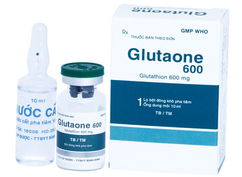
Nhóm thuốc: Thuốc cấp cứu và giải độc
Dạng bào chế:Bột đông khô pha tiêm
Đóng gói:Hộp 1 lọ + 1 ống nước cất pha tiêm 3ml
Thành phần:
Glutathion 600mg
SĐK:VD-15116-11
Nhà sản xuất:Công ty Dược & trang thiết bị Y tế Bình Định (BIDIPHAR) – VIỆT NAMNhà đăng ký:Công ty Dược & trang thiết bị Y tế Bình Định (BIDIPHAR)Nhà phân phối:
Chỉ định:
Hỗ trợ làm giảm độc tính trên thần kinh của xạ trị và của các hóa chất điều trị ung thư bao gồm cisplatin, cyclophosphamid, oxaplatin, 5-fìuorouracil, carboplatin: Tiêm tĩnh mạch glutathion ngay trước khi tiến hành xạ trị và trước phác đò hóa trị liệu của các hóa chất trên.
Hỗ trợ điều trị ngộ độc thủy ngân: Phối hợp các thuốc điều trị ngộ độc thủy ngân đặc hiệu như 2,3- dimercaptoprnpan-l-suỉ/onat và meso-2,3- dimercaptosuccinic acid vói tiêm truyền glutathion và vitamin C liều cao làm giảm nồng độ thủy ngân trong máu.
Hỗ trợ trong điều trị xơ gan do rượu, xơ gan, viêm gan do virus B, c, D và gan nhiễm mỡ: Giúp cải thiện thế trạng của bệnh nhân và các chỉ số sinh hóa như bilirubin, GOT, GPT, GT cũng như giảm MDA và tổn thương tế bào gan rõ rệt.
Hỗ trợ trong điều trị các bệnh lý liên quan đến rổi loạn mạch ngoại vi, mạch vành và các rối loạn huyết học: Cải thiện các thông số huyết động của hệ tuần hoàn lớn và nhỏ, giúp kéo dài khoảng cách đi bộ không cảm thấy đau ở các bệnh nhân bị tắc động mạch chi dưới; Cải thiện đáp ứng vận mạch với các thuốc giãn mạch vành như acetylcholin, nitroglycerin ở những bệnh nhân có các yếu tố nguy cơ bệnh mạch vành; Cải thiện tình trạng thiếu máu ở các bệnh nhân lọc máu do suy thận mãn: Tiêm truyền glutathion cuối mỗi chu kỳ lọc máu giúp làm giảm liều erythropoietin đến 50%.
Hỗ trợ điều trị chảy máu dưới nhện: Giúp cải thiện triệu chứng chảy máu dưới nhện.
Hỗ trợ trong điều trị đái tháo đường không phụ thuộc insulin: Giúp làm tăng nhạy cảm với insulin ở các bệnh nhân này.
Hỗ trợ trong điều trị viêm tụy cấp: glutathion có thể có hiệu quả trong việc bảo tồn các chức năng của các cơ quan khỏi sự tấn công của chất trung gian hóa học của phản ứng viêm.
DƯỢC LỰC HỌC
Glutathion là một tripeptid nội sinh và có mặt trong các tế bào của tất cả các cơ quan và bộ máy của cơ thể. Sự có mặt rộng rãi này có liên quan đến sự đa dạng trong chức năng sinh học của glutathion; bao gồm cả các vai trò quan trọng của nó trong nhiều quá trình sinh hóa và trao đổi chất.
Nhóm sulfridilic của cystein trong glutathion rất ái nhân và do đó nó dễ dàng phản ứng với các chất hóa học hoặc của các chất chuyển hóa khác theo cơ chế ái điện tử, kết quả là làm bất hoạt các chất ngoại sinh có thể gây độc. Hơn nữa,glutathiondạng khử, khi phản ứng với một lượng lớn các chất chuyển hóa thông qua phản ứng oxy hóa sẽ tạo ra các phức hợp kém độc hơn và có thế dễ dàng bị chuyến hóa và bài tiết ra dưới dạng acid mercaptan.
Nhờ vậy, glutathion có thể được ứng dụng trong điều trị nhiễm độc có liên quan đến các cơ chế trên, ví dụ như nhiễm độc gan do rượu ethylic hoặc do thuốc, hoặc do các tác nhân hóa trị liệu chuyên biệt…
DƯỢC ĐỘNG HỌC
Sau khi được đưa vào tĩnh mạch, glutathion nằm phần lớn trong hồng cầu, trong khi ở huyết tương nó bị phân hủy nhanh chóng bởi gamma-glutamyl- transpeptidase và gamma-glutamyl-cyclotransferase. Do đó, nồng độ đỉnh của glutathion dạng khử trong huyết tương là rất nhỏ, mặc dù dùng ở liều cao (nồng độ đỉnh trong huyết tương chỉ đạt khoảng 1nmol/ml sau khi 600mg được đưa vào tĩnh mạch); trong khi các mức độ trao đổi chất cystein là lớn hơn nhiều (nồng độ đỉnh trong huyết tương là 17 nmol/ml). Nồng độ trong máu thì lại ngược lại, sau 5-10 phút đưa 600mg glutathion vào qua đường tĩnh mạch, nồng độ đã đạt khoảng 100nmol/ml. Sau đó nồng độ trong máu giảm dần và đạt nồng độ ổn định sau 60 phút thuốc được đưa vào cơ thể.
Liều lượng – Cách dùng
– Truyền tĩnh mạch: 600 mg/ngày.
– Các tình trạng nghiêm trọng hơn: 600-1200 mg/ngày.
1. Dùng theo đường tiêm truyền tĩnh mạch:
1.1. Hỗ trợ làm giảm độc tính trên thần kinh của xạ trị và của các hoá chất điều trị ung thư:
+ Tiêm truyền tĩnh mạch chậm gluthation ngay trước khi tiến hành xạ trị 15 phút: Liều dùng 1200 mg.
+ Tiêm truyền tĩnh mạch chậm gluthation trong 15 phút trước phác đồ hóa trị liệu của các hóa chất (cisplatin, cyclophosphamid, oxaplatin, 5 fluorouracil, carboplatin): Liều dùng 1500 mg – 2400 mg. Lặp lại liều 900 mg – 1200 mg sau ngày thứ 2 và thứ 5 của đợt điều trị. Có thể lặp lại hàng tuần liều 1200 mg.
1.2. Hỗ trợ trong điều trị ngộ độc thuỷ ngân: Phối hợp các thuốc điều trị ngộ độc thủy ngân đặc hiệu như 2,3 – dimercaptopropan – 1- sulfonat và meso – 1,3 – dimercaptosuccinic acid với tiêm truyền gluthation và vitamin C liều cao làm giảm nồng độ thủy ngân trong máu. Liều dùng trong đợp cấp 1200 – 1800 mg/ngày. Liều duy trì 600 mg/ngày cho đến khi hồi phục.
1.3. Hỗ trợ trong điều trị xơ gan do rượu, xơ gan, viêm gan do vi rút B,C,D và gan nhiễm mỡ:
+ Hỗ trợ điều trị xơ gan do rượu: Liều dùng 600 mg – 1200 mg/ngày, tiêm tĩnh mạch chậm.
+ Hỗ trợ điều trị xơ gan, viêm gan do virus B, C, D và gan nhiễm mỡ: 600 mg – 1200mg/ngày, tiêm tĩnh mạch chậm cho đến khi hồi phục.
1.4. Hỗ trợ điều trị trong các bệnh lý liên quan đến rối loạn mạch ngoại vi, mạch vành và các rối loạn huyết học:
– Rối loạn mạch ngoại vi: 600 mg/lần, 2 lần/ngày, truyền tĩnh mạch.
– Bệnh mạch vành: truyền tĩnh mạch 1200 mg – 3000 mg hoặc truyền trực tiếp vào động mạch vành trái 300mg (50 mg – 2 mL/phút).
– Bệnh nhân lọc máu do suy thận mãn: Tiêm truyền gluthation 1200 mg/ngày cuối mỗi chu kỳ lọc máu giúp làm giảm liều erythropoietin đến 50%.
1.5. Hỗ trợ điều trị chảy máu dưới nhện: Truyền tĩnh mạch chậm 600 mg glutathion ngay sau phẫu thuật, lặp lại liều trên sau mỗi 6 giờ trong khoảng 14 ngày hoặc hơn.
1.6. Hỗ trợ trong điều trị đái tháo đường không phụ thuộc insulin: 600 mg – 1200 mg/ngày, tiêm tĩnh mạch chậm liên tục trong một tuần, sau đó dùng mỗi tuần 2 – 3 lần, mỗi lần 0,6 g.
1.7. Hỗ trợ trong điều trị viêm tuỵ cấp: 600 mg – 1200 mg/ngày, tiêm tĩnh mạch chậm.
2. Dùng theo đường tiêm bắp:
Hỗ trợ trong điều trị vô sinh ở nam giới: 600 mg -1200 mg/ngày, tiêm bắp liên tục trong 2 tháng.
Chú ý:
Dung dịch sau khi pha tiêm ổn định trong khoảng 2 giờ ờ nhiệt độ phòng (25°C) và khoảng 8 giờ ở 0°c đến 5°c.
Hướng dẫn cách dùng thuốc:
Truyền tĩnh mạch: Hoàn nguyên lọ thuốc bột 1200 mg với 4ml nước cất pha tiêm, sau đó pha loãng với ít nhất 20 ml dung dịch tiêm truyền: dextrose 5%, dextrose 10%, natri clorid 0,9%, Lactated Ringer, natri bicarbonat 1,4%,…. Truyền tĩnh mạch trong 30 phút
Chống chỉ định:
Quá mẫn với glutathion hoặc một trong các thành phần khác của thuốc.
Tác dụng phụ:
Số ít các trường hợp buồn nôn, nôn, đau đầu đã được ghi nhận. Có thế gây nổi mẩn da và sẽ hết khi ngừng thuốc.
Thông báo cho bác sĩ những tác dụng không mong muốn gặp phải khi sử dụng thuốc.
Chú ý đề phòng:
Khi được đưa vào cơ thể qua đường tĩnh mạch, thuốc phải được hòa tan hoàn toàn trong nước pha tiêm cho dung dịch trong suốt, không màu và tiêm chậm.
PHỤ NỮ CÓ THAI VÀ CHO CON BÚ
Mặc dù những nghiên cứu thử nghiệm cho thấy rằng không có bằng chứng về độc tính của glutathion lên phôi bào, loại thuốc này, cũng giống như tất cả các loại thuốc mới khác, không được khuyên dùng trong thời kì mang thai và cho con bú.
TÁC ĐỘNG CỦA THUỐC KHI LÁI TÀU XE VÀ VẬN HÀNH MÁY MÓC
Với các tác dụng không mong muốn được ghi nhận khi dùng thuốc, thuốc có thể ảnh hưởng lên việc lái xe và vận hành máy móc, và điều này nên được lưu ý.
Thông tin thành phần Glutathione
Dược lực:
Glutathione (GSH ) là một chất chống oxy hóa trong thực vật , động vật , nấm và một số vi khuẩn. Glutathione là một tripeptide nội sinh (được xem như kho dự trữ các chất chống oxy hóa) có mặt trong tất cả các tế bào động vật, được tổng hợp từ tế bào bằng 3 amin gồm cysteine, glutamic và glycine, chúng được tạo ra trong gan và sau đó phân bố khắp cơ thể.
Glutathione tồn tại ở cả hai trạng thái khử (Reduced Glutathione -GSH) và oxy hóa (oxidized – GSSG ). Ở trạng thái khử, nhóm cysteine của thiol có thể tặng một chất khử tương đương (H + + e – ) cho các phân tử khác, chẳng hạn như các loại oxy phản ứng để trung hòa chúng, hoặc cho cystein protein để duy trì dạng khử của chúng. Với việc tặng một điện tử, chính glutathione trở nên phản ứng và dễ dàng phản ứng với một glutathione phản ứng khác để tạo thành glutathione disulfide (GSSG). Phản ứng như vậy có thể xảy ra do nồng độ glutathione tương đối cao trong các tế bào (lên đến 7 mM trong gan).
Tác dụng :
Glutathione làm tăng khả năng miễn dịch của cơ thể. Glutathione là trung tâm của hệ thống phòng thủ chống oxy hoá cho cơ thể, hệ thống này bảo vệ các tế bào chống lại sự thiệt hại của các tác nhân vật lý, sự ô nhiễm, độc tố, sự truyền nhiễm, tia UV. Mức độ glutathione giảm theo tuổi và sự suy tàn này có liên hệ với tấn công của các loại bệnh như bệnh của Alheimer, bệnh đục thuỷ tinh thể, bệnh Parkinson và bệnh xơ cứng động mạch.
Glutathione giúp tái sinh năng lượng, giảm stress;
Kích thích tăng sinh Collagen, làm chậm quá trình lão hóa, giảm sự xuất hiện nếp nhăn, đường nhăn;
Hỗ trợ giải độc tế bào, duy trì làn da sáng khỏe từ bên trong;
Tăng cường sức khỏe, giảm nguy cơ các vấn đề bệnh về tim và các bệnh khác.
Chỉ định :
Thuốc tiêm:
Phụ trị tình trạng nhiễm độc do rượu ethylic, tác nhân phospho hữu cơ, acetaminophen, tác nhân hóa trị liệu chuyên biệt, thuốc độc tế bào hay thuốc trị lao, thuốc có tác động trên tâm thần, thuốc an thần hay thuốc chống trầm cảm.
Phòng ngừa & điều trị tổn thương do phóng xạ.
Bệnh gan.
Viên uống:
Giúp chống oxy hóa, trung hòa các gốc tự do;
Giúp tăng cường hệ miễn dịch, chống oxy hóa;
Giúp cải thiện sức khỏe làn da;
Liều lượng – cách dùng:
Thuốc tiêm:
Tiêm IM, IV chậm hay truyền IV 1-2 ống TAD 300 (300-600 mg)/ngày.
Các tình trạng nghiêm trọng hơn: 1-2 lọ TAD 600 (600-1200 mg)/ngày.
Viên uống:
250-500 mg/ ngày;
Chống chỉ định :
Quá mẫn với thuốc.
Tác dụng phụ
Thỉnh thoảng: nổi mẩn da (sẽ hết khi ngưng dùng thuốc).
Lưu ý: Dùng thuốc theo chỉ định của Bác sĩ
Nguồn tham khảo drugs.com, medicines.org.uk, webmd.com và TraCuuThuocTay.com tổng hợp.
Cần tư vấn thêm về Thuốc Glutaone 600 tác dụng, liều dùng, giá bao nhiêu? bình luận cuối bài viết.
Tuyên bố miễn trừ trách nhiệm y tế
Nội dung của TraCuuThuocTay.com chỉ nhằm mục đích cung cấp thông tin về Thuốc Glutaone 600 tác dụng, liều dùng, giá bao nhiêu? và không nhằm mục đích thay thế cho tư vấn, chẩn đoán hoặc điều trị y tế chuyên nghiệp.
Chúng tôi miễn trừ trách nhiệm y tế nếu bệnh nhân tự ý sử dụng thuốc mà không tuân theo chỉ định của bác sĩ.
Vui lòng liên hệ với bác sĩ hoặc phòng khám, bệnh viện gần nhất để được tư vấn.
Đánh giá 5* post
The post Thuốc Glutaone 600 tác dụng, liều dùng, giá bao nhiêu? appeared first on Tra Cứu Thuốc Tây.
from Tra Cứu Thuốc Tây https://ift.tt/2CkHbVo
Bác sĩ Nguyễn Quang Huy
Dẫn nguồn từ Tra Cứu Thuốc Tây https://tracuuthuoctay.com/thuoc-glutaone-600-tac-dung-lieu-dung-gia-bao-nhieu/
from Bác sĩ Nguyễn Quang Huy https://ift.tt/3kGV6GE
Dẫn nguồn từ Bác sĩ Nguyễn Quang Huy https://bsquanghuy.blogspot.com/2020/08/thuoc-glutaone-600-tac-dung-lieu-dung.html
0 notes
Text
Prospective Serum Metabolomic Profiling of Lethal Prostate Cancer
Impaired metabolism may play an important role in the pathogenesis of lethal prostate cancer, yet there is a paucity of evidence regarding the association. We conducted a large prospective serum metabolomic analysis of lethal prostate cancer in 523 cases and 523 matched controls nested within the Alpha‐Tocopherol, Beta‐Carotene Cancer Prevention (ATBC) Study. Median time from baseline fasting serum collection to prostate cancer death was 18 years (maximum 30 years). We identified 860 known biochemicals through an ultrahigh‐performance LC‐MS/MS platform. Conditional logistic regression models estimated odds ratios (OR) and 95% confidence intervals of risk associated with 1‐standard deviation (s.d.) increases in log‐metabolite signals. We identified 34 metabolites associated with lethal prostate cancer with a false discovery rate (FDR)<0.15. Notably, higher serum thioproline, and thioproline combined with two other cysteine‐related amino acids and redox metabolites, cystine and cysteine, were associated with reduced risk (1‐s.d. OR=0.75 and 0.71, respectively; P≤8.2×10‐5). By contrast, the dipeptide leucylglycine (OR=1.36, P=8.2×10‐5), and three gamma‐glutamyl amino acids (OR=1.28‐1.30, P≤4.6×10‐4) were associated with increased risk of lethal prostate cancer. Cases with metastatic disease at diagnosis (N=179) showed elevated risk for several lipids, including especially the ketone body 3‐hydroxybutyrate (BHBA), acyl carnitines, and dicarboxylic fatty acids (1.37≤OR≤1.49, FDR<0.15). These findings provide a prospective metabolomic profile of lethal prostate cancer characterized by altered biochemicals in the redox, dipeptide, pyrimidine, and gamma‐glutamyl amino acid pathways, whereas ketone bodies and fatty acids were associated specifically with metastatic disease.
This article is protected by copyright. All rights reserved.
http://bit.ly/2DUvx09
0 notes
Text
Nearly Half of American Adults Have Cardiovascular Disease
According to the latest statistics1 by the American Heart Association (AHA), nearly half (48 percent) of all American adults have some form of cardiovascular disease (CVD) — a classification that includes high blood pressure, coronary heart disease, heart failure and stroke — and deaths from CVD are again on the rise, after decades of being on the decline.
In 2016, there were 840,678 recorded deaths from CVD in the U.S., up from 836,546 the year before. The rise in prevalence of CVD, however, is primarily driven by updated blood pressure guidelines, which as of 2017 identify a blood pressure over 130/80 mm Hg as hypertensive, whereas before the cutoff was 140/90 mm Hg.
According to the AHA, about 80 percent of CVD cases could be prevented by maintaining a healthy lifestyle, which includes lowering high blood pressure and high cholesterol, controlling Type 2 diabetes, avoiding smoking, exercising and maintaining a healthy weight.
To this, the AHA has now also added the recommendation to get a minimum of seven hours of sleep each night. Blood pressure remains a primary focus, however. Volunteer president of the AHA said in a press release:2
“As one of the most common and dangerous risk factors for heart disease and stroke, this overwhelming presence of high blood pressure can’t be dismissed from the equation in our fight against cardiovascular disease.
Research has shown that eliminating high blood pressure could have a larger impact on CVD deaths than the elimination of all other risk factors among women and all except smoking among men.”
Cholesterol Is Not a Culprit in CVD
While I agree with four of the AHA’s suggestions, high cholesterol has been repeatedly found to not be a risk factor for cardiovascular disease, and higher cholesterol may actually be healthier than lower levels. Three to some degree interrelated factors that have a far greater influence on your cardiovascular health are:
High iron3
Insulin resistance
Chronic inflammation
Elevated iron levels will significantly contribute to inflammation, but even if your iron is normal, chronic inflammation can be caused by a wide range of factors, starting with your diet. Your diet is also the key factor at play when it comes to your insulin level, and can worsen the effects of iron overload.
High Iron Significantly Raises Your Risk of CVD
Most people, including doctors, fail to recognize that excess iron causes significant biological harm. When iron reacts with hydrogen peroxide, which is produced as a normal part of energy production in your mitochondria, hydroxyl free radicals are formed. These are among the most damaging free radicals known, causing severe mitochondrial dysfunction, which in turn is at the heart of most chronic degenerative diseases.
Importantly, elevated ferritin has been linked to dysfunctional glucose metabolism,4 raising the risk of diabetes fivefold in men and fourfold in women, a magnitude of correlation similar to that of obesity.5 High ferritin also doubles your risk of metabolic syndrome,6 a condition associated with an increased risk of high blood pressure and heart disease.
If you eat excessive processed foods and net carbs (total carbs minus fiber) the situation is further exacerbated, as burning carbs as your primary fuel can add another 30 to 40 percent more reactive oxygen species on top of the hydroxyl free radicals generated by the presence of high iron.
A meta-analysis7 published in 2013 found that 27 of 55 published studies demonstrated a positive relationship between iron and CVD, with higher iron levels being linked to higher risk of disease. Twenty of the studies found no significant relationship, and only eight reported a negative relationship, with higher iron levels being associated with lower risk of disease.
For example, a Scandinavian study found elevated ferritin levels raised men’s risk of heart attack two- to threefold. In another, people with high ferritin were five times more likely to suffer a heart attack than those with normal levels.
A third found elevated ferritin doubled the risk of heart attack. Importantly, in this study they found that each 1 percent increase in ferritin raised the risk of heart attack by 4 percent, and the only risk factor that weighed heavier than ferritin was smoking.
Canadian scientists have also evaluated the link between serum iron (opposed to serum ferritin) to heart attack risk, as ferritin is not a perfect marker for iron status. They too found that higher iron raised the risk of heart attack in men twofold, and fivefold in women.
Two Tests That Reveal More About Your CVD Risk Than Cholesterol
Rather than focusing on cholesterol, two tests that are far more important for assessing your CVD risk are the serum ferritin and gamma-glutamyl transpeptidase (GGT) tests. The GGT test can be used as a screening marker for excess free iron and is a great indicator of your sudden cardiac death risk. The recommended, ideal levels, of ferritin and GGT are as follows:
• Ferritin — Adult men and nonmenstruating women: 30 to 40 nanograms per milliliter (ng/mL) or 75 to 100 nanomoles per liter (nmol/L8).
The most commonly used threshold for iron deficiency in clinical studies is 12 to 15 ng/mL (30 to 37 nmol/L).9 You do not want to be below 20 ng/mL (50 nmol/L) or above 80 ng/mL (200 nmol/L). High iron during pregnancy is also problematic; having a level of 60 or 70 ng/mL (150 or 175 nmol/L) is associated with greater odds of poor pregnancy outcomes.
• GGT — Below 16 units per liter (U/L) for men and below 9 U/L for women. Above 25 U/L for men and 18 U/L for women, your risk of chronic disease increases significantly.
Ferritin and GGT are interactive, and low GGT tends to be protective against higher ferritin. So, if your GGT is low, you’re largely protected even if your ferritin is a bit higher than ideal. Still, it would still be wise to take steps to lower your ferritin to a more ideal level. On the other hand, even if your ferritin is low, having elevated GGT levels is cause for concern and needs to be addressed.
Other tests that can help you evaluate your CVD risk include an NMR LipoProfile, high-sensitivity C-reactive protein, fasting insulin, fasting blood sugar, your HDL/cholesterol ratio and triglyceride/HDL ratio. For more information about these tests, “Cholesterol Does Not Cause Heart Disease.”
How to Lower Your Iron and GGT
If your iron level is too high, the easiest way to lower it is to donate blood two or three times a year. If you have severe overload you may need to do more regular phlebotomies. Regular sauna use, which is an effective form of detoxification, is also helpful, as is avoiding red meat.
To lower your GGT, you’ll need to implement strategies that boost glutathione, as GGT is inversely related to glutathione. As your GGT level rises, your glutathione goes down. The amino acid cysteine, found in whey protein, poultry and eggs, plays an important role in your body’s production of glutathione. Red meat, which does not contain cysteine, will tend to raise GGT, as will alcohol, so both should be avoided.10
Research also suggests eating at least 10 servings of fruits and vegetables rich in vitamin C, fiber, beta-carotene, anthocyanins and folate per week can help reduce GGT.11
General detoxification is another important component if your GGT is high, as your liver’s job is to remove toxins from your body. The fact that your GGT is elevated means your liver is under stress. For additional tips on how to lower chronic inflammation, see “Cholesterol Isn’t the Problem in Heart Disease; Inflammation Is.”
The Role Insulin Plays in Heart Disease
youtube
According to Dr. Thomas Dayspring, a lipidologist (expert on cholesterol), most heart attacks are due to insulin resistance.In the video above, biochemical engineer Ivor Cummins explains the role insulin resistance plays in heart disease, and why cholesterol is not the problem.12
In simple layman’s terms Cummins demonstrates the connection between the metabolic functionality of adipose fat — which actually acts as a signaling organ — and insulin sensitivity, and how and why:
A metabolically healthy normal weight (MHNW) person who has good insulin sensitivity has a low risk level for cardiovascular disease (CVD)
A metabolically obese yet normal weight (MONW) individual who is insulin resistant has a high risk
A metabolically unhealthy obese (MUO) individual who is insulin resistant also has a high risk
But a metabolically healthy obese (MHO) individual who has good insulin sensitivity is at low risk for CVD
In other words, there’s healthy body fat and unhealthy body fat, or put another way, fat that protects your health and fat that promotes disease. The key difference is the presence or absence of insulin sensitivity. The higher your insulin resistance, the worse markers such as fasting insulin, triglyceride-HDL ratio and HbA1c will be, suggesting you’re at increased risk for diseases such as diabetes and heart disease.
Recent research has shown that two specific metrics — circulating adiponectin and macrophages — can with near 100 percent accuracy predict your obese phenotype, meaning whether you’re obese insulin sensitive or obese insulin resistant. But what makes one person insulin sensitive and another insulin resistant? This is where your diet comes into play.
More often than not, excessive amounts of glucose from net carbs (total carbohydrates minus fiber) are what set the disease process into motion by causing your insulin level to spike. When repeated over time, your adipose fat tissue begins to lose its systemic signaling capabilities, precipitating insulin resistance.
Eventually, the high sugar load will cause your pancreas to diminish its production of insulin, and the hyperinsulinemia that prevented lipolysis of triglycerides in your fat cells will cease. Subsequently, your liver will begin to output glucose even when you’re not eating, and this is when your blood glucose finally begins to skyrocket.
Prior to this, the elevated insulin actually kept the blood glucose in check. But as insulin production drops, there’s nothing to prevent the blood glucose from rising anymore.
Eighty Percent of Americans Are Insulin Resistant to Some Degree
youtube
The late Dr. Joseph Kraft, former chairman of the department of clinical pathology and nuclear medicine at St. Joseph’s Hospital in Chicago, wrote the book “Diabetes Epidemic and You: Should Everyone Be Tested?” In it, he presents data that suggests 80 percent of Americans are in fact insulin resistant, or have “diabetes in situ.”
Based on data from 14,000 patients,13 Kraft developed a powerful predictive test for diabetes.14 He would have the patient drink 75 grams of glucose, and then measure their insulin response over time, at half-hour intervals for up to five hours.
He noticed five distinctive patterns suggesting that a vast majority of people were already diabetic, even though their fasting glucose was normal. Only 20 percent of patients had healthy post-prandial insulin sensitivity and low diabetes risk. According to Kraft, “Those with cardiovascular disease not identified with diabetes … are simply undiagnosed.”
One of the take-home messages here is that insulin resistance and hyperinsulinemia (a condition marked by excess insulin in your blood relative to your level of glucose) are two sides of the same coin, as they drive and promote each other. In other words, if you have hyperinsulinemia, you are essentially insulin resistant and on your way toward developing Type 2 diabetes.
In summary, both insulin resistance and hyperinsulinemia promote fatty liver and high blood glucose, and both of those in turn promote atherosclerosis. High blood pressure is another side effect of insulin resistance that drives atherosclerosis by placing stress on your arteries. As noted by Cummins, most idiopathic hypertension (high blood pressure with no known cause) is now thought to be caused by hyperinsulinemia.
Hyperinsulinemia/insulin resistance promotes inflammation, causing your visceral fat to release inflammatory cytokines and systemic signaling molecules. Over time, your visceral fat becomes increasingly resistant as well, causing the systemic signaling to falter. Taken as a whole, this cascade of events drives atherogenic dyslipidemia, characterized by the now familiar culprits: high LDL cholesterol, oxidized LDL and triglycerides, and low HDL.
According to Cummins, while high LDL is a very erratic marker for heart disease risk, an elevated LDL “particle count” is actually a very good marker for insulin resistance. Thus, the LDL metrics should be more thought of as indicative of inflammatory issues, and not as the LDL itself being the problem. In their entirety, all of these factors are what flag the development of heart disease.
Three Underlying Causes of Heart Attacks
High cholesterol and blocked arteries are also the conventional explanation for why heart attacks occur.
Alas, there’s plenty of evidence refuting these notions. In his 2004 book, “The Etiopathogenesis of Coronary Heart Disease,”15 the late Dr. Giorgio Baroldi wrote that the largest study done on heart attack incidence revealed only 41 percent of people who have a heart attack actually have a blocked artery and, of those, 50 percent of the blockages occur after the heart attack, not prior to it.
This means at least 80 percent of heart attacks are not associated with blocked arteries at all. According to Dr. Thomas Cowan, a practicing physician, founding board member of the Weston A. Price Foundation and author of “Human Heart, Cosmic Heart,” three of the core, underlying issues that cause heart attacks are:
1. Decreased parasympathetic tone followed by sympathetic nervous system activation — Common causes for this include chronic stress, poor sleep, high blood pressure, diabetes, a high-sugar, low-fat type of diet, smoking and factors that contribute to low mitochondrial function. (In my book, “Fat for Fuel,” I address a number of factors that suppress mitochondrial function, thereby leading to low sympathetic tone.)
2. Collateral circulation failure (lack of microcirculation to the heart) — To understand how the blood flows to and through your heart, check out the Riddle’s Solution section on heartattacknew.com’s FAQ page.16 There, you’ll find detailed images of what the actual blood flow looks like.
Contrary to popular belief, blood flow is not restricted to just two, three or four coronary arteries (opinions differ on the actual number). Rather, you have a multitude of smaller blood vessels — capillaries — feeding blood into your heart, and if one or more of your main arteries get blocked, your body will automatically sprout new blood vessels to make up for the reduced flow.
In other words, your body performs its own bypass. According to Cowan, your body is “perfectly capable of bringing the blood to whatever area of the heart it needs, and as long as your capillary network is intact, you will be protected from having a heart attack.”
Not surprisingly, the same factors that cause low sympathetic tone also lead to loss of microcirculation. For example, smoking has a corrosive effect on microcirculation, not just in your extremities but also your heart. A high-sugar, low-fat diet, prediabetes and diabetes, and chronic inflammation also reduce microcirculation.
One of the most effective ways to encourage and improve microcirculation is physical movement, so chronic inactivity will also deteriorate your body’s ability to maintain healthy microcirculation.
Another highly effective and noninvasive treatment option that will help improve microcirculation to your heart is enhanced external counterpulsation (EECP). It’s a Medicare insurance-approved therapy, and studies show EECP alone can relieve about 80 percent of angina. EECP works by inflating compression cuffs on your thighs and calves that are synchronized with your EKG.
When your heart is in diastole (relaxed), the balloons inflate, forcing blood toward your heart, thereby forcing the growth of new capillaries. It’s a really powerful and safe alternative to coronary bypass surgery for most people. Rather than bypassing one or two large arteries, you create thousands of new capillary beds that supply even more blood than the bypassed vessels. To find a provider, visit EECP.com.17
3. Lactic acid buildup in the heart muscle due to impaired mitochondrial function — In essence, angina is a symptom of poor mitochondrial function, causing a buildup of lactic acid that triggers cramps and pain. When this pain and cramping occurs in your heart, it’s called angina. The lactic acid buildup also restricts blood flow and makes the tissue more toxic.
Eventually, as the lactic acid continues to build up, it eventually starts interfering with the ability of calcium to get into the heart muscle. This in turn renders your heart unable to contract, which is exactly what you see on a stress echo or a nuclear thallium scan.
Statins Do More Harm Than Good for Your Heart
Unfortunately, we’re wasting tens of billions of dollars on ineffective treatments and surgical procedures for heart disease in the U.S. Among them are statin drugs to lower cholesterol.
While these drugs may decrease the frequency of mild heart attacks, they will not necessarily lower your risk of heart disease or death from a major heart attack because of the damage they cause to your muscles, including your heart muscle.
Importantly, statins deplete your body of CoQ10, vitamin K2, dolichol and selenium, thereby threatening your heart and overall health even further. Statins’ ability to lower the risk of minor heart attacks may actually be related to their ability to lower C-reactive protein, far more so than the lowering of cholesterol.
However, according to Dr. Duane Graveline, who himself was a victim of statin side effects and ultimately died from complications related to statin use, you only need one-tenth of the dosage, say 2 milligrams (mg) rather than 20 mg to get this anti-inflammatory benefit, and there are far safer and more effective ways to lower inflammation than taking a statin, even at a low dosage.
Statistics Reveal Stents Are Poor Solutions for CVD
youtube
Stents, a commonly performed surgical procedure used to remediate damage from coronary artery disease, are another often ill-advised “remedy” for heart disease. Three studies18,19,20 published in 2017 and 2018 reveal just how ineffective this procedure is. There are a number of parameters that are crucial for evaluating the efficacy of a treatment for heart disease, including:
Mortality — Will the patient actually live longer as a result of that intervention?
The risk of heart attack as a result of the intervention
Alleviation of angina (chest pain)
Earlier research had already dismissed the use of percutaneous interventions (PCI) for most of these parameters, showing the use of stents had no impact on long-term rates of death, nonfatal myocardial infarctions (MI) or hospitalization rates for acute coronary syndrome.
The sole indication left for the use of stents was angina, as some of the findings showed it helped reduce prevalence of chest pain. Alas, these studies show even this parameter is unaffected by stent placement.
In one of these studies,21 200 participants with severe single vessel blockage were selected. During the initial six weeks, all patients underwent an exercise test followed by intensive medical treatment, after which they were randomly assigned to two groups.
The first underwent a PCI during which coronary angioplasty was performed and a stent was placed. The second group also underwent a PCI procedure with an angiogram but without a balloon angioplasty or stent placement. At the conclusion of the six weeks, patients again underwent an exercise test and were questioned about their symptoms.
Lo and behold, there was no difference in chest pain (angina) between the treatment group and the sham group. Both groups experienced nearly identical improvements in exercise tolerance and no difference in reported improvements of their symptoms.
How to Protect Yourself Against CVD
In summary, to protect yourself against CVD, you’ll want to implement strategies that:
Lower your insulin resistance and restore your insulin sensitivity
Increase your parasympathetic tone and deactivate your sympathetic nervous system
Improve microcirculation to your heart
Improve your mitochondrial function
Here are a number of suggestions that can help you accomplish these things:
Avoid environmental pollutants and toxins, including smoking, vaping, heavy metals, herbicides and pesticides, especially glyphosate.
Minimize your exposure to electromagnetic fields and wireless radiation from cellphones, Wi-Fi, routers, smart meters and more, as this kind of radiation has been shown to cause serious free radical damage and mitochondrial dysfunction.
Eat an unprocessed whole food-based diet low in net carbs and high in healthy fats. A ketogenic diet — which is very low in net carbohydrates and high in healthy fats — is key for boosting mitochondrial function.
When your body is able to burn fat for fuel, your liver creates water-soluble fats called ketones that burn far more efficiently than carbs, thereby creating fewer reactive oxygen species and secondary free radicals. Ketones also decrease inflammation and improve glucose metabolism.22
Eat nitrate-rich foods to help normalize your blood pressure. Good sources include arugula, cilantro, rhubarb, butter leaf lettuce, mesclun mixed greens, beet greens, fresh beet juice, kvass (fermented beet juice) and fermented beet powder.
Get plenty of nonexercise movement each day; walk more and incorporate higher intensity exercise as your health allows.
Intermittently fast. After you’ve become accustomed to intermittently fasting for 16 to 18 hours, you can try a stricter fast once or twice a week, when you eat a 300- to 800-calorie meal loaded with detox supporting nutrients, followed by a 24-hour fast. So, in essence, you’re then only eating one 300- to 800-calorie meal in 42 hours.
If you have heart disease, consider EECP. To find a provider, see EECP.com.23
If you have heart disease, you may also consider taking g-strophanthin, an adrenal hormone that helps create more parasympathetic nervous system neurotransmitters, thereby supporting your parasympathetic nervous system. It also helps flush out lactic acid. Strophanthus is the name of the plant, the active ingredient of which is called g-strophanthin in Europe, and ouabain in the United States.
Get sensible sun exposure to optimize your vitamin D status and/or take an oral vitamin D3 supplement with magnesium and vitamin K2.
Implement heart-based wellness practices such as connecting with loved ones and practicing gratitude.
Some of these strategies are also part of Dr. Ornish’s Program for Reversing Heart Disease — a lifestyle-based program that can be boiled down to “Eat well, move more, stress less and love more.” This highly effective program is approved for reimbursement under Medicare’s intensive cardiac rehabilitation program and many insurance companies.
Ornish details the program in his book, “Undo It! How Simple Lifestyle Changes Can Reverse Most Chronic Diseases.” If you would like further guidance, you can find a listing of all the sites that have been trained and certified to teach the program on Ornish.com, along with support groups you can attend free of charge. At present, there are facilities offering the program in 18 states.
from Articles http://articles.mercola.com/sites/articles/archive/2019/02/12/cardiovascular-disease-risk-factors.aspx
source https://niapurenaturecom.tumblr.com/post/182753206621
0 notes
Text
Nearly Half of American Adults Have Cardiovascular Disease
According to the latest statistics1 by the American Heart Association (AHA), nearly half (48 percent) of all American adults have some form of cardiovascular disease (CVD) — a classification that includes high blood pressure, coronary heart disease, heart failure and stroke — and deaths from CVD are again on the rise, after decades of being on the decline.
In 2016, there were 840,678 recorded deaths from CVD in the U.S., up from 836,546 the year before. The rise in prevalence of CVD, however, is primarily driven by updated blood pressure guidelines, which as of 2017 identify a blood pressure over 130/80 mm Hg as hypertensive, whereas before the cutoff was 140/90 mm Hg.
According to the AHA, about 80 percent of CVD cases could be prevented by maintaining a healthy lifestyle, which includes lowering high blood pressure and high cholesterol, controlling Type 2 diabetes, avoiding smoking, exercising and maintaining a healthy weight.
To this, the AHA has now also added the recommendation to get a minimum of seven hours of sleep each night. Blood pressure remains a primary focus, however. Volunteer president of the AHA said in a press release:2
"As one of the most common and dangerous risk factors for heart disease and stroke, this overwhelming presence of high blood pressure can't be dismissed from the equation in our fight against cardiovascular disease.
Research has shown that eliminating high blood pressure could have a larger impact on CVD deaths than the elimination of all other risk factors among women and all except smoking among men."
Cholesterol Is Not a Culprit in CVD
While I agree with four of the AHA's suggestions, high cholesterol has been repeatedly found to not be a risk factor for cardiovascular disease, and higher cholesterol may actually be healthier than lower levels. Three to some degree interrelated factors that have a far greater influence on your cardiovascular health are:
High iron3
Insulin resistance
Chronic inflammation
Elevated iron levels will significantly contribute to inflammation, but even if your iron is normal, chronic inflammation can be caused by a wide range of factors, starting with your diet. Your diet is also the key factor at play when it comes to your insulin level, and can worsen the effects of iron overload.
High Iron Significantly Raises Your Risk of CVD
Most people, including doctors, fail to recognize that excess iron causes significant biological harm. When iron reacts with hydrogen peroxide, which is produced as a normal part of energy production in your mitochondria, hydroxyl free radicals are formed. These are among the most damaging free radicals known, causing severe mitochondrial dysfunction, which in turn is at the heart of most chronic degenerative diseases.
Importantly, elevated ferritin has been linked to dysfunctional glucose metabolism,4 raising the risk of diabetes fivefold in men and fourfold in women, a magnitude of correlation similar to that of obesity.5 High ferritin also doubles your risk of metabolic syndrome,6 a condition associated with an increased risk of high blood pressure and heart disease.
If you eat excessive processed foods and net carbs (total carbs minus fiber) the situation is further exacerbated, as burning carbs as your primary fuel can add another 30 to 40 percent more reactive oxygen species on top of the hydroxyl free radicals generated by the presence of high iron.
A meta-analysis7 published in 2013 found that 27 of 55 published studies demonstrated a positive relationship between iron and CVD, with higher iron levels being linked to higher risk of disease. Twenty of the studies found no significant relationship, and only eight reported a negative relationship, with higher iron levels being associated with lower risk of disease.
For example, a Scandinavian study found elevated ferritin levels raised men's risk of heart attack two- to threefold. In another, people with high ferritin were five times more likely to suffer a heart attack than those with normal levels.
A third found elevated ferritin doubled the risk of heart attack. Importantly, in this study they found that each 1 percent increase in ferritin raised the risk of heart attack by 4 percent, and the only risk factor that weighed heavier than ferritin was smoking.
Canadian scientists have also evaluated the link between serum iron (opposed to serum ferritin) to heart attack risk, as ferritin is not a perfect marker for iron status. They too found that higher iron raised the risk of heart attack in men twofold, and fivefold in women.
Two Tests That Reveal More About Your CVD Risk Than Cholesterol
Rather than focusing on cholesterol, two tests that are far more important for assessing your CVD risk are the serum ferritin and gamma-glutamyl transpeptidase (GGT) tests. The GGT test can be used as a screening marker for excess free iron and is a great indicator of your sudden cardiac death risk. The recommended, ideal levels, of ferritin and GGT are as follows:
• Ferritin — Adult men and nonmenstruating women: 30 to 40 nanograms per milliliter (ng/mL) or 75 to 100 nanomoles per liter (nmol/L8).
The most commonly used threshold for iron deficiency in clinical studies is 12 to 15 ng/mL (30 to 37 nmol/L).9 You do not want to be below 20 ng/mL (50 nmol/L) or above 80 ng/mL (200 nmol/L). High iron during pregnancy is also problematic; having a level of 60 or 70 ng/mL (150 or 175 nmol/L) is associated with greater odds of poor pregnancy outcomes.
• GGT — Below 16 units per liter (U/L) for men and below 9 U/L for women. Above 25 U/L for men and 18 U/L for women, your risk of chronic disease increases significantly.
Ferritin and GGT are interactive, and low GGT tends to be protective against higher ferritin. So, if your GGT is low, you're largely protected even if your ferritin is a bit higher than ideal. Still, it would still be wise to take steps to lower your ferritin to a more ideal level. On the other hand, even if your ferritin is low, having elevated GGT levels is cause for concern and needs to be addressed.
Other tests that can help you evaluate your CVD risk include an NMR LipoProfile, high-sensitivity C-reactive protein, fasting insulin, fasting blood sugar, your HDL/cholesterol ratio and triglyceride/HDL ratio. For more information about these tests, "Cholesterol Does Not Cause Heart Disease."
How to Lower Your Iron and GGT
If your iron level is too high, the easiest way to lower it is to donate blood two or three times a year. If you have severe overload you may need to do more regular phlebotomies. Regular sauna use, which is an effective form of detoxification, is also helpful, as is avoiding red meat.
To lower your GGT, you'll need to implement strategies that boost glutathione, as GGT is inversely related to glutathione. As your GGT level rises, your glutathione goes down. The amino acid cysteine, found in whey protein, poultry and eggs, plays an important role in your body's production of glutathione. Red meat, which does not contain cysteine, will tend to raise GGT, as will alcohol, so both should be avoided.10
Research also suggests eating at least 10 servings of fruits and vegetables rich in vitamin C, fiber, beta-carotene, anthocyanins and folate per week can help reduce GGT.11
General detoxification is another important component if your GGT is high, as your liver's job is to remove toxins from your body. The fact that your GGT is elevated means your liver is under stress. For additional tips on how to lower chronic inflammation, see "Cholesterol Isn't the Problem in Heart Disease; Inflammation Is."
The Role Insulin Plays in Heart Disease
youtube
According to Dr. Thomas Dayspring, a lipidologist (expert on cholesterol), most heart attacks are due to insulin resistance. In the video above, biochemical engineer Ivor Cummins explains the role insulin resistance plays in heart disease, and why cholesterol is not the problem.12
In simple layman's terms Cummins demonstrates the connection between the metabolic functionality of adipose fat — which actually acts as a signaling organ — and insulin sensitivity, and how and why:
A metabolically healthy normal weight (MHNW) person who has good insulin sensitivity has a low risk level for cardiovascular disease (CVD)
A metabolically obese yet normal weight (MONW) individual who is insulin resistant has a high risk
A metabolically unhealthy obese (MUO) individual who is insulin resistant also has a high risk
But a metabolically healthy obese (MHO) individual who has good insulin sensitivity is at low risk for CVD
In other words, there's healthy body fat and unhealthy body fat, or put another way, fat that protects your health and fat that promotes disease. The key difference is the presence or absence of insulin sensitivity. The higher your insulin resistance, the worse markers such as fasting insulin, triglyceride-HDL ratio and HbA1c will be, suggesting you're at increased risk for diseases such as diabetes and heart disease.
Recent research has shown that two specific metrics — circulating adiponectin and macrophages — can with near 100 percent accuracy predict your obese phenotype, meaning whether you're obese insulin sensitive or obese insulin resistant. But what makes one person insulin sensitive and another insulin resistant? This is where your diet comes into play.
More often than not, excessive amounts of glucose from net carbs (total carbohydrates minus fiber) are what set the disease process into motion by causing your insulin level to spike. When repeated over time, your adipose fat tissue begins to lose its systemic signaling capabilities, precipitating insulin resistance.
Eventually, the high sugar load will cause your pancreas to diminish its production of insulin, and the hyperinsulinemia that prevented lipolysis of triglycerides in your fat cells will cease. Subsequently, your liver will begin to output glucose even when you're not eating, and this is when your blood glucose finally begins to skyrocket.
Prior to this, the elevated insulin actually kept the blood glucose in check. But as insulin production drops, there's nothing to prevent the blood glucose from rising anymore.
Eighty Percent of Americans Are Insulin Resistant to Some Degree
youtube
The late Dr. Joseph Kraft, former chairman of the department of clinical pathology and nuclear medicine at St. Joseph's Hospital in Chicago, wrote the book "Diabetes Epidemic and You: Should Everyone Be Tested?" In it, he presents data that suggests 80 percent of Americans are in fact insulin resistant, or have "diabetes in situ."
Based on data from 14,000 patients,13 Kraft developed a powerful predictive test for diabetes.14 He would have the patient drink 75 grams of glucose, and then measure their insulin response over time, at half-hour intervals for up to five hours.
He noticed five distinctive patterns suggesting that a vast majority of people were already diabetic, even though their fasting glucose was normal. Only 20 percent of patients had healthy post-prandial insulin sensitivity and low diabetes risk. According to Kraft, "Those with cardiovascular disease not identified with diabetes … are simply undiagnosed."
One of the take-home messages here is that insulin resistance and hyperinsulinemia (a condition marked by excess insulin in your blood relative to your level of glucose) are two sides of the same coin, as they drive and promote each other. In other words, if you have hyperinsulinemia, you are essentially insulin resistant and on your way toward developing Type 2 diabetes.
In summary, both insulin resistance and hyperinsulinemia promote fatty liver and high blood glucose, and both of those in turn promote atherosclerosis. High blood pressure is another side effect of insulin resistance that drives atherosclerosis by placing stress on your arteries. As noted by Cummins, most idiopathic hypertension (high blood pressure with no known cause) is now thought to be caused by hyperinsulinemia.
Hyperinsulinemia/insulin resistance promotes inflammation, causing your visceral fat to release inflammatory cytokines and systemic signaling molecules. Over time, your visceral fat becomes increasingly resistant as well, causing the systemic signaling to falter. Taken as a whole, this cascade of events drives atherogenic dyslipidemia, characterized by the now familiar culprits: high LDL cholesterol, oxidized LDL and triglycerides, and low HDL.
According to Cummins, while high LDL is a very erratic marker for heart disease risk, an elevated LDL "particle count" is actually a very good marker for insulin resistance. Thus, the LDL metrics should be more thought of as indicative of inflammatory issues, and not as the LDL itself being the problem. In their entirety, all of these factors are what flag the development of heart disease.
Three Underlying Causes of Heart Attacks
High cholesterol and blocked arteries are also the conventional explanation for why heart attacks occur.
Alas, there's plenty of evidence refuting these notions. In his 2004 book, "The Etiopathogenesis of Coronary Heart Disease,"15 the late Dr. Giorgio Baroldi wrote that the largest study done on heart attack incidence revealed only 41 percent of people who have a heart attack actually have a blocked artery and, of those, 50 percent of the blockages occur after the heart attack, not prior to it.
This means at least 80 percent of heart attacks are not associated with blocked arteries at all. According to Dr. Thomas Cowan, a practicing physician, founding board member of the Weston A. Price Foundation and author of "Human Heart, Cosmic Heart," three of the core, underlying issues that cause heart attacks are:
1. Decreased parasympathetic tone followed by sympathetic nervous system activation — Common causes for this include chronic stress, poor sleep, high blood pressure, diabetes, a high-sugar, low-fat type of diet, smoking and factors that contribute to low mitochondrial function. (In my book, "Fat for Fuel," I address a number of factors that suppress mitochondrial function, thereby leading to low sympathetic tone.)
2. Collateral circulation failure (lack of microcirculation to the heart) — To understand how the blood flows to and through your heart, check out the Riddle's Solution section on heartattacknew.com's FAQ page.16 There, you'll find detailed images of what the actual blood flow looks like.
Contrary to popular belief, blood flow is not restricted to just two, three or four coronary arteries (opinions differ on the actual number). Rather, you have a multitude of smaller blood vessels — capillaries — feeding blood into your heart, and if one or more of your main arteries get blocked, your body will automatically sprout new blood vessels to make up for the reduced flow.
In other words, your body performs its own bypass. According to Cowan, your body is "perfectly capable of bringing the blood to whatever area of the heart it needs, and as long as your capillary network is intact, you will be protected from having a heart attack."
Not surprisingly, the same factors that cause low sympathetic tone also lead to loss of microcirculation. For example, smoking has a corrosive effect on microcirculation, not just in your extremities but also your heart. A high-sugar, low-fat diet, prediabetes and diabetes, and chronic inflammation also reduce microcirculation.
One of the most effective ways to encourage and improve microcirculation is physical movement, so chronic inactivity will also deteriorate your body's ability to maintain healthy microcirculation.
Another highly effective and noninvasive treatment option that will help improve microcirculation to your heart is enhanced external counterpulsation (EECP). It's a Medicare insurance-approved therapy, and studies show EECP alone can relieve about 80 percent of angina. EECP works by inflating compression cuffs on your thighs and calves that are synchronized with your EKG.
When your heart is in diastole (relaxed), the balloons inflate, forcing blood toward your heart, thereby forcing the growth of new capillaries. It's a really powerful and safe alternative to coronary bypass surgery for most people. Rather than bypassing one or two large arteries, you create thousands of new capillary beds that supply even more blood than the bypassed vessels. To find a provider, visit EECP.com.17
3. Lactic acid buildup in the heart muscle due to impaired mitochondrial function — In essence, angina is a symptom of poor mitochondrial function, causing a buildup of lactic acid that triggers cramps and pain. When this pain and cramping occurs in your heart, it's called angina. The lactic acid buildup also restricts blood flow and makes the tissue more toxic.
Eventually, as the lactic acid continues to build up, it eventually starts interfering with the ability of calcium to get into the heart muscle. This in turn renders your heart unable to contract, which is exactly what you see on a stress echo or a nuclear thallium scan.
Statins Do More Harm Than Good for Your Heart
Unfortunately, we're wasting tens of billions of dollars on ineffective treatments and surgical procedures for heart disease in the U.S. Among them are statin drugs to lower cholesterol.
While these drugs may decrease the frequency of mild heart attacks, they will not necessarily lower your risk of heart disease or death from a major heart attack because of the damage they cause to your muscles, including your heart muscle.
Importantly, statins deplete your body of CoQ10, vitamin K2, dolichol and selenium, thereby threatening your heart and overall health even further. Statins' ability to lower the risk of minor heart attacks may actually be related to their ability to lower C-reactive protein, far more so than the lowering of cholesterol.
However, according to Dr. Duane Graveline, who himself was a victim of statin side effects and ultimately died from complications related to statin use, you only need one-tenth of the dosage, say 2 milligrams (mg) rather than 20 mg to get this anti-inflammatory benefit, and there are far safer and more effective ways to lower inflammation than taking a statin, even at a low dosage.
Statistics Reveal Stents Are Poor Solutions for CVD
youtube
Stents, a commonly performed surgical procedure used to remediate damage from coronary artery disease, are another often ill-advised "remedy" for heart disease. Three studies18,19,20 published in 2017 and 2018 reveal just how ineffective this procedure is. There are a number of parameters that are crucial for evaluating the efficacy of a treatment for heart disease, including:
Mortality — Will the patient actually live longer as a result of that intervention?
The risk of heart attack as a result of the intervention
Alleviation of angina (chest pain)
Earlier research had already dismissed the use of percutaneous interventions (PCI) for most of these parameters, showing the use of stents had no impact on long-term rates of death, nonfatal myocardial infarctions (MI) or hospitalization rates for acute coronary syndrome.
The sole indication left for the use of stents was angina, as some of the findings showed it helped reduce prevalence of chest pain. Alas, these studies show even this parameter is unaffected by stent placement.
In one of these studies,21 200 participants with severe single vessel blockage were selected. During the initial six weeks, all patients underwent an exercise test followed by intensive medical treatment, after which they were randomly assigned to two groups.
The first underwent a PCI during which coronary angioplasty was performed and a stent was placed. The second group also underwent a PCI procedure with an angiogram but without a balloon angioplasty or stent placement. At the conclusion of the six weeks, patients again underwent an exercise test and were questioned about their symptoms.
Lo and behold, there was no difference in chest pain (angina) between the treatment group and the sham group. Both groups experienced nearly identical improvements in exercise tolerance and no difference in reported improvements of their symptoms.
How to Protect Yourself Against CVD
In summary, to protect yourself against CVD, you'll want to implement strategies that:
Lower your insulin resistance and restore your insulin sensitivity
Increase your parasympathetic tone and deactivate your sympathetic nervous system
Improve microcirculation to your heart
Improve your mitochondrial function
Here are a number of suggestions that can help you accomplish these things:
Avoid environmental pollutants and toxins, including smoking, vaping, heavy metals, herbicides and pesticides, especially glyphosate.
Minimize your exposure to electromagnetic fields and wireless radiation from cellphones, Wi-Fi, routers, smart meters and more, as this kind of radiation has been shown to cause serious free radical damage and mitochondrial dysfunction.
Eat an unprocessed whole food-based diet low in net carbs and high in healthy fats. A ketogenic diet — which is very low in net carbohydrates and high in healthy fats — is key for boosting mitochondrial function.
When your body is able to burn fat for fuel, your liver creates water-soluble fats called ketones that burn far more efficiently than carbs, thereby creating fewer reactive oxygen species and secondary free radicals. Ketones also decrease inflammation and improve glucose metabolism.22
Eat nitrate-rich foods to help normalize your blood pressure. Good sources include arugula, cilantro, rhubarb, butter leaf lettuce, mesclun mixed greens, beet greens, fresh beet juice, kvass (fermented beet juice) and fermented beet powder.
Get plenty of nonexercise movement each day; walk more and incorporate higher intensity exercise as your health allows.
Intermittently fast. After you've become accustomed to intermittently fasting for 16 to 18 hours, you can try a stricter fast once or twice a week, when you eat a 300- to 800-calorie meal loaded with detox supporting nutrients, followed by a 24-hour fast. So, in essence, you're then only eating one 300- to 800-calorie meal in 42 hours.
If you have heart disease, consider EECP. To find a provider, see EECP.com.23
If you have heart disease, you may also consider taking g-strophanthin, an adrenal hormone that helps create more parasympathetic nervous system neurotransmitters, thereby supporting your parasympathetic nervous system. It also helps flush out lactic acid. Strophanthus is the name of the plant, the active ingredient of which is called g-strophanthin in Europe, and ouabain in the United States.
Get sensible sun exposure to optimize your vitamin D status and/or take an oral vitamin D3 supplement with magnesium and vitamin K2.
Implement heart-based wellness practices such as connecting with loved ones and practicing gratitude.
Some of these strategies are also part of Dr. Ornish's Program for Reversing Heart Disease — a lifestyle-based program that can be boiled down to "Eat well, move more, stress less and love more." This highly effective program is approved for reimbursement under Medicare's intensive cardiac rehabilitation program and many insurance companies.
Ornish details the program in his book, "Undo It! How Simple Lifestyle Changes Can Reverse Most Chronic Diseases." If you would like further guidance, you can find a listing of all the sites that have been trained and certified to teach the program on Ornish.com, along with support groups you can attend free of charge. At present, there are facilities offering the program in 18 states.
from HealthyLife via Jake Glover on Inoreader http://articles.mercola.com/sites/articles/archive/2019/02/12/cardiovascular-disease-risk-factors.aspx
0 notes
Text
Nearly Half of American Adults Have Cardiovascular Disease
According to the latest statistics1 by the American Heart Association (AHA), nearly half (48 percent) of all American adults have some form of cardiovascular disease (CVD) — a classification that includes high blood pressure, coronary heart disease, heart failure and stroke — and deaths from CVD are again on the rise, after decades of being on the decline.
In 2016, there were 840,678 recorded deaths from CVD in the U.S., up from 836,546 the year before. The rise in prevalence of CVD, however, is primarily driven by updated blood pressure guidelines, which as of 2017 identify a blood pressure over 130/80 mm Hg as hypertensive, whereas before the cutoff was 140/90 mm Hg.
According to the AHA, about 80 percent of CVD cases could be prevented by maintaining a healthy lifestyle, which includes lowering high blood pressure and high cholesterol, controlling Type 2 diabetes, avoiding smoking, exercising and maintaining a healthy weight.
To this, the AHA has now also added the recommendation to get a minimum of seven hours of sleep each night. Blood pressure remains a primary focus, however. Volunteer president of the AHA said in a press release:2
"As one of the most common and dangerous risk factors for heart disease and stroke, this overwhelming presence of high blood pressure can't be dismissed from the equation in our fight against cardiovascular disease.
Research has shown that eliminating high blood pressure could have a larger impact on CVD deaths than the elimination of all other risk factors among women and all except smoking among men."
Cholesterol Is Not a Culprit in CVD
While I agree with four of the AHA's suggestions, high cholesterol has been repeatedly found to not be a risk factor for cardiovascular disease, and higher cholesterol may actually be healthier than lower levels. Three to some degree interrelated factors that have a far greater influence on your cardiovascular health are:
High iron3
Insulin resistance
Chronic inflammation
Elevated iron levels will significantly contribute to inflammation, but even if your iron is normal, chronic inflammation can be caused by a wide range of factors, starting with your diet. Your diet is also the key factor at play when it comes to your insulin level, and can worsen the effects of iron overload.
High Iron Significantly Raises Your Risk of CVD
Most people, including doctors, fail to recognize that excess iron causes significant biological harm. When iron reacts with hydrogen peroxide, which is produced as a normal part of energy production in your mitochondria, hydroxyl free radicals are formed. These are among the most damaging free radicals known, causing severe mitochondrial dysfunction, which in turn is at the heart of most chronic degenerative diseases.
Importantly, elevated ferritin has been linked to dysfunctional glucose metabolism,4 raising the risk of diabetes fivefold in men and fourfold in women, a magnitude of correlation similar to that of obesity.5 High ferritin also doubles your risk of metabolic syndrome,6 a condition associated with an increased risk of high blood pressure and heart disease.
If you eat excessive processed foods and net carbs (total carbs minus fiber) the situation is further exacerbated, as burning carbs as your primary fuel can add another 30 to 40 percent more reactive oxygen species on top of the hydroxyl free radicals generated by the presence of high iron.
A meta-analysis7 published in 2013 found that 27 of 55 published studies demonstrated a positive relationship between iron and CVD, with higher iron levels being linked to higher risk of disease. Twenty of the studies found no significant relationship, and only eight reported a negative relationship, with higher iron levels being associated with lower risk of disease.
For example, a Scandinavian study found elevated ferritin levels raised men's risk of heart attack two- to threefold. In another, people with high ferritin were five times more likely to suffer a heart attack than those with normal levels.
A third found elevated ferritin doubled the risk of heart attack. Importantly, in this study they found that each 1 percent increase in ferritin raised the risk of heart attack by 4 percent, and the only risk factor that weighed heavier than ferritin was smoking.
Canadian scientists have also evaluated the link between serum iron (opposed to serum ferritin) to heart attack risk, as ferritin is not a perfect marker for iron status. They too found that higher iron raised the risk of heart attack in men twofold, and fivefold in women.
Two Tests That Reveal More About Your CVD Risk Than Cholesterol
Rather than focusing on cholesterol, two tests that are far more important for assessing your CVD risk are the serum ferritin and gamma-glutamyl transpeptidase (GGT) tests. The GGT test can be used as a screening marker for excess free iron and is a great indicator of your sudden cardiac death risk. The recommended, ideal levels, of ferritin and GGT are as follows:
• Ferritin — Adult men and nonmenstruating women: 30 to 40 nanograms per milliliter (ng/mL) or 75 to 100 nanomoles per liter (nmol/L8).
The most commonly used threshold for iron deficiency in clinical studies is 12 to 15 ng/mL (30 to 37 nmol/L).9 You do not want to be below 20 ng/mL (50 nmol/L) or above 80 ng/mL (200 nmol/L). High iron during pregnancy is also problematic; having a level of 60 or 70 ng/mL (150 or 175 nmol/L) is associated with greater odds of poor pregnancy outcomes.
• GGT — Below 16 units per liter (U/L) for men and below 9 U/L for women. Above 25 U/L for men and 18 U/L for women, your risk of chronic disease increases significantly.
Ferritin and GGT are interactive, and low GGT tends to be protective against higher ferritin. So, if your GGT is low, you're largely protected even if your ferritin is a bit higher than ideal. Still, it would still be wise to take steps to lower your ferritin to a more ideal level. On the other hand, even if your ferritin is low, having elevated GGT levels is cause for concern and needs to be addressed.
Other tests that can help you evaluate your CVD risk include an NMR LipoProfile, high-sensitivity C-reactive protein, fasting insulin, fasting blood sugar, your HDL/cholesterol ratio and triglyceride/HDL ratio. For more information about these tests, "Cholesterol Does Not Cause Heart Disease."
How to Lower Your Iron and GGT
If your iron level is too high, the easiest way to lower it is to donate blood two or three times a year. If you have severe overload you may need to do more regular phlebotomies. Regular sauna use, which is an effective form of detoxification, is also helpful, as is avoiding red meat.
To lower your GGT, you'll need to implement strategies that boost glutathione, as GGT is inversely related to glutathione. As your GGT level rises, your glutathione goes down. The amino acid cysteine, found in whey protein, poultry and eggs, plays an important role in your body's production of glutathione. Red meat, which does not contain cysteine, will tend to raise GGT, as will alcohol, so both should be avoided.10
Research also suggests eating at least 10 servings of fruits and vegetables rich in vitamin C, fiber, beta-carotene, anthocyanins and folate per week can help reduce GGT.11
General detoxification is another important component if your GGT is high, as your liver's job is to remove toxins from your body. The fact that your GGT is elevated means your liver is under stress. For additional tips on how to lower chronic inflammation, see "Cholesterol Isn't the Problem in Heart Disease; Inflammation Is."
The Role Insulin Plays in Heart Disease
According to Dr. Thomas Dayspring, a lipidologist (expert on cholesterol), most heart attacks are due to insulin resistance. In the video above, biochemical engineer Ivor Cummins explains the role insulin resistance plays in heart disease, and why cholesterol is not the problem.12
In simple layman's terms Cummins demonstrates the connection between the metabolic functionality of adipose fat — which actually acts as a signaling organ — and insulin sensitivity, and how and why:
A metabolically healthy normal weight (MHNW) person who has good insulin sensitivity has a low risk level for cardiovascular disease (CVD)
A metabolically obese yet normal weight (MONW) individual who is insulin resistant has a high risk
A metabolically unhealthy obese (MUO) individual who is insulin resistant also has a high risk
But a metabolically healthy obese (MHO) individual who has good insulin sensitivity is at low risk for CVD
In other words, there's healthy body fat and unhealthy body fat, or put another way, fat that protects your health and fat that promotes disease. The key difference is the presence or absence of insulin sensitivity. The higher your insulin resistance, the worse markers such as fasting insulin, triglyceride-HDL ratio and HbA1c will be, suggesting you're at increased risk for diseases such as diabetes and heart disease.
Recent research has shown that two specific metrics — circulating adiponectin and macrophages — can with near 100 percent accuracy predict your obese phenotype, meaning whether you're obese insulin sensitive or obese insulin resistant. But what makes one person insulin sensitive and another insulin resistant? This is where your diet comes into play.
More often than not, excessive amounts of glucose from net carbs (total carbohydrates minus fiber) are what set the disease process into motion by causing your insulin level to spike. When repeated over time, your adipose fat tissue begins to lose its systemic signaling capabilities, precipitating insulin resistance.
Eventually, the high sugar load will cause your pancreas to diminish its production of insulin, and the hyperinsulinemia that prevented lipolysis of triglycerides in your fat cells will cease. Subsequently, your liver will begin to output glucose even when you're not eating, and this is when your blood glucose finally begins to skyrocket.
Prior to this, the elevated insulin actually kept the blood glucose in check. But as insulin production drops, there's nothing to prevent the blood glucose from rising anymore.
Eighty Percent of Americans Are Insulin Resistant to Some Degree
The late Dr. Joseph Kraft, former chairman of the department of clinical pathology and nuclear medicine at St. Joseph's Hospital in Chicago, wrote the book "Diabetes Epidemic and You: Should Everyone Be Tested?" In it, he presents data that suggests 80 percent of Americans are in fact insulin resistant, or have "diabetes in situ."
Based on data from 14,000 patients,13 Kraft developed a powerful predictive test for diabetes.14 He would have the patient drink 75 grams of glucose, and then measure their insulin response over time, at half-hour intervals for up to five hours.
He noticed five distinctive patterns suggesting that a vast majority of people were already diabetic, even though their fasting glucose was normal. Only 20 percent of patients had healthy post-prandial insulin sensitivity and low diabetes risk. According to Kraft, "Those with cardiovascular disease not identified with diabetes … are simply undiagnosed."
One of the take-home messages here is that insulin resistance and hyperinsulinemia (a condition marked by excess insulin in your blood relative to your level of glucose) are two sides of the same coin, as they drive and promote each other. In other words, if you have hyperinsulinemia, you are essentially insulin resistant and on your way toward developing Type 2 diabetes.
In summary, both insulin resistance and hyperinsulinemia promote fatty liver and high blood glucose, and both of those in turn promote atherosclerosis. High blood pressure is another side effect of insulin resistance that drives atherosclerosis by placing stress on your arteries. As noted by Cummins, most idiopathic hypertension (high blood pressure with no known cause) is now thought to be caused by hyperinsulinemia.
Hyperinsulinemia/insulin resistance promotes inflammation, causing your visceral fat to release inflammatory cytokines and systemic signaling molecules. Over time, your visceral fat becomes increasingly resistant as well, causing the systemic signaling to falter. Taken as a whole, this cascade of events drives atherogenic dyslipidemia, characterized by the now familiar culprits: high LDL cholesterol, oxidized LDL and triglycerides, and low HDL.
According to Cummins, while high LDL is a very erratic marker for heart disease risk, an elevated LDL "particle count" is actually a very good marker for insulin resistance. Thus, the LDL metrics should be more thought of as indicative of inflammatory issues, and not as the LDL itself being the problem. In their entirety, all of these factors are what flag the development of heart disease.
Three Underlying Causes of Heart Attacks
High cholesterol and blocked arteries are also the conventional explanation for why heart attacks occur.
Alas, there's plenty of evidence refuting these notions. In his 2004 book, "The Etiopathogenesis of Coronary Heart Disease,"15 the late Dr. Giorgio Baroldi wrote that the largest study done on heart attack incidence revealed only 41 percent of people who have a heart attack actually have a blocked artery and, of those, 50 percent of the blockages occur after the heart attack, not prior to it.
This means at least 80 percent of heart attacks are not associated with blocked arteries at all. According to Dr. Thomas Cowan, a practicing physician, founding board member of the Weston A. Price Foundation and author of "Human Heart, Cosmic Heart," three of the core, underlying issues that cause heart attacks are:
1. Decreased parasympathetic tone followed by sympathetic nervous system activation — Common causes for this include chronic stress, poor sleep, high blood pressure, diabetes, a high-sugar, low-fat type of diet, smoking and factors that contribute to low mitochondrial function. (In my book, "Fat for Fuel," I address a number of factors that suppress mitochondrial function, thereby leading to low sympathetic tone.)
2. Collateral circulation failure (lack of microcirculation to the heart) — To understand how the blood flows to and through your heart, check out the Riddle's Solution section on heartattacknew.com's FAQ page.16 There, you'll find detailed images of what the actual blood flow looks like.
Contrary to popular belief, blood flow is not restricted to just two, three or four coronary arteries (opinions differ on the actual number). Rather, you have a multitude of smaller blood vessels — capillaries — feeding blood into your heart, and if one or more of your main arteries get blocked, your body will automatically sprout new blood vessels to make up for the reduced flow.
In other words, your body performs its own bypass. According to Cowan, your body is "perfectly capable of bringing the blood to whatever area of the heart it needs, and as long as your capillary network is intact, you will be protected from having a heart attack."
Not surprisingly, the same factors that cause low sympathetic tone also lead to loss of microcirculation. For example, smoking has a corrosive effect on microcirculation, not just in your extremities but also your heart. A high-sugar, low-fat diet, prediabetes and diabetes, and chronic inflammation also reduce microcirculation.
One of the most effective ways to encourage and improve microcirculation is physical movement, so chronic inactivity will also deteriorate your body's ability to maintain healthy microcirculation.
Another highly effective and noninvasive treatment option that will help improve microcirculation to your heart is enhanced external counterpulsation (EECP). It's a Medicare insurance-approved therapy, and studies show EECP alone can relieve about 80 percent of angina. EECP works by inflating compression cuffs on your thighs and calves that are synchronized with your EKG.
When your heart is in diastole (relaxed), the balloons inflate, forcing blood toward your heart, thereby forcing the growth of new capillaries. It's a really powerful and safe alternative to coronary bypass surgery for most people. Rather than bypassing one or two large arteries, you create thousands of new capillary beds that supply even more blood than the bypassed vessels. To find a provider, visit EECP.com.17
3. Lactic acid buildup in the heart muscle due to impaired mitochondrial function — In essence, angina is a symptom of poor mitochondrial function, causing a buildup of lactic acid that triggers cramps and pain. When this pain and cramping occurs in your heart, it's called angina. The lactic acid buildup also restricts blood flow and makes the tissue more toxic.
Eventually, as the lactic acid continues to build up, it eventually starts interfering with the ability of calcium to get into the heart muscle. This in turn renders your heart unable to contract, which is exactly what you see on a stress echo or a nuclear thallium scan.
Statins Do More Harm Than Good for Your Heart
Unfortunately, we're wasting tens of billions of dollars on ineffective treatments and surgical procedures for heart disease in the U.S. Among them are statin drugs to lower cholesterol.
While these drugs may decrease the frequency of mild heart attacks, they will not necessarily lower your risk of heart disease or death from a major heart attack because of the damage they cause to your muscles, including your heart muscle.
Importantly, statins deplete your body of CoQ10, vitamin K2, dolichol and selenium, thereby threatening your heart and overall health even further. Statins' ability to lower the risk of minor heart attacks may actually be related to their ability to lower C-reactive protein, far more so than the lowering of cholesterol.
However, according to Dr. Duane Graveline, who himself was a victim of statin side effects and ultimately died from complications related to statin use, you only need one-tenth of the dosage, say 2 milligrams (mg) rather than 20 mg to get this anti-inflammatory benefit, and there are far safer and more effective ways to lower inflammation than taking a statin, even at a low dosage.
Statistics Reveal Stents Are Poor Solutions for CVD
Stents, a commonly performed surgical procedure used to remediate damage from coronary artery disease, are another often ill-advised "remedy" for heart disease. Three studies18,19,20 published in 2017 and 2018 reveal just how ineffective this procedure is. There are a number of parameters that are crucial for evaluating the efficacy of a treatment for heart disease, including:
Mortality — Will the patient actually live longer as a result of that intervention?
The risk of heart attack as a result of the intervention
Alleviation of angina (chest pain)
Earlier research had already dismissed the use of percutaneous interventions (PCI) for most of these parameters, showing the use of stents had no impact on long-term rates of death, nonfatal myocardial infarctions (MI) or hospitalization rates for acute coronary syndrome.
The sole indication left for the use of stents was angina, as some of the findings showed it helped reduce prevalence of chest pain. Alas, these studies show even this parameter is unaffected by stent placement.
In one of these studies,21 200 participants with severe single vessel blockage were selected. During the initial six weeks, all patients underwent an exercise test followed by intensive medical treatment, after which they were randomly assigned to two groups.
The first underwent a PCI during which coronary angioplasty was performed and a stent was placed. The second group also underwent a PCI procedure with an angiogram but without a balloon angioplasty or stent placement. At the conclusion of the six weeks, patients again underwent an exercise test and were questioned about their symptoms.
Lo and behold, there was no difference in chest pain (angina) between the treatment group and the sham group. Both groups experienced nearly identical improvements in exercise tolerance and no difference in reported improvements of their symptoms.
How to Protect Yourself Against CVD
In summary, to protect yourself against CVD, you'll want to implement strategies that:
Lower your insulin resistance and restore your insulin sensitivity
Increase your parasympathetic tone and deactivate your sympathetic nervous system
Improve microcirculation to your heart
Improve your mitochondrial function
Here are a number of suggestions that can help you accomplish these things:
Avoid environmental pollutants and toxins, including smoking, vaping, heavy metals, herbicides and pesticides, especially glyphosate.
Minimize your exposure to electromagnetic fields and wireless radiation from cellphones, Wi-Fi, routers, smart meters and more, as this kind of radiation has been shown to cause serious free radical damage and mitochondrial dysfunction.
Eat an unprocessed whole food-based diet low in net carbs and high in healthy fats. A ketogenic diet — which is very low in net carbohydrates and high in healthy fats — is key for boosting mitochondrial function.
When your body is able to burn fat for fuel, your liver creates water-soluble fats called ketones that burn far more efficiently than carbs, thereby creating fewer reactive oxygen species and secondary free radicals. Ketones also decrease inflammation and improve glucose metabolism.22
Eat nitrate-rich foods to help normalize your blood pressure. Good sources include arugula, cilantro, rhubarb, butter leaf lettuce, mesclun mixed greens, beet greens, fresh beet juice, kvass (fermented beet juice) and fermented beet powder.
Get plenty of nonexercise movement each day; walk more and incorporate higher intensity exercise as your health allows.
Intermittently fast. After you've become accustomed to intermittently fasting for 16 to 18 hours, you can try a stricter fast once or twice a week, when you eat a 300- to 800-calorie meal loaded with detox supporting nutrients, followed by a 24-hour fast. So, in essence, you're then only eating one 300- to 800-calorie meal in 42 hours.
If you have heart disease, consider EECP. To find a provider, see EECP.com.23
If you have heart disease, you may also consider taking g-strophanthin, an adrenal hormone that helps create more parasympathetic nervous system neurotransmitters, thereby supporting your parasympathetic nervous system. It also helps flush out lactic acid. Strophanthus is the name of the plant, the active ingredient of which is called g-strophanthin in Europe, and ouabain in the United States.
Get sensible sun exposure to optimize your vitamin D status and/or take an oral vitamin D3 supplement with magnesium and vitamin K2.
Implement heart-based wellness practices such as connecting with loved ones and practicing gratitude.
Some of these strategies are also part of Dr. Ornish's Program for Reversing Heart Disease — a lifestyle-based program that can be boiled down to "Eat well, move more, stress less and love more." This highly effective program is approved for reimbursement under Medicare's intensive cardiac rehabilitation program and many insurance companies.
Ornish details the program in his book, "Undo It! How Simple Lifestyle Changes Can Reverse Most Chronic Diseases." If you would like further guidance, you can find a listing of all the sites that have been trained and certified to teach the program on Ornish.com, along with support groups you can attend free of charge. At present, there are facilities offering the program in 18 states.
from
http://articles.mercola.com/sites/articles/archive/2019/02/12/cardiovascular-disease-risk-factors.aspx
source http://niapurenaturecom.weebly.com/blog/nearly-half-of-american-adults-have-cardiovascular-disease
0 notes
Link
#gamma glutamyl cysteine supplement#gamma glutamylcysteine#Gamma Glutamyl Cysteine#glutathione boosting supplements#what is acute inflammation and chronic inflammation#what is the difference between acute and chronic inflammation#glutathione and chronic inflammation#chronic glutathione depletion#glutathione & inflammation#glutathione and the immune system#Glutathione and Homeostasis#Glutathione Homeostasis#Glutathione and the human body
0 notes
Text
7 Onion Benefits
7 Onion Benefits
1. Helps Fights Cancer
According to many clinical studies looking at onion nutrition benefits, onions help reduce the risk of developing colon, ovarian and mouth cancers through their rich supply of antioxidants that prevent cell damage. Onion’s sulfur compounds have been found in studies to prevent the growth of tumors and cancer development by protecting cells from mutation and inducing apoptosis.
Even consuming onions just several times per week has been linked to cancer protective benefits. But, of course, the more you consume onions, the more cancer protection you receive. For example, large studies from southern European populations published in the American Journal of Clinical Nutrition show an inverse association between the frequency of consuming onions and other allium vegetables and the risk of several common cancers. (5)
2. Protects Heart Health
Onions have fibrinolytic benefits, meaning they provide cardiovascular protection by reducing the risk of blood clot formation. Additionally, they can protect against “bad” LDL cholesterol. They do this by limiting the activity of harmful free radicals within blood vessels, therefore lowering oxidative stress and improving blood circulation and blood pressure levels.
3. Helps Maintain Strong Bones
Onions nutrition has positive benefits for bone health because onions can help foster greater bone mineral density, which lowers the risk for fractures. A study done by the Department of Family Medicine at the University of South Carolina found that bone density increased in women as the frequency of onion consumption increased.
Women who consumed onions once a day or more had an overall bone density that was 5 percent greater than individuals who consumed onions once a month or less. Researchers concluded that women who consume onions most frequently may decrease their risk of hip fractures by more than 20 percent versus those who never consume onions. (6)
One possible mechanism responsible for the bone-building benefits of onion nutrition might be onions’ GPCS substances (gamma-L-glutamyl-trans-S-1-propenyl-L-cysteine sulfoxides), which help inhibit the breakdown of bone and prevent osteoporosis and reverse corticosteroid-induced bone loss.
4. Can Help Prevent or Manage Diabetes
A large meta-analysis done by the Plant Resources Research Institute in Korea found that onion extract can help fight diabetes because onion intake may be effective for lowering plasma glucose concentrations and body weight. (7) Onions are an effective, natural way to control the level of blood sugar released into the bloodstream and prevent insulin resistance.
Onion nutrition research also shows that onions supply chromium, which is beneficial for controlling blood glucose and may be beneficial for preventing diabetes.
5. Lowers Risk of Arthritis and Asthma
Because onions as a great anti-inflammatory food, they’re one of the best vegetable choices if you suffer from painful inflammatory diseases like arthritis or asthma. According to the National Arthritis Foundation, quercetin found in onions may be especially beneficial for arthritis sufferers because it helps inhibit inflammation-causing leukotrienes, prostaglandins and histamines that worsen pain and swelling. (8)
6. Defend Against Respiratory Infections
The next time you’re suffering from a cold or respiratory illness, try consuming more onions as a natural way to beat that cold. Experts believe that certain onion nutrition phyonutrients can increase immune defense; fight inflammation; reduce mucus in the nasal passages, lungs and respiratory system; and can help you feel better quicker.
7. May Help Improve Fertility
Antioxidants have a big impact on sperm health parameters, so onions may be one natural way to improve fertility. When researchers from Azad University in Iran investigated the effects of onions on fertility of rats, they found that total testosterone significantly increased, as did sperm concentration, viability and mobility in the rats who received high levels of onions over the course of 20 days.
http://musicbanter.com/song-writing-lyrics-poetry/79770-ghaw2007s-lyrics-collection.html http://futureproducers.com/forums/production-techniques/songwriting-and-lyricism/ghaw2007s-lyrics-523656 http://musesongwriters.com/forums/index.php?/topic/65827-ghaw2007s-lyrics http://boards.soapoperanetwork.com/topic/55799-ghaw2007s-lyrics http://justusboys.com/forum/threads/435561-ghaw2007-s-Lyrics http://gayheaven.org/showthread.php?t=536605 http://allthelyrics.com/forum/showthread.php?t=159439 http://writerscafe.org/ghaw/writing http://songwriterforum.co.uk/index.php?topic=11560.0
0 notes
Photo

#تعرف_على_فوائد_البصل 👇👇 نبات البصل أوراقه انبوبية غضة، قواعدها تحت سطح التربة متشحمة مهمتها خزن المواد الغذائية، له ساقاً قرصية، أسفل قواعد الأوراق الشحمية، تنتهي بجذور ليفية، عند نهاية موسم النمو الخضري يخرج منه شمراخ (ساق كاذبة) يحمل في نهايته نورة تتكون من عدة أزهار نجمية الشكل لونها أبيض، وللبصل رائحة نفادة مميزة، ويرجع ذلك لكثرة المواد الكبريتية به. يوجد منه اكثر من نوع : هم البصل الأبيض و البصل الأحمر والاصفر والبنى . البصل هو من أقدم الخضراوات المعروفة لدى الإنسان. استعمل في أغلب حضارات العالم. يتوفر البصل حالياً طازجاً، مجمداً، معلباً، مخللاً أو مجففاً. يستخدم البصل عادة مقطعاً أو شرائح في أغلب أنواع المأكولات مثل الأكلات المطبوخة أو بعض السلطات، يختلف مذاق البصل حيث يكون حاداً وحاراً ذو نكهة قوية أو حلواً وذو نكهة معتدلة. في بعض المناطق يؤكل البصل المخلل كوجبة خفيفة، في الهند، يعتبر البصل من الأصناف الأساسية في الأكل، لذلك فهو أساسي في المطبخ الهندي. وهو عادة ما يستخدم كأساس للكاري. النسيج الموجود في البصل استخدم مؤخراً في الدراسة العلمية للبرهنة على استخدامات المجهر، حيث يوجد في نسيج البصل خلاياً كبيرة سهلة الملاحظة حتى في حالات تكبير صغيرة. يستخدم البصل في مناطق متعددة في العالم لعلاج البثور والتقرحات وفي الطب البديل يستخدم البصل لعلاج الزكام وحمى القش. البصل مثله مثل الثوم من الفصيلة الثومية وكلاهما غني بالمركبات القوية المحتوية على الكبريت المسؤولة عن الرائحة النفاذة والعديد من آثارهم الصحية. هو من النباتات الغنية بالماء وتحتوي على مواد قلوية ويعتبر امن النباتات المدرة والتي تمنع تكون الحصى والملينة المفيدة لتمشية الأمعاء لما تحويه من ألياف، كما أنه غني بفيتامين C ويحوي على القليل من فيتامين A و B كم يحوي أيضا على الأملاح المعدنية الهامة وللازمة لبناء الجسم مثل الصوديوم والكالسيوم والفسفور والمغنيسيوم. يحتوي البصل على ثنائي كبريتيد بروبيل الأليل (بالإنجليزية: allyl propyl disulphide) وهو غني جدا بالكروم الذي يساعد الخلايا على الاستجابة للأنسولين، بالإضافة إلى فيتامين جيم (سي) والعديد من الفلافونيدات وكما سبق الذكر أهمها الكورسيتن. يؤدي الاستهلاك المنتظم للبصل إلى تقليل مستويات الكولسترول المرتفعة وضغط الدم المرتفع، وهذا يمنع تصلب الشرايين ويقلل خطر النوبات القلبية والسكتات القلبية. هذه التأثيرات المفيدة مصدرها وجود مركبات الكبريت والكروم وفيتامين بي6 والذي يساعد على منع الأمراض القلبية عن طريق خفض مستويات الهوموسستئين العالية وهو عامل مسبب للنوبات والسكتات القلبية. البصل هو واحد من الخضروات والفواكه القليلة التي ساهمت في خفض مخاطر أمراض القلب وذلك طبقا لسبع دراسات. يقلل الكورسيتن مضاد الأكسدة من حجم وعدد البقع القبل سرطانية (بالإنجليزية: precancerous lesions) في معدة الإنسان. اكتشف حديثا في البصل مركب يسمى GPCS gamma-L-glutamyl-trans-S-1-propenyl-L-cysteine sulfoxide والذي يثبط الخلايا الهادمة للعظم osteoclasts. وكلما أعطي الجيبيسيإس أكثر للحيوانات في التجربة كلما قل معدل هدم العظم وبالتالي يقلل من خطر هشاشة العظام. وجود العديد من مضادات الالتهاب في البصل تجعله مفيدا في علاج أعراض الالتهاب المصاحبة لالتهاب المفاصل الروماتويدي والالتهاب الناتج عن الربو والاحتقان المصاحب للبرد. يحتوي كل من البصل والثوم على مركبات تثبط lipoxygenase و cyclooxygenase وهي الانزيمات التي تنتج البروستاجلندينات والثرومبوكسينات prostaglandins و thromboxanes وذلك يؤدي إلى تقليل الالتهاب. كما سبق الذكر فإن البصل مصدر جيد جدا للكروم وهو المكون المعدني في عامل احتمال الغلوكوز، وهو جزيء يساعد الخلايا على الاستجابة الملائمة للأنسولين. أظهرت الدراسات الطبية لمرض السكر أن الكروم يستطيع أن يقلل مستويات غلوكوز الدم في حالة الصيام وتحسين احتمال الجلوكوز وخفض مستويات الأنسولين وخفض المستويات الكلية للكولسترول وثلاثي الجلسريدات triglyceride، بينما يرفع مستويات الليبوبروتين عالي الكثافة أو ما يسمى الكولسترول الجيد. كلما يزيد استهلاك الجسم من البصل، كلما قل مستوى الغلوكوز الموجود في اختبارات احتمال الغلوكوز (بالإنجليزية: glucose tolerance tests) وهو اختبار يهدف منه التعرف على سرعة ازالة الغلوكوز من الدم ويستدل منه على الإصابة بالسكر ومقاومة الأنسولين وأحيانا يستخدم للكشف عن نقص سكر الدم (بالإنجليزية: Hypoglycemia). تقترح الأدلة الطبية والتجريبية أن ثنائي كبريتيد بروبيل الأليل هو المسؤول عن هذا التأثير ويقلل سكر الدم عن طريق زيادة كمية الأنسولين الحر المتاح. يقوم ثنائي كبريتيد بروبيل الأليل بمنافسة الأنسولين وهو ثنائي كبريتيد أيضاً على أخذ المواقع في الكبد حيث يكون الأنسولين غير منشط. ذلك يؤدي إلى زيادة كمية الأنسولين المتاحة لتوجيه الغلوكوز للخلايا مسببا انخفاض سكر الدم. تحتوي كل حبة بصل متوسطة الحجم (110غ) ، بحسب وزارة الزراعة الأميركية على المعلومات الغذائية التالية : السعرات الحرارية: 44 الدهون: 0.11 الدهون المشبعة: 0 الكاربوهيدرات: 10.27 الألياف: 1.9 البروتينات: 1.21 الكولسترول: 0
0 notes
Text
Thuốc Tad tác dụng, liều dùng, giá bao nhiêu? | Tracuuthuoctay
TraCuuThuocTay.com chia sẻ: Thuốc Tad điều trị bệnh gì?. Tad công dụng, tác dụng phụ, liều lượng.
BÌNH LUẬN cuối bài để biết: Thuốc Tad giá bao nhiêu? mua ở đâu? Tp HCM, Hà Nội, Cần Thơ, Bình Dương, Đồng Nai, Đà Nẵng. Vui lòng tham khảo các chi tiết dưới đây.
Tad
Tác giả: Dược sĩ Hạnh Nguyễn
Tham vấn y khoa nhóm biên tập.
ngày cập nhật: 16/1/2012

Nhóm thuốc: Thuốc cấp cứu và giải độc
Dạng bào chế:Bột pha tiêm
Đóng gói:Hộp 5 lọ bột + 5 ống nước cất pha tiêm
Thành phần:
Glutathione sodium
Hàm lượng:
600mg Glutathione kh
SĐK:VN-15217-12
Nhà sản xuất: Biomedica Foscama Industria Chimico Farmaceutica S.P.A – Ý Nhà đăng ký: Austin Pharma Specialities Company Nhà phân phối:
Chỉ định:
Hỗ trợ làm giảm độc tính trên thần kinh của xạ trị và của các hóa chất điều trị ung thư bao gồm cisplatin, cyclophosphamid, oxaplatin, 5-fìuorouracil, carboplatin: Tiêm tĩnh mạch glutathion ngay trước khi tiến hành xạ trị và trước phác đò hóa trị liệu của các hóa chất trên.
Hỗ trợ điều trị ngộ độc thủy ngân: Phối hợp các thuốc điều trị ngộ độc thủy ngân đặc hiệu như 2,3- dimercaptoprnpan-l-suỉ/onat và meso-2,3- dimercaptosuccinic acid vói tiêm truyền glutathion và vitamin C liều cao làm giảm nồng độ thủy ngân trong máu.
Hỗ trợ trong điều trị xơ gan do rượu, xơ gan, viêm gan do virus B, c, D và gan nhiễm mỡ: Giúp cải thiện thế trạng của bệnh nhân và các chỉ số sinh hóa như bilirubin, GOT, GPT, GT cũng như giảm MDA và tổn thương tế bào gan rõ rệt.
Hỗ trợ trong điều trị các bệnh lý liên quan đến rổi loạn mạch ngoại vi, mạch vành và các rối loạn huyết học: Cải thiện các thông số huyết động của hệ tuần hoàn lớn và nhỏ, giúp kéo dài khoảng cách đi bộ không cảm thấy đau ở các bệnh nhân bị tắc động mạch chi dưới; Cải thiện đáp ứng vận mạch với các thuốc giãn mạch vành như acetylcholin, nitroglycerin ở những bệnh nhân có các yếu tố nguy cơ bệnh mạch vành; Cải thiện tình trạng thiếu máu ở các bệnh nhân lọc máu do suy thận mãn: Tiêm truyền glutathion cuối mỗi chu kỳ lọc máu giúp làm giảm liều erythropoietin đến 50%.
Hỗ trợ điều trị chảy máu dưới nhện: Giúp cải thiện triệu chứng chảy máu dưới nhện.
Hỗ trợ trong điều trị đái tháo đường không phụ thuộc insulin: Giúp làm tăng nhạy cảm với insulin ở các bệnh nhân này.
Hỗ trợ trong điều trị viêm tụy cấp: glutathion có thể có hiệu quả trong việc bảo tồn các chức năng của các cơ quan khỏi sự tấn công của chất trung gian hóa học của phản ứng viêm.
DƯỢC LỰC HỌC
Glutathion là một tripeptid nội sinh và có mặt trong các tế bào của tất cả các cơ quan và bộ máy của cơ thể. Sự có mặt rộng rãi này có liên quan đến sự đa dạng trong chức năng sinh học của glutathion; bao gồm cả các vai trò quan trọng của nó trong nhiều quá trình sinh hóa và trao đổi chất.
Nhóm sulfridilic của cystein trong glutathion rất ái nhân và do đó nó dễ dàng phản ứng với các chất hóa học hoặc của các chất chuyển hóa khác theo cơ chế ái điện tử, kết quả là làm bất hoạt các chất ngoại sinh có thể gây độc. Hơn nữa,glutathiondạng khử, khi phản ứng với một lượng lớn các chất chuyển hóa thông qua phản ứng oxy hóa sẽ tạo ra các phức hợp kém độc hơn và có thế dễ dàng bị chuyến hóa và bài tiết ra dưới dạng acid mercaptan.
Nhờ vậy, glutathion có thể được ứng dụng trong điều trị nhiễm độc có liên quan đến các cơ chế trên, ví dụ như nhiễm độc gan do rượu ethylic hoặc do thuốc, hoặc do các tác nhân hóa trị liệu chuyên biệt…
DƯỢC ĐỘNG HỌC
Sau khi được đưa vào tĩnh mạch, glutathion nằm phần lớn trong hồng cầu, trong khi ở huyết tương nó bị phân hủy nhanh chóng bởi gamma-glutamyl- transpeptidase và gamma-glutamyl-cyclotransferase. Do đó, nồng độ đỉnh của glutathion dạng khử trong huyết tương là rất nhỏ, mặc dù dùng ở liều cao (nồng độ đỉnh trong huyết tương chỉ đạt khoảng 1nmol/ml sau khi 600mg được đưa vào tĩnh mạch); trong khi các mức độ trao đổi chất cystein là lớn hơn nhiều (nồng độ đỉnh trong huyết tương là 17 nmol/ml). Nồng độ trong máu thì lại ngược lại, sau 5-10 phút đưa 600mg glutathion vào qua đường tĩnh mạch, nồng độ đã đạt khoảng 100nmol/ml. Sau đó nồng độ trong máu giảm dần và đạt nồng độ ổn định sau 60 phút thuốc được đưa vào cơ thể.
Liều lượng – Cách dùng
– Truyền tĩnh mạch: 600 mg/ngày.
– Các tình trạng nghiêm trọng hơn: 600-1200 mg/ngày.
1. Dùng theo đường tiêm truyền tĩnh mạch:
1.1. Hỗ trợ làm giảm độc tính trên thần kinh của xạ trị và của các hoá chất điều trị ung thư:
+ Tiêm truyền tĩnh mạch chậm gluthation ngay trước khi tiến hành xạ trị 15 phút: Liều dùng 1200 mg.
+ Tiêm truyền tĩnh mạch chậm gluthation trong 15 phút trước phác đồ hóa trị liệu của các hóa chất (cisplatin, cyclophosphamid, oxaplatin, 5 fluorouracil, carboplatin): Liều dùng 1500 mg – 2400 mg. Lặp lại liều 900 mg – 1200 mg sau ngày thứ 2 và thứ 5 của đợt điều trị. Có thể lặp lại hàng tuần liều 1200 mg.
1.2. Hỗ trợ trong điều trị ngộ độc thuỷ ngân: Phối hợp các thuốc điều trị ngộ độc thủy ngân đặc hiệu như 2,3 – dimercaptopropan – 1- sulfonat và meso – 1,3 – dimercaptosuccinic acid với tiêm truyền gluthation và vitamin C liều cao làm giảm nồng độ thủy ngân trong máu. Liều dùng trong đợp cấp 1200 – 1800 mg/ngày. Liều duy trì 600 mg/ngày cho đến khi hồi phục.
1.3. Hỗ trợ trong điều trị xơ gan do rượu, xơ gan, viêm gan do vi rút B,C,D và gan nhiễm mỡ:
+ Hỗ trợ điều trị xơ gan do rượu: Liều dùng 600 mg – 1200 mg/ngày, tiêm tĩnh mạch chậm.
+ Hỗ trợ điều trị xơ gan, viêm gan do virus B, C, D và gan nhiễm mỡ: 600 mg – 1200mg/ngày, tiêm tĩnh mạch chậm cho đến khi hồi phục.
1.4. Hỗ trợ điều trị trong các bệnh lý liên quan đến rối loạn mạch ngoại vi, mạch vành và các rối loạn huyết học:
– Rối loạn mạch ngoại vi: 600 mg/lần, 2 lần/ngày, truyền tĩnh mạch.
– Bệnh mạch vành: truyền tĩnh mạch 1200 mg – 3000 mg hoặc truyền trực tiếp vào động mạch vành trái 300mg (50 mg – 2 mL/phút).
– Bệnh nhân lọc máu do suy thận mãn: Tiêm truyền gluthation 1200 mg/ngày cuối mỗi chu kỳ lọc máu giúp làm giảm liều erythropoietin đến 50%.
1.5. Hỗ trợ điều trị chảy máu dưới nhện: Truyền tĩnh mạch chậm 600 mg glutathion ngay sau phẫu thuật, lặp lại liều trên sau mỗi 6 giờ trong khoảng 14 ngày hoặc hơn.
1.6. Hỗ trợ trong điều trị đái tháo đường không phụ thuộc insulin: 600 mg – 1200 mg/ngày, tiêm tĩnh mạch chậm liên tục trong một tuần, sau đó dùng mỗi tuần 2 – 3 lần, mỗi lần 0,6 g.
1.7. Hỗ trợ trong điều trị viêm tuỵ cấp: 600 mg – 1200 mg/ngày, tiêm tĩnh mạch chậm.
2. Dùng theo đường tiêm bắp:
Hỗ trợ trong điều trị vô sinh ở nam giới: 600 mg -1200 mg/ngày, tiêm bắp liên tục trong 2 tháng.
Chú ý:
Dung dịch sau khi pha tiêm ổn định trong khoảng 2 giờ ờ nhiệt độ phòng (25°C) và khoảng 8 giờ ở 0°c đến 5°c.
Hướng dẫn cách dùng thuốc:
Truyền tĩnh mạch: Hoàn nguyên lọ thuốc bột 1200 mg với 4ml nước cất pha tiêm, sau đó pha loãng với ít nhất 20 ml dung dịch tiêm truyền: dextrose 5%, dextrose 10%, natri clorid 0,9%, Lactated Ringer, natri bicarbonat 1,4%,…. Truyền tĩnh mạch trong 30 phút
Chống chỉ định:
Quá mẫn với glutathion hoặc một trong các thành phần khác của thuốc.
Tác dụng phụ:
Số ít các trường hợp buồn nôn, nôn, đau đầu đã được ghi nhận. Có thế gây nổi mẩn da và sẽ hết khi ngừng thuốc.
Thông báo cho bác sĩ những tác dụng không mong muốn gặp phải khi sử dụng thuốc.
Chú ý đề phòng:
Khi được đưa vào cơ thể qua đường tĩnh mạch, thuốc phải được hòa tan hoàn toàn trong nước pha tiêm cho dung dịch trong suốt, không màu và tiêm chậm.
PHỤ NỮ CÓ THAI VÀ CHO CON BÚ
Mặc dù những nghiên cứu thử nghiệm cho thấy rằng không có bằng chứng về độc tính của glutathion lên phôi bào, loại thuốc này, cũng giống như tất cả các loại thuốc mới khác, không được khuyên dùng trong thời kì mang thai và cho con bú.
TÁC ĐỘNG CỦA THUỐC KHI LÁI TÀU XE VÀ VẬN HÀNH MÁY MÓC
Với các tác dụng không mong muốn được ghi nhận khi dùng thuốc, thuốc có thể ảnh hưởng lên việc lái xe và vận hành máy móc, và điều này nên được lưu ý.
Thông tin thành phần Glutathione
Dược lực:
Glutathione (GSH ) là một chất chống oxy hóa trong thực vật , động vật , nấm và một số vi khuẩn. Glutathione là một tripeptide nội sinh (được xem như kho dự trữ các chất chống oxy hóa) có mặt trong tất cả các tế bào động vật, được tổng hợp từ tế bào bằng 3 amin gồm cysteine, glutamic và glycine, chúng được tạo ra trong gan và sau đó phân bố khắp cơ thể.
Glutathione tồn tại ở cả hai trạng thái khử (Reduced Glutathione -GSH) và oxy hóa (oxidized – GSSG ). Ở trạng thái khử, nhóm cysteine của thiol có thể tặng một chất khử tương đương (H + + e – ) cho các phân tử khác, chẳng hạn như các loại oxy phản ứng để trung hòa chúng, hoặc cho cystein protein để duy trì dạng khử của chúng. Với việc tặng một điện tử, chính glutathione trở nên phản ứng và dễ dàng phản ứng với một glutathione phản ứng khác để tạo thành glutathione disulfide (GSSG). Phản ứng như vậy có thể xảy ra do nồng độ glutathione tương đối cao trong các tế bào (lên đến 7 mM trong gan).
Tác dụng :
Glutathione làm tăng khả năng miễn dịch của cơ thể. Glutathione là trung tâm của hệ thống phòng thủ chống oxy hoá cho cơ thể, hệ thống này bảo vệ các tế bào chống lại sự thiệt hại của các tác nhân vật lý, sự ô nhiễm, độc tố, sự truyền nhiễm, tia UV. Mức độ glutathione giảm theo tuổi và sự suy tàn này có liên hệ với tấn công của các loại bệnh như bệnh của Alheimer, bệnh đục thuỷ tinh thể, bệnh Parkinson và bệnh xơ cứng động mạch.
Glutathione giúp tái sinh năng lượng, giảm stress;
Kích thích tăng sinh Collagen, làm chậm quá trình lão hóa, giảm sự xuất hiện nếp nhăn, đường nhăn;
Hỗ trợ giải độc tế bào, duy trì làn da sáng khỏe từ bên trong;
Tăng cường sức khỏe, giảm nguy cơ các vấn đề bệnh về tim và các bệnh khác.
Chỉ định :
Thuốc tiêm:
Phụ trị tình trạng nhiễm độc do rượu ethylic, tác nhân phospho hữu cơ, acetaminophen, tác nhân hóa trị liệu chuyên biệt, thuốc độc tế bào hay thuốc trị lao, thuốc có tác động trên tâm thần, thuốc an thần hay thuốc chống trầm cảm.
Phòng ngừa & điều trị tổn thương do phóng xạ.
Bệnh gan.
Viên uống:
Giúp chống oxy hóa, trung hòa các gốc tự do;
Giúp tăng cường hệ miễn dịch, chống oxy hóa;
Giúp cải thiện sức khỏe làn da;
Liều lượng – cách dùng:
Thuốc tiêm:
Tiêm IM, IV chậm hay truyền IV 1-2 ống TAD 300 (300-600 mg)/ngày.
Các tình trạng nghiêm trọng hơn: 1-2 lọ TAD 600 (600-1200 mg)/ngày.
Viên uống:
250-500 mg/ ngày;
Chống chỉ định :
Quá mẫn với thuốc.
Tác dụng phụ
Thỉnh thoảng: nổi mẩn da (sẽ hết khi ngưng dùng thuốc).
Lưu ý: Dùng thuốc theo chỉ định của Bác sĩ
Nguồn tham khảo drugs.com, medicines.org.uk, webmd.com và TraCuuThuocTay.com tổng hợp.
Cần tư vấn thêm về Thuốc Tad tác dụng, liều dùng, giá bao nhiêu? bình luận cuối bài viết.
Tuyên bố miễn trừ trách nhiệm y tế
Nội dung của TraCuuThuocTay.com chỉ nhằm mục đích cung cấp thông tin về Thuốc Tad tác dụng, liều dùng, giá bao nhiêu? và không nhằm mục đích thay thế cho tư vấn, chẩn đoán hoặc điều trị y tế chuyên nghiệp.
Chúng tôi miễn trừ trách nhiệm y tế nếu bệnh nhân tự ý sử dụng thuốc mà không tuân theo chỉ định của bác sĩ.
Vui lòng liên hệ với bác sĩ hoặc phòng khám, bệnh viện gần nhất để được tư vấn.
Đánh giá 5* post
The post Thuốc Tad tác dụng, liều dùng, giá bao nhiêu? appeared first on Tra Cứu Thuốc Tây.
Dẫn nguồn từ Tra Cứu Thuốc Tây https://tracuuthuoctay.com/thuoc-tad-tac-dung-lieu-dung-gia-bao-nhieu/
0 notes
Link
#Glutathione Reporter#Glutathione and it’s importance to Life#The importance of glutathione#importance of glutathione in the body#How do cells produce Glutathione?#gamma-glutamylcysteine#Measuring cellular Glutathione concentration#What is Gamma-Glutamylcysteine#Gamma-Glutamylcysteine (GGC)#GGC supplement#gamma glutamyl cysteine supplement#gamma glutamylcysteine#Gamma Glutamyl Cysteine#glutathione boosting supplements#what is acute inflammation and chronic inflammation#what is acute and chronic inflammation#what is the difference between acute and chronic inflammation#glutathione and chronic inflammation
0 notes
Link
#Glutathione Reporter#Glutathione and it’s importance to Life#The importance of glutathione#importance of glutathione in the body#How do cells produce Glutathione?#gamma-glutamylcysteine#Measuring cellular Glutathione concentration#What is Gamma-Glutamylcysteine#Gamma-Glutamylcysteine (GGC)#GGC supplement#gamma glutamyl cysteine supplement#gamma glutamylcysteine#Gamma Glutamyl Cysteine#glutathione boosting supplements
0 notes
Link
#Glutathione Reporter#Glutathione and it’s importance to Life#The importance of glutathione#importance of glutathione in the body#How do cells produce Glutathione?#gamma-glutamylcysteine#Measuring cellular Glutathione concentration#What is Gamma-Glutamylcysteine#Gamma-Glutamylcysteine (GGC)#GGC supplement#gamma glutamyl cysteine supplement#gamma glutamylcysteine
0 notes
Last Seen Blogs


