#Juniper Publishers in USA
Text
Role of Micro Nutrients Bio- Fortification in Agriculture: A Review
Abstract
Calcium, Zinc and iron are in the list of essential plant nutrients for growth and development. Bio fortification or foliar fertilization is very important strategy for crop management, which is very useful for obtaining maximum crop yield and quality. Bio fortification is used as a way of providing additional doses of macro- and micro-nutrients, stimulants, plant hormones and other favorable elements. The present review describes the role of calcium, zinc and iron in agricultural crop production and impact of bio-fortification of nutrients on crops.
Read More about this Article: https://juniperpublishers.com/ijesnr/IJESNR.MS.ID.556141.php
Read More Juniper Publishers Google Scholar: https://scholar.google.com/citations?view_op=view_citation&hl=en&user=4WXzQFMAAAAJ&citation_for_view=4WXzQFMAAAAJ:4fKUyHm3Qg0C
#Juniper Publishers Google Scholar#Juniper Publishers in USA#earth science#waste management#sustainable development#environmental engineering
3 notes
·
View notes
Text
Manufacturing and Evaluation of High-Quality Composites using Out-of-Autoclave Prepregs
Abstract
Carbon fiber-reinforced thermoset polymers have become popular in a wide variety of applications such as primary aerospace structures, sporting goods and wind energy systems. Autoclave processing has been the preferred method for fabricating high performance composites. However, the need for low-cost, high-performance composites prompted researchers and industries to develop new techniques such as vacuum aided resin transfer molding (VARTM) and vacuum-bag-only cure out-of-autoclave (OOA). Manufacturing parts with less than 1% void content, on the other hand, remains a difficulty. In the present study, the OOA technique was used to create high-quality (less than 0.25 percent void content) carbon/epoxy composites. The phases in the processing that result in good quality are described. Physical, mechanical, and fatigue properties of the manufactured composites were evaluated.
Keywords: Fiber-reinforced; Polymers; Carbon; Composites; Vacuum
Introduction
In spite of numerous application possibilities, the usage of composites has been limited because of high costs. While the material costs sum to 8-10% of the total costs, manufacturing and processing costs contribute to the majority of the overall costs of the composites [1]. Cost savings of up to 75% have been achieved by using low-cost composite manufacturing techniques and by making integral parts [2]. A reduction in man-hours by 70-85% was also reported when implementing automated composite tape layers [3]. Hence, several studies have been devoted over the past few decades in developing non-autoclave manufacturing techniques that can significantly reduce the manufacturing costs of composites [4-10]. Bond et al. [11] presented a comparative summary of the physical, mechanical, and thermal performance of composites manufactured using different non-autoclave processes developed in the past few decades. In addition to huge capital and tool cost-savings, non-autoclave composite manufacturing processes offer several advantages such as scalability to large parts, and flexibility to manufacture hybrid, complex-shaped parts [5]. The out-of-autoclave (OOA) process is a vacuum-bag-only cure process that uses engineered prepregs that can be cured in regular ovens instead of an autoclave. Centea et al. [12] conducted a literature review on the processing of vacuum- bag-only prepreg and their effect on composite quality. They also presented the development and defining properties of vacuum bag only prepreg. The cost and environmental performance are also discussed in their study. The OOA process not only results in less energy consumption but the lower capital and tooling costs, fewer coefficient of thermal expansion issues, and the scalability to larger and integral parts made it a competitive alternative to the autoclave process. Developing low-cost advanced composites will allow to fully utilize the advantages of composites and to advance the usage of composites in several applications. And the improved performance of the composites is directly related to the fiber, resin, and especially void content. While void content less than 2% is typically desirable in aerospace composites, OOA has to produce composites with less than 1% to truly deliver advanced composite products that are comparable to autoclave composites.
Park et al. [13] utilized vacuum-bag-only to manufacture carbon/epoxy composites and investigated curing techniques for producing high-performance composites with low void content. They stated that improving the resin flow may allow for producing parts with minimal void content (1.3%). The composite laminates generated by their recommended technique showed a slight decrease in compressive strength compared to autoclave curing.
The compressive strength decreased by 6.5% for [0/90]₄ stacking sequence and 7.6% for [0/452/90]s stacking sequence. The inplane shear strength increased by 3% compared to laminates obtained by autoclave curing. In the present study, high-quality composites with a void content of less than 0.25% have been consistently manufactured employing the OOA manufacturing process. The manufacturing process utilized in accomplishing the high-quality composites is presented. Physical, mechanical, and fatigue tests have been conducted to evaluate the performance of the manufactured composites. Low-velocity impact tests were performed on the manufactured composite panels. Residual compressive strength of the impacted panels was evaluated. The effect of impact on the fatigue life of the composites was studied.
Materials
MTM45-1/CF2412 carbon prepregs obtained from Cytec Engineered Materials Inc., NJ, USA have been used for the present study. These prepregs contain 6K 5HS AS4C carbon fabric impregnated with MTM 45-1, a variable cure temperature, highperformance toughened epoxy resin.
Manufacturing
Flat composite panels have been manufactured using the OOA manufacturing process. The schematic of the bagging procedure employed for the OOA process is shown in Figure 1. The manufacturing procedure includes laying up the prepregs that were cut to the required dimensions and orientations onto an aluminum mold free from surface defects and already coated with Frekote release agent. Hand pressure and rollers were used to press the prepregs over the mold starting from one side of the prepreg and moving progressively towards the rest of the surface. This process is repeated for all the prepregs to remove entrapped air bubbles as well as folds or wrinkles. Thin glass strings, FEP release film, breather, and vacuum outlets were placed and sealed with a vacuum bag. A vacuum line was connected to the vacuum pump and checked for any leaks. A two-stage vacuum pump with a capacity of 5 L s-1and an ultimate vacuum of 0.013 Pa has been used to manufacture these panels. The set-up was maintained under vacuum for 12 hours. The lay-up is heated to 180oF and held for 4.5 hours. The temperature is then increased to 250oF and held for 4.5 hrs. The part is then cooled down to room temperature, demolded, and post-cured at 350oF for 2 hrs.
Characterization
Fiber volume fraction testing using sulphuric acid digestion method
Fiber volume fraction tests were conducted on the manufactured OOA composite panels using sulphuric acid digestion method. Four specimens each weighing from 0.50 to 2 gm. were cut from the panel. The edges of the specimens were polished thoroughly to facilitate accurate density measurements. The samples were dried in an oven for 1 hour at 120°C to remove any surface moisture, weighed, and tested for density. Table 1 shows the density, fiber volume fraction, and void content of the composite samples. The samples had an average fiber volume fraction of 53.99 %, and void content of 0.21 %. According to published studies, the amount of voids in a material has a direct impact on its mechanical properties [13-15]. For interlaminar shear strength, flexural strength, and flexural modulus, Ghiorse et al. reported reductions of 9.7%, 10.3%, and 5.3 percent per percent void, respectively [14]. Sergio et al. discovered that increasing void content had a significant negative impact on the fatigue life of composite constructions [15]. As a result, lowering the void percentage from 1% to around 0.25 percent should improve mechanical qualities.
Tensile Tests
Static tensile tests were performed on the OOA composites to evaluate the ultimate tensile strength required for fatigue testing. Samples of 2.286 mm (0.09 in.) thickness (6 layers) with 25.4 mm (1 in.) and 12.7 mm (0.5 in.) width either slipped or failed in the grips. Hence the thickness of the samples was decreased to 0.064 in (4 layers). While samples with 25.4 mm (1 in.) width failed in the grips without slipping, 12.7 mm (0.5 in.) wide samples failed in the middle. The test results obtained for 12.7 mm (0.5 in.) width and 1.626 mm (0.064 in.) thickness were reported below. Composites coupons were cut from the panels were manufactured using 4 layers of MTM45-1/CF2412 OOA prepregs. Tests were conducted on the coupons using Instron 4204 testing machine in accordance with ASTM D 3039. Samples were tested at a crosshead speed of 12.7 mm/min. (0.05 in./min). The ultimate tensile strength, modulus, and failure strain are tabulated in Table 2. The samples had an average tensile modulus, strength, and strain to failure of 824.79 MPa, 65.20 GPa, and 1.27% respectively. Hence the post-impact fatigue tension tests were performed at three stress levels – 50% Sult = 413.68 MPa, 65% Sult = 53.78 MPa and 75% Sult = 62.05 MPa (Sult -ultimate strength). When the samples fail at these levels during the fatigue tests, the stress level values were further dropped down.
Flexure Tests
Static flexure tests were performed on the OOA composites to evaluate the bending properties. Samples of 0.09 in thickness (6 layers) with 12.7 mm (0.5 in.) width manufactured from 6 layers of MTM45-1/CF2412 prepregs were used as test specimens. Tests were conducted on an Instron testing machine according to ASTM D790-03. A span to depth ratio of 40:1 was used to avoid failure by shear. Six specimens were tested at a crosshead speed of 6.096 mm/ min. (0.24 in./min.) The ultimate flexural strength, modulus, and strain to failure values are tabulated in Table 3.
Low-Velocity Impact Tests
Low-velocity impact tests have been performed on the composite panels manufactured using Out-of-Autoclave (OOA) process. A Dynatup Instron Model 9250 Impact Testing Machine with impulse control and a data system was used to carry out the low-velocity impact tests. Three different energy levels of 10J, 20J, and 25J were considered. The hemi-spherical impactor had a mass of 6.88 Kg and a diameter of 12.7 mm (0.5 in). The energytime history, load vs. displacement, and velocity-time history plots are shown in Figures 2 & 4 respectively. The impactor penetrated the samples at 30J of energy.
Compression-After-Impact (CAI) Tests
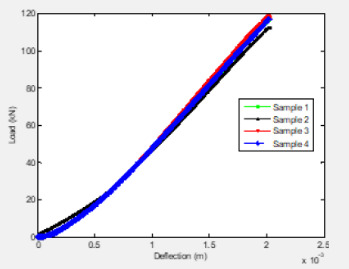
CAI tests have been conducted on MTM45-1/CF2412 composites manufactured using the OOA process. The tests were performed according to ASTM D7137. Four specimens of size 152.4 mm (6 in.) x 152.4 mm (6 in.) were first subjected to low-velocity impact tests and then machined to 152.4 mm (6 in.) x 101.6 mm (4 in.) for the CAI tests. Laminate construction consists of 12 fabric plies with a stacking sequence of [(+45/-45)/(0/90)]3S. Impact energy per unit thickness of 6672 J/m, an industry standard for evaluating thick, quasi-isotropic laminates was selected. Just clearly visible impact damage (VID) has been observed at 32J. The CAI test fixture is edge-loaded between the flat platens. Loads were applied at a cross-head speed of 1.27 mm/min. (0.05 in./ min). Compression load vs. deflection curves are shown in Figure 5. The ultimate compression-after-impact strength values of the specimens are tabulated in Table 4. The front view of the tested samples is shown in Figure 6.
Tension Fatigue Tests
Fatigue tests have been conducted on unimpacted OOA composites in an MTS 810 closed-loop servo-hydraulic testing system. Tests were performed on 12.7 mm (0.5 in.) wide x 254 mm (10 in.) long x 1.6256 mm (0.064 in.) thick MTM45-1/CF2412 samples. Fatigue behavior of the coupons at sinusoidal tension – tension loadings of 80%, 85%, 86%, 88%, and 90% of the ultimate strength (827.37 MPa) or ultimate tensile load of 15.57 kN (3500 lb) have been observed. A frequency of 2Hz and a load ratio of R = 0.1 (R = σmin/ σmax) have been used. Failure of samples in the grips was not observed with an increase in grip pressure to 5.75 MPa. The fatigue life of the coupons at different loadings is presented in Table 5. Figure 7 shows the S-N curve of an unimpacted sample under tension–tension (T-T) loading. Since the gap between the fatigue life at 88% and 90% stress levels is huge, more fatigue tests will be conducted at 89% of the ultimate load and other stress levels as needed. Figure 8 shows the failed specimens. Both fiber fracture and delaminations throughout the length of the specimen were observed. Post-impact compression fatigue tests are in progress.
Post-Impact Compression Fatigue Tests
CAI fatigue tests have being conducted on MTM45-1/CF2412 composites manufactured using the OOA process. The laminate construction consists of 8 fabric plies with a stacking sequence of [(+45/-45)/(0/90)]2S. Samples had a thickness of 3.35 mm (0.132 in). The 152.4 mm (6 in.) x 101.6 mm (4 in.) panels were subjected to the 15J of impact energy according to ASTM D7136. The CAI test fixture is edge-loaded between the flat platens as shown in Figure 9. Fatigue behavior of the coupons in sinusoidal compression–compression loadings of 60%, 65%, 70%, 75%, 80%, and 90% of the ultimate strength (224 MPa) or ultimate compressive load of 76.02 kN (17,090 lb) has been used. Initially, panels of 12 fabric plies have been constructed. The ultimate compressive failure load of the specimens was 115.65 kN (26,000 lb). Due to the load cell limits of the available test machines, panels with lower thickness have been chosen. A frequency of 2Hz and a load ratio of R = 10 (R = σmin/ σmax) have been used. The fatigue life of the composites at different load percentiles is given in Table 6. The fatigue curves are shown in Figures 10 & 11. The samples did not fail at 60% of loading even after 700,000 cycles and the tests were stopped.
Conclusion
A low-cost OOA vacuum-bag-only cure prepreg technology was successfully used to produce high-quality carbon fiber composites with void content less than 0.25 percent. The processing stages that lead to high-quality parts are shown. The fabricated panels were put through tensile, flexure, impact, compression-after-impact, and tensile fatigue tests. The effects of post-impact compression fatigue were studied. The results reveal that the OOA method is capable of producing parts with quality and performance comparable to those produced by the autoclave process at a fraction of the cost.
For more Open Access Journals in Juniper Publishers please click on: https://juniperpublishers.com/
For more articles in Academic Journal of Polymer Science please click on: https://juniperpublishers.com/ajop/index.php
#polymer#rubber#juniper publishers in USA#copolymer#juniper publisher journals#juniper open access journals
0 notes
Text
A Randomized, Double-Blind, Placebo- Controlled Study Trial to Evaluate the Potential Effects of Naticol®, Fish Collagen Peptides on Symptoms of Sarcopenia in the Elderly
Abstract
Background: Previous research has shown the potential effects of different doses of specific fish collagen peptides (Naticol®) on muscle mass and muscle function. In addition to these benefits, clinical studies have suggested that ingestion of specific fish collagen peptides (Naticol®) might also have beneficial effects on joint health such as osteoarthritis. Joint health, loss of muscle mass, and loss of muscle function are all symptoms experienced by elderly adults, and especially elderly adults suffering from sarcopenia, suggesting a possible role for Naticol® to help to reduce these symptoms in this vulnerable population.
Aim: The aim of this study was to determine the effect of 24 weeks’ supplementation of Naticol® on symptoms of sarcopenia in an elderly population.
Methods: In a randomized, double blind, placebo-controlled, clinical trial 28 elderly adults consumed one 15g sachet of either Naticol® or the Placebo product (maltodextrin) mixed into 20cl of cold water daily before breakfast, for 24 weeks. Symptoms of sarcopenia were assessed using dual x-ray absorptiometry (DXA) to measure lean body mass, the Short Physical Performance Battery to assess physical performance, the handgrip strength assessment to assess upper body muscle function, and the chair stand test to assess lower body muscle function.
Results: This study showed that 24 weeks of supplementation with Naticol® significantly improved symptoms of sarcopenia compared to placebo, by increasing lean muscle mass and increasing muscular function in the handgrip strength assessment, Short Physical Performance Battery, and Chair Stand Test.
Conclusion: The results of this study demonstrated that daily supplementation of Naticol® (containing fish collagen peptides) in elderly adults can improve symptoms of Sarcopenia, increasing lean muscle mass and increasing both upper and lower body muscle function.
Read More about this Article: https://juniperpublishers.com/jojcs/JOJCS.MS.ID.555850.php
Read More Juniper Publishers Google Scholar: https://scholar.google.com/citations?view_op=view_citation&hl=en&user=rp_7-igAAAAJ&citation_for_view=rp_7-igAAAAJ:LkGwnXOMwfcC
#Juniper Publishers Review#Juniper Publishers in USA#Journal of Case Studies#molecular biology#Biotechnology#caesarean section#Cardiology#blood transfusion
0 notes
Text
Pandanus Conoideus Lamk Protects Inflammation by Regulating Reactive Oxygen Species and Nuclear Factor Kappa B in Lps-Induced Murine Macrophages
Abstract
Background: Pandanus conoideus Lamk (Red fruit) is a Papuan traditional food which has been used to treat various diseases. Despite these various effects of Red fruit, little is known about the physiological mechanism. Aims: The aim of this study was to investigate the anti-inflammatory properties of Red fruit oil (RFO) and establish the signal pathway of leading compounds.
Methods: Raw 264.7 murine macrophage cells were used with lipopolysaccharide (LPS). Cell viability and the pro-inflammatory factors were investigated using MTT assay, real time PCR, western blot analysis, and Enzyme linked immuno-sorbent assay (ELISA). The quantification of leading compounds in RFO was performed using high performance liquid chromatography (HPLC).
Results: RFO did not affect cell viability. RFO significantly reduced the production of nitric oxide (NO) and prostaglandin E2 (PGE2), and both the protein level and mRNA level of iNOS in LPS-induced macrophages. RFO also regulated the reactive oxygen species (ROS) in LPS-induced macrophages. RFO attenuated the translocation of NF-κB p65 subunit, phosphorylation of I-κB, extracellular signal-regulated kinase (ERK), and c-Jun N-terminal kinase (JNK) in a dose-dependent manner. HPLC analysis determined that 1 g of RFO had 14.05±0.8 mg of β-cryptoxanthin and 7.4±0.7 mg of β-carotene.
Conclusion: RFO provides an anti-inflammatory effect by regulating ROS and NF-κB through MAPK due to the antioxidant activity.
Keywords: Pandanus conoideus Lamk; Macrophages; Anti-inflammation; ROS; NF-κB; β-cryptoxanthin
Abbreviations: RFO: Red fruit (Pandanus conoideus Lamk ) oil; LPS: Lipopolysaccharide; NO: Nitric oxide; iNOS: Inducible NO synthase; IL: Interleukin; ROS: Reactive oxygen species; ELISA: Enzyme linked immuno-sorbent assay; HPLC: High performance liquid chromatography; COX-2: Cyclooxygenase-2; PGE2: Prostaglandin E2; ERK: Extracellular signal-regulated kinase; JNK: c-Jun N-terminal kinase; MAPK: Mitogen-activated protein kinase; DMEM: Dulbecco’s modified Eagle medium; FBS: Fetal bovine serum; DCFH-DA: 2’7’-dichlorofluorescein diacetate; MTT: 3-(4,5-dimethylthiazol-2-yl)-2,5-diphenyltetrazolium bromide; RT- PCR: Real time polymerase chain reaction
Introduction
The inflammation process is tightly regulated by both initiation and maintenance signals and considered to be a major risk factor in the pathogenesis of chronic diseases where the macrophages are important immune cells which regulate inflammation producing expression of inflammatory proteins and pro-inflammatory chemokines, cytokines, and nitric oxide (NO) [1,2]. Macrophages are highly sensitive to initiators of inflammation as lipopolysaccharide (LPS) which respond by the release of mediators not only interleukins (ILs) and cytokines, but also inducible NO synthase (iNOS) and reactive oxygen species (ROS), which inducing the inflammatory gene expression where each is associated somehow with the pathophysiological of the inflammation [3-5]. Because macrophages produce a wide range of biologically active molecules participated in both beneficial and detrimental outcomes in inflammation, modulation of macrophage activation is a good strategy to prevent this diseases. Red fruit (Pandanus conoideus Lamk) is Papuan traditional food which has been used to treat various diseases such as cancer [6] preeclampsia [7], hepatitis [8], liver cirrhosis [9], diabetes mellitus [10], and sinusitis [11]. This bioavailability of red fruit has been due to unsaturated fatty acids such as palmitoleic acid, oleic acid, linoleic acid, linolenic acid and some carotenoids [10,12]. Despite these many biological effects, few researches were reported on the mechanism of red fruit oil (RFO). β-cryptoxanthin is a typical carotenoid found abundantly in persimmon, papaya, paprika, and carrot. β-cryptoxanthin has been reported to possess several beneficial functions, such as antioxidant, cancer-preventive effects, and anti-metabolic syndrome effects [13-16]. In present study, we hypothesized that the cause of this anti-chronic inflammation and anti-cancer effect is due to antioxidant function of RFO, and evaluated the anti-inflammatory effect of RFO on LPSstimulated RAW 264.7 macrophage cells. We also investigated the mechanism of inflammatory effect of reduced ROS by RFO in LPS-stimulated macrophages and investigated the component of β-cryptoxanthin in RFO.
Materials and Methods
Chemicals and reagents
RFO (APOTEK®) was supplied from Smile international Co., Ltd (Seoul, Korea). Dulbecco’s modified Eagle medium (DMEM), fetal bovine serum (FBS), and penicillin–streptomycin was purchased from Corning (Oneonta, NY, USA). 2’7’-dichlorofluorescein diacetate (DCFH-DA) and anti-iNOS antibody were purchased from BD (San Jose, CA, USA). Peroxidase-conjugated secondary antibodies and TriZol were purchased from Life technologies (Grand island, NY, USA). Phosphor-JNK, phosphor-ERK, phosphor-p38, phosphor-IκB and NF-κB antibodies were purchased from Cell Signaling Technology Inc. (Beverly, MA, USA). The enzyme immunoassay kit used for prostagladin E2 (PGE2) was obtained from R&D Systems (Minneapolis, MN, USA). The ECL detection reagents were purchased from GE Healthcare (Buckinghamshire, UK). LPS (Escherichia coli 0111: B5) was purchased from Creative Biolabs (Shirley, NY, USA). β-actin, 3-(4,5-dimethylthiazol-2-yl)-2,5-diphenyltetrazolium bromide (MTT), and other chemicals were purchased from Sigma–Aldrich (St. Louis, MO, USA).
Cell culture
RAW 264.7, the murine macrophage cell line was purchased from American Type Culture Collection and maintained in DMEM supplement with 1 mg/mL glucose, 10% FBS, 100 mg/mL penicillin-streptomycin at 37 °C with 5% CO2
Cell viability assay
The cytotoxic effect of RFO against RAW264.7 cell lines was evaluated by MTT assay. Briefly, cells were seeded at a density of 5 × 10³ cells/well in a 96-well plate for 24 h. Then, the cells were treated with at various concentrations of fractions with or without 1 μg/mL LPS. After 24 h, 2 mg/mL MTT was added onto each well, then incubated until formazan was constituted for 3h. The formazan was dissolved in dimethyl sulfoxide (DMSO) and the absorbance at 550 nm was measured using microplate reader (Molecular Devices, Sunnyvale, CA). Cell viability was calculated as a percentage of viable cells in drugs treated group versus untreated control. Each experiment was repeated three times.
Nitrite assay
Cells were treated with various concentrations of RFO for 30 min and incubated with 1 μg/mL LPS for 24 h. Because NO production is reflected in the accumulation of nitrite in the cell culture medium, 50 μL of supernatants were removed and mixed with the same volume of Greiss reagent (Promega, Madison, WI). After incubation for 10 min, the absorbance of mixture at 450 nm was measured using a spectrophotometer (TECAN, Austria). The nitrite levels were estimated as the percentage of absorbance of the sample to the respective controls.
Cyclooxygenase2 (COX-2) assay
Cells were treated with various concentrations of RFO for 30 min and incubated with 1 μg/mL LPS for 24 h. After incubation, the supernatants were removed and followed COX-2 measurement. The COX-2 concentrations were evaluated using a specific enzyme immunoassay (EIA) kit (Cayman, Ann Arbor, MI) according to the manufacturer’s instructions.
Prostaglandin E2 assay
Cells were treated with various concentrations of RFO for 30 min and incubated with 1 μg/mL LPS for 24 h. After incubation, the supernatants were removed and followed PGE2 measurement. The PGE2 concentrations were evaluated using a specific enzyme immunoassay (EIA) kit (Cayman, Ann Arbor, MI) according to the manufacturer’s instructions.
iNOS gene measurement by real-time PCR
The cells from the supernatants had been removed were subjected to RNA isolation. RNA isolation was performed using TRIzol reagent according to the manufacturer’s instructions. cDNA was synthesized using hyperscript RT master mix (GeneAll, Daejeon, Korea). The primers were described as; iNOS forward: 5′-ATGTCCGAAGCAAACATCAC-3′, reverse: 5′-TAATGTCCAGGAAGTAGGTG-3′, and GAPDH forward: 5′-TGTGATGGTGGGAATGGGTCAG-3′, reverse: 5′-TTTGATGTCAC GCACGATTTCC-3′. The PCR was amplified using ABI 7500 and Taqman gene expression master mix (Applied Biosystems, Waltham, MA, USA). The quantitative analysis was performed to compare the Δ Δ Ct after the normalization by GAPDH as an internal control. After analysis, PCR products were electrophoresed on 3% agrose gel and images were taken by cybergreen detection using Kodak imagestation FX® (Kodak, Rochester, NY, USA)
Analysis of ROS by flowcytometry
Cells were treated with various concentrations of RFO for 30 min and incubated with 1 μg/mL LPS for 24 h. Cells were followed by the addition of 10 mg/mL DCFH-DA). The suspensions were washed with PBS after incubation for 20 min. The suspensions were then assayed with a flowcytometer (C6 Accuri, BD, Bedford, MA, USA) according to Rhee et al. [4].
Western blot analysis
Cells were treated as described previously, then total lysates were prepared with lysis buffer (50 mM Tris (pH 7.4), 300 mM NaCl, 5 mM EDTA (pH 8.0), 0.5 % Triton X-100, 1 mM aprotinin, 1 mM leupeptin, 1mM pepstatin, 10mM iodoacetamide, and 2 mM phenylmethylsulfonyl fluoride (PMSF). Meanwhile, each nucleus extracts and cytosol extracts were isolated using a NE-PER nuclear and cytoplasmic extraction reagent kit (Pierce, Rockford, IL). Briefly, cells were washed with PBS, and were prepared with ice-cold extraction buffers sequentially. After centrifugation at 16,000xg, the cytoplasmic protein and nuclear extract were separated. Total lysates and nuclear fractions were estimated with Bio-Rad dye reagent concentrate (Bio-Rad Laboratories, Hercules, CA), then resolved on a 10% SDS-PAGE. After electrophoresis, the proteins were electro transferred to a PVDF membrane, blocked with 1% BSA, and probed with anti-iNOS (1:1,000), phospho- JNK (1:1,000), phospho-ERK (1:1,000), phospho-p38 (1:1,000), phospho-IκB (1:1,000), and NF-κB (1:500) antibodies at 4 °C overnight. The blot was washed, exposed to HRP-conjugated secondary antibodies for 2 h, and finally developed through enhanced chemiluminescence. For ß-actin detection, previously used membranes were soaked in stripping buffer (62.5 mM Tris- HCl, pH 6.8, 150 mM NaCl, 2% SDS, 100 mM ß-mercaptoethanol) at 65 ℃ for 30 min and hybridized with anti-ß-actin. The relative protein expression was densitometerically quantified using the BioRad GS-670 densitometer (BioRad, Hercules, CA) and normalized to β-actin.
High performance liquid chromatography (HPLC)
To determine the content of β-cryptoxanthin in RFO, we performed HPLC analysis according to previous studies [17]. HPLC analysis was performed using Agilent 1100 model with a pump (G1311C), auto sampler (G1329B), column, and diode array detector purchased from Agilent (Santa Clara, CA, USA). The analysis conditions are described in Table 1.
Statistical analysis
All results are presented as mean ± S.D. and are representing three or more independent experiments. Data were compared using the one-way ANOVA using Prism® (GraphPad, La Jolla, CA, USA) with p-values less than 0.05 considered statistically significant.
Results
RFO did not affect cell viability
Figure 1A showed the effect of RFO on viability of RAW 264.7 with or without LPS. Cell viability was not affected against 10- 1,000 μg/mL of RFO with or without LPS.
RFO reduced NO in LPS-induced macrophages
To assess the effects of RFO on NO production in LPSinduced RAW 264.7 macrophages, cells were treated with various concentrations of RFO for 30 min, then incubated with 1 μg/mL LPS for 24 h. NO release was elevated 224 ± 19.24% (p < 0.001) following LPS treatment, which was reduced 224 ± 19.24% at 10 μg/mL (p < 0.05), 161.38 ± 21.81% at 25 μg/mL (p < 0.001), and 136.16 ± 30.56% at 50 μg/mL (p < 0.001) with RFO combination (Figure 1B).
RFO decreased COX-2 production in LPS-induced macrophages
COX-2 production was significantly increased from 33.17 ± 5.23 ng/mL to 86.25 ± 1.88 ng/mL (p < 0.001) following LPS treatment. However, it was reduced 60.52 ± 12.49 ng/mL at 10 μg/mL (p < 0.05), 32.16 ± 8.85 pg/mL at 25 μg/mL (p < 0.001), and 13.27 ± 1.67 ng/mL at 50 μg/mL (p < 0.001) with RFO combination (Figure 1C).
RFO also decreased PGE2 production in LPS-induced macrophages
Meanwhile, PGE2 production was significantly increased 440.6 ± 35.36 pg/mL (p < 0.001) following LPS treatment, which was reduced 227.5 ± 13.6 pg/mL at 10 μg/mL (p < 0.001), 180.77 ± 48.95 pg/mL at 25 μg/mL (p < 0.001), and 103.27 ± 51.67 pg/ mL at 50 μg/mL (p < 0.001) with RFO combination (Figure 1D).
RFO suppressed both mRNA and protein levels of iNOS in LPS-induced macrophages
To determine the inhibitory effects of RFO on proinflammatory mediator NO, COX-2, and PGE2 production, the biosynthesis of transcriptional levels of iNOS was performed with semi-quantitative reverse-transcription PCR and western blot analysis on LPS-induced RAW 264.7 macrophages. Figure 1D indicates that both mRNA level and protein level of iNOS were significantly decreased by treatment of RFO (p < 0.001). Consistent with the findings shown in Figure 1E, RFO had a significant concentration-dependent inhibitory effect on the inflammation through pro-inflammatory mediator NO in LPSinduced RAW 264.7 macrophages.
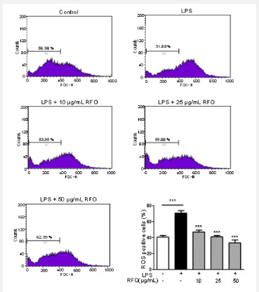
RFO attenuated ROS in LPS-induced macrophages
The excess ROS is known to be injured intracellular proteins, lipids and nucleic acids and induce inflammation [18]. Thus, we investigated the ROS production in response to LPS using flowcytometry. DCFH-DA binds ROS produced cells. Figure 2 showed the DCFH-DA positive cells were increased following LPS treatment from 40.71 ± 2.11% to 70.87 ± 3.09%. However, ROS production was also significantly inhibited by RFO with a dose dependent manner; 47.08 ± 2.45% at 10 μg/mL (p < 0.001),41.34 ± 1.41% at 25 μg/mL (p < 0.001), and 33.76 ± 3.56% at 50 μg/mL (p < 0.001).
RFO suppressed nuclear translocation of the NF-κB p65 subunit via MAPKinase
Since p65 is a major component of NF-κB activated by LPS in macrophages, we evaluated the levels of p65 in nuclear extracts by western blotting analysis. Phosphorylation of IκB results in degradation and release of NF-κB, which then translocates to the nucleus. Therefore, we examined whether RFO could prevent phosphorylation of IκB induced by LPS treatment. Figure 3A shows that IκB phosphorylation was increased by treatment with LPS alone in cytosol level, but that such phosphorylation was significantly inhibited in the presence of RFO, similar to results for the nuclear translocation of p65. Taken together, these data suggest that the inhibitory effect of RFO on the LPS-induced translocation of p65 might be involved in the suppression of IκB phosphorylation. To further investigate whether the inhibition of pro-inflammatory mediators by RFO is modulated through the MAPK pathway, we evaluated the effects of RFO on the LPSinduced phosphorylation of p38, ERK, and JNK (Figure 3B). RFO suppressed LPS-induced phosphorylation of p38, ERK and JNK. These results suggest that RFO blocks MAPK pathways to suppress the inflammatory response in LPS-induced RAW 264.7 macrophages.
HPLC analysis of RFO
Table 2 showed the HPLC analysis of RFO. HPLC revealed that 1 g of RFO had 14.05±0.8 mg of β-cryptoxanthin and 7.4 ± 0.7 mg of β-carotene.
Discussion
Inflammation is an immune response that protects our body against host response to infection and injury [19,20]. All inflammatory responses act through mononuclear cells, macrophages, and lymphocytes. Macrophages play on important innate immune effectors and increase pro-inflammatory factors including nitric oxide (NO), prostaglandin E2 (PGE2) cytokines.
The excessive amounts of NO and PGE2 produced by activation of iNOS and COX-2 in response to LPS play an important role in inflammation [21,22]. The overproduction of iNOS-derived NO is involved in the pathology of various inflammatory disorders and tissue damage conditions. A change in the NO level through the inhibition of iNOS enzyme activity or iNOS induction provides a means of assessing the effect of these agents on the inflammatory process. iNOS is implicated in the synthesis of prostaglandin H2 starting of arachidonic acid, which is a precursor of PGE2, in activated macrophages with LPS [23]. In addition, iNOS leads to overproduction of NO, PGE2, and COX-2 which results in the production of inflammatory diseases. Thus, modulation of iNOS and NO expressions could be one of the strategies to reduce inflammatory diseases. The production of inflammatory cytokines is a crucial part of regulating inflammation and tumor progression. The key signaling pathway mediating the inflammatory response, the NF-κB transcription factor, has been well-established in various inflammatory diseases and cancers [24,25]. It is also well known that NF-κB is a significant role factor regulating the expression of inflammation-associated enzymes and cytokine genes, such as iNOS, COX-2, TNF-α and IL-1β, which contain NF-κB binding motifs within their respective promoters [1,26]. Therefore, this signaling pathway is a good target for anti-cancer and antiinflammatory drug development. Many of the upstream kinases and downstream substrates are the same for the each of the major cascades. Our results revealed that anti-inflammatory activities of RFO are mediated through the inhibition of IκB phosphorylation and nuclear translocation of the NF-κB p65 subunit. Besides, these results also indicate that the inhibitory effects of RFO on MAPK and NF-κB signaling are related to a decrease in ROS. It is well known that oxidative stress stimulates ROS production in RAW 264.7 cell line [11,27]. Our data showed the pretreatment with RFO significantly decreased ROS production in LPS-induced RAW264.7 cells using DCFH-DA staining which demonstrated that RFO had a potent to reduce the oxidative stress. We also suggested that RFO regulated MAPK and NF-κB signaling of inflammation operate through oxidative stress. These results demonstrated that RFO could act as scavenging agents or acting on redox state of the cell and other acting as scavenging agents. In previous study, we already demonstrated that RFO regulated the cellular senescence through ROS modulation in H2O2-induced endothelial cells [5].
Carotenoids such as β-cryptoxanthin, β-carotene are one of the antioxidants which are not produced in the human body that must be ingested from outside. Many studies indicated that healthy people had the higher level of β-cryptoxanthin in blood [28-31]. β-cryptoxanthin is the only provitamin A component of carotenoid-based xanthophylls [14,32]. Carotenoids are lipid soluble components that must be ingested with fat to absorb completely in the body. Carotenoids affect the inflammation levels in blood as strong antioxidants and helps purify the blood. Park et al. showed that the daily oral administration of β-cryptoxanthin prevented the progression of osteoarthritis and inhibited proinflammatory cytokines in mice [33]. Therefore, we examined the effects of RFO on the production of several inflammatory mediators and on the expression levels of iNOS in LPS-induced RAW 264.7 macrophage cells. Our results demonstrated that RFO inhibited the expression of iNOS as well as the production of NO and PGE2 and the mechanisms underlying the suppression of the inflammatory response of the NF-κB and ROS. According to the US USDA database, β-carotene content of RFO was significantly higher at 335 times of blackberry, 119 times of broccoli, 13.9 times of pumpkin, and 5.2 times for carrot [34,36]. In addition, β-cryptoxanthin content of RFO was significantly higher at 76 times of orange and 15 times of papaya [30,37]. These findings suggested that RFO might be a beneficial therapeutic agent in the treatment of a variety of inflammatory diseases.
Conclusion
RFO is Papuan traditional food and had been used to treat various disease for long time. In this study, we suggested RFO had an anti-inflammatory effect through regulating inflammatory mediators such as iNOS, COX-2, PGE2, and excessive ROS for the first time. These physiological benefits of RFO may be attributed by regulation of NF-κB transcription. HPLC indicated that large number of carotenoids such as β-cryptoxanthin, β-carotene. This finding may be a synergistic adjuvant therapy for inflammatory diseases by acting as a radical scavenger, ROS inhibitor.
To Know More About Nutrition and Food Science International Journal
Please click on: https://juniperpublishers.com/nfsij/index.php
For more Open Access Journals in Juniper Publishers
please click on: https://juniperpublishers.com/index.php
#food safety#food microbiology#food preservation#Food Engineering#Juniper publishers USA#open access journals
0 notes
Text

Last Love by Melissa Schroeder is now live!

Welcome back to Juniper Springs, home of gay ducks, Nerdvana, and the LOLS.
Okay, so here’s the thing. I’m Liv, the normal O’Bryan sister. I have two kids I’m raising on my own, and I even have what people think is a boring job. Going to Vegas with my insane sisters is out of character for me, but I take that chance because of my new job. And let’s face it. I lost my husband over five years ago and I just haven’t…well…there’s been no one.
But an altercation with some drunken idiots has me almost falling on my butt, until a set of very strong hands save me.
Mason Spencer is younger, beyond sexy, and interested in me. So, I give in for one night. Can’t hurt, right? He goes above and beyond my wildest expectations. The next morning, I say thank you and head back to my life.
Until I move in next door to him, and my insane dog Houdini keeps showing up on his doorstep so I can’t even avoid him.
Worse, Mason tells me he wants more than that one night stand. Worser, he seems to fit right in with my kids and me and I discover that being with him makes me happier than I’ve been in years.
But remember, I’m the normal sister, and there’s nothing normal about an older woman with two kids snatching up a young man like Mason.
Of course, I’ve realized that there is nothing normal about the town, so maybe I would fit right in.
Author Note: Yep, it’s that time again to jump back into Juniper Springs. Home of Little Old Ladies, the Juniper Springs Express, and a seriously younger man with his mind set on his sexy neighbor. There’s a rescue dog with a mohawk who lives up to his name, and a meddling younger sister determined to help Liv find love.

Download today!
Amazon: https://amzn.to/3CazyvE
Apple Books: https://apple.co/3gagiHJ
Nook: https://bit.ly/3UEBfcS
Kobo: https://bit.ly/3ExQFd7
Google Play: https://bit.ly/3g8zvtq
Goodreads: https://bit.ly/3tATSlX

Meet Melissa Schroeder

From an early age, USA Today Bestselling author Melissa loved to read. First, it was the books her mother read to her including her two favorites, Winnie the Pooh and the Beatrix Potter books. She cut her preteen teeth on Trixie Belden and read and reviewed To Kill a Mockingbird in middle school. It wasn't until she was in college that she tried to write her first stories, which were full of angst and pain, and really not that fun to read or write. After trying several different genres, she found romance in a Linda Howard book.
Since her first published book, Grace Under Pressure, Mel has had over 60 short stories, novellas, and novels published. She has written in genres ranging from historical to contemporary to futuristic and has worked with 8 publishers although she handles most of her publishing herself. She is best known for her Harmless and Santini series.
After years of following her military husband around the country and world, Mel happily lives with her family in horse and wine country in Northern Virginia.
Connect with Melissa
Website | www.melissaschroeder.net
Goodreads | https://bit.ly/3uiGfbx
Amazon | https://amzn.to/3iBJJ6A
Facebook | https://bit.ly/3FvVGDT
Facebook Group | https://bit.ly/3H6Icj4
Instagram | https://bit.ly/3OY8QMN
TikTok | https://bit.ly/3XphnMg
Twitter | https://bit.ly/3OXmIaa
Bookbub | https://bit.ly/3uiGeUX
Pinterest | https://bit.ly/3FlUWAO
Newsletter | https://bit.ly/3FkhHW2
0 notes
Text
Aftereffect of Exogenous Adriamycin Management throughout Critically Ill Individuals in Delirium along with Snooze: A new Randomized Manipulated Trial
This kind of produces spatial heterogeneity within an environment high quality that giant herbivores must respond to in manners forecasted by simply excellent free submitting principle. We watched free-ranging bison to try whether, (One) controlled environments provide high quality look for food as compared to environments throughout undisturbed rangeland, (Two) buffalo respond through alterations in #Link# pack composition or perhaps exercise in order to variations environment top quality, as well as (Three or more) burned up and automatically handled environments offer comparable look features. Many of us learned that home sorts used up just like Ten years ago always produce good quality look as verified simply by buffalo undigested And awareness (15.4g kilograms(-1) dried up mass) than wide open (12.A few h kilograms(-1)), closed (12.6 g kilograms(-1)), as well as routinely altered habitats (12.7 grams #Link# kilograms(-1)). Bison pack make up along with action didn't vary throughout habitat sorts inside conditions, despite several between-season variance inside total group composition using sex segregation staying most evident prior to mid-summer. With regard to semi-arid rangelands encroached using woody plant life (electronic.grams. pifion-juniper from the western USA) our own evidence coming from free-ranging bison points too burning up ends in top quality look for food when compared with occurs in equally robotically manipulated as well as undisturbed habitats. Buffalo roam commonly from drinking water, sample obtainable crops continually, and they are long-lived gregarious pets that will learn to take advantage of your spatiotemporal heterogeneity inside their large home varies. Bison have similar diet programs to be able to cattle and so, wherever bison as well as cow are allowed to comingle, we suggest the actual looking parameters regarding free-ranging bison are impressive ecological signs of rangeland top quality both for bison along with cow. (Chemical) 2015 The particular Authors. Published by Elsevier Limited.Track record: Cognitive-behavioural remedy (CBT) pertaining to years as a child anxiety attacks is assigned to modest outcomes in the context of parental panic. Goals: This research evaluated set up results of CBT for kids using anxiety attacks in the context of mother's panic disorders has been enhanced from the inclusion of (we) treatments for maternal dna anxiety attacks, as well as (ii) treatment #Link# devoted to maternal dna answers. Your slow cost-effectiveness in the additional therapies was also evaluated. Layout: Participants have been randomised to receive (my partner and i) youngster cognitive-behavioural treatments (CCBT); (ii) CCBT along with CBT to a target maternal anxiety attacks [ CCBT + maternal cognitive-behavioural treatment (MCBT)]; as well as (3) CCBT by having an involvement to target mother-child connections (MCIs) (CCBT + MCI). Placing: A new NHS university hospital throughout Berkshire, British. Participants: 200 as well as 14 kids with a primary panic, whose mums also acquired an anxiety dysfunction. Interventions: Just about all households obtained ten times of human CCBT. Mums in the CCBT + MCBT arm furthermore received ten classes regarding CBT concentrating on their unique anxiety disorders.
0 notes
Text
In Vitro Radiosensitivity of Adamantinomatous Craniopharyngiomas
Authored by Elfar úlfarsson

Abstract
Background and purpose: Improvements in radiation treatment for craniopharyngiomas are needed. No clear in vivo data exists on radiosensitivity of craniopharyngiomas and in vitro data is lacking. The purpose of this study was to assess the radiosensitivity of adamantinomatous craniopharyngioma in vitro.
Materials and methods: Craniopahryngioma cells from 7 individuals (two different passage number) were seeded in triplicate wells and exposed to 8 photon doses in the range 0-10 Gy in a 137Cs irradiation chamber. The radiosensitivity was measured as clonogenic cell survival. The surviving fraction at 2 Gy (SF2), the mean inactivation dose ( ) and the α/β ratios were calculated from the dose response curve fitted with the Linear Quadratic model.
Results: The SF2, and α/β values for the cell strains ranged from 0.31 to 0.47, 1.65 to 2.44 and 10 to 30 Gy, respectively. The mean, standard deviation and coefficient of variation of the SF2, and α/β values were 0.40 ± 0.07, 17%, 2.04 ± 0.30, 15% and 19 ± 6 Gy, 33%, respectively.
Conclusion: The high α/β value indicates that adamantinomatous craniopharyngioma is among early responding tissues. This supports the clinical praxis of fractionated radiation for those tumors.
Keywords: Craniopharyngioma; Radiosensitivity, Clonogenic cell survival, Alpha/beta ratio, radiation treatment
Go to
Introduction
Craniopharyngioma is a benign neoplasm of epithelial origin located in the sellar and suprasellar region. The adamantinomatous craniopharyngioma represents around 2/3 of all craniopharyngiomas and is the most common pathology in this region in children [1,2]. Other histological subtypes are the squamous papillary and the xanthogranulomatous type [3]. The former occurs almost exclusively in adults. The main treatment options are surgery and conventional radiotherapy (CRT). Surgical cure is hampered by the eloquent surrounding structures and is accomplished in few cases. The acknowledgement of the devastating effect of hypothalamus damage on quality of life has led to more conservative surgery and increased use of adjuvant RT in children [4,5]. CRT has proved its value in controlling craniopharyngiomas; however considerable irradiation to the surrounding tissues is unavoidable by this technique and not without sacrifices.
The decline in intellectual function in children [6], inability to treat the youngest patient group, radiation induced malignant gliomas [7] and 10-years recurrent free survival rates of 32 % - 49 % [8,9] in children stresses the limits in the current CRT.
Gamma Knife radiosurgery (GKRS) has been used as an alternative to CRT to reduce the radiation load on the surrounding tissues. Reports on low morbidity from the anterior visual pathways using this technique, 3.1%(10), underlines the power of photon beam focusing. The main drawbacks are the target volume restrictions and higher risk of geometrical misses compared to CRT. The ultimate limitation of the single session GKRS is that the most effective GKRS-dose, 11.7 - 12.7 Gy, is also the critical toxicity dose for the anterior visual pathway [10,11].
The majority of craniopharyngioma patients receive radiation treatment during their diseased life and around one third of them experience tumor progress [12]. The hazard of repeated irradiation precludes further radiation in most cases and the danger of repeated surgical trauma is well known. The gravity of the situation of late recurrences is reflected in a very high mortality after salvage treatment [13,14]. Thus, improvement in the radiation treatment is clearly needed. No clear data exists on the radiosensitivity of craniopharyngiomas and the optimal dose has not been defined. The lack of detailed dose-response data and standardized way of reporting endpoints disables a thorough interpretation of radiobiological data from the literature. Furthermore in vitro radiosensitivity data, that could be useful in designing new fractionation regimens, is lacking.
The purpose of this study is first to assess the in vitro radiosensitivity of adamantinomatous craniopharyngiomas and second to address the alpha beta ratio (α/β) specifically from a clinical perspective.
Go to
Methods and Materials
Cell cultures
Primary cultures of human craniopharyngioma cells were isolated and prepared from tumour samples in a similar manner as for keratinocytes according to the methods described elsewhere [15,16]. The obtained cultures used for the (irradiation) experiments were plated without feeder cells in passages between 3 and 9 (median 5). All patients harboured histological verified adamantinomtous type of craniopharyngioma. The procedures were in accordance with the ethical standards of the institutional committee on human experimentation and with the Helsinki Declaration of 1975, as revised in 2000.
Clonogenic survival
Appropriate cell numbers were plated for survival using the clonogenic assay technique described previously [17]. The single-cell suspensions were plated into 35 mm plastic petri dishes (Corning, New York) in triplicates to a final medium volume of 3 ml and then left in the incubator for 3-4 h to attach before irradiation.
The cells were irradiated at room temperature with doses of 0-10 Gy at a dose rate of 0.5 Gy/min from a 137Cs-source (Scanditronix, Uppsala, Sweden). The cultures were then incubated for 10-14 days, with a change of medium after 5-7 days. Thereafter colonies were fixed, stained and counted. Radiation survival curves were constructed from two independent experiments. The mean PE for un-irradiated cells was 43, 55, 85, 12, 10, 42 and 36% for case 1 to 7, respectively.
Dose-response models for clonogenic cell survival. The LQ model(18) was used to fit the data with the Maximum Likelihood method, where the probability for clonogenic cell survival S at a dose D is given by [19]:
S = e-(αD+βD2 ) (1)
The surviving fraction at 2 Gy (SF2) was computed from the whole survival curve and was used as a unique measure of cellular radiation sensitivity. The mean activation dose, D , was calculated according to Taylor [20]. The D was chosen since it has been shown to keep the scattering of data smaller than for other parameters such as SF2 and D0 [21].
Assessment of α/β in vivo
From in vivo data, the LQ model can be used to calculate the α/β from isoeffective fractionation regimens. This is based on the assumption that the biological effective dose (BED) is the same if two fractionation regimens result in an equivalent clinical effect [22]. In this study, the same tumour control rates were assumed with 2.0 Gy daily fractions with a total dose of 50 Gy [23] and 11.5 Gy prescription dose at the 50% isodose line using GKRS [10]. For the GKRS, it was assumed as a first reasonable approximation, that the tumor response was related to the mean dose to the tumor which is generally 30 - 50% higher than the dose to the periphery. Thus for a prescribed dose of 11.5 Gy, the mean dose will be in the range from 15 Gy to 17 Gy. Calculation were done for two cases; 1) without- and with correction for repopulation during the time of fractionated treatment, in the second case, two values of Tk (the time after start of the treatment when repopulation starts) were used; for keratinocyte-related malignancies such as head and neck tumors (Tk=21 days) and craniopharyngioma related normal tissues such as mucosa (Tk =7 days) [24]. For each of the two Tk values, the cell doubling time (Tp) was estimated in patient C1 and C2using the formula
Tp=0.693*t/ln(Nt/N0).
Go to
Results
The clonogenic cell survival curves and the radiosensitivity parameters from the seven craniopharyngioma cell strains are presented in Figure 1 and Table 1, respectively. The mean values and the coefficient of variation (CV) for SF2 and D were 0.401 (SE +/- 0.021) and 2.04 (SD +/- 0.08) and 17% and 15%, respectively. The α/β value ranged from 10.3 to 29.9 Gy with CV of 33%. The mean value was 19 Gy (SE +/- 2.4). Acknowledging the fact that other survival models will fit the data in the lower dose region better, we used the entire dose range when fitting the data. This was done since doses in the range of 8-13Gy are being used for these types of tumors.
In Figure 2, α/β values are given as a function of the mean dose to the tumor in GKRS Estimated mean doses to the tumor, for a prescription dose of 11.5 Gy will be in the range from 15 Gy to 17 Gy. Results are shown, without correction for repopulation, as well as corrected with two Tk values, with two sets of Tp and α values.
Go to
Discussion
In this study we present a novel data set on radiosensitivity of adamantinomatous craniopharyngioma cell strains, based on clonogenic survival assays. More than 25 years ago Fertil et al. [25] pointed out a correlation between the radiosensitivity of human cancer cell lines in vitro and the radiocurability of the corresponding tumor types in vivo. Since then histological groups of human cell lines have been characterized by an intrinsic radiosensitivity in vitro [26,27] and this data has been shown to be useful in predicting tumour response to RT [28].
The high variation of α/β within specific tumor group as for adamantinomatous craniopharyngiomas in this study is commonly noted in reports for different human cancer lines in vitro. Taghian et al. [29] reported data on glioblastoma, which is among the most radioresistant tumours. The α/β ranged from 3.7 to 48 Gy. Weichselbaum et al. [30] reported mean α/β for human cancer cell lines with large SD values, reflecting a high variation within each tumour group.
According to SF2 and Dcraniopharyngiomas cell strains are slightly more radiosensitive than the average radiosensitive cancer cell line [27] and the variation is not higher than reported in other cancer cell lines [21,27]. Comparative in vitro data for other benign intracranial tumours is lacking. However, from in vivo data, and with the method used here α/β for meningiomas and vestibularis schwannomas have been calculated to be 3.3 Gy and 2-3 Gy, respectively [31]. In this study, the α/β will be in the range of 4-6 Gy for adamantinomatous craniopharyngiomas, without correction for repopulation and from 6 Gy to more than 20 Gy if reasonable parameters for correction for repopulation are applied (Figure 2). Those values are still much lower than the in vitro values in this study. The reason for this discrepancy could be explained by a lack of correlation between the in vivo and in vitro data, by paucity of reliable in vivo data or both.
Arguments for craniopharyngiomas having high α/β can be found when growth kinetics is considered. It is known that the growth kinetics of the tissue irradiated dominates the radiation response. A higher proliferation indices [32-35] and more dramatic response to irradiation [10,36-39] compared to meningiomas and acustic neurinomas support our findings of a high α/β for craniopharyngiomas. The α/β for cranipharyngioma related tissues, such as mucosa and epithelium is in the range for early responding tissues or 7 - 15 Gy [40-42]. Since there is often a close similarity between the mean lethal dose of tumour and the normal tissues from which they arise, it is likely that α/β for cranipharyngioma is in this high range. If this is the case, one should consider the repopulation time factor in calculating the α/β from clinical data. This results in higher α/β values (Figure 2) [43,44].
The above estimations of α/β values based on in vivo data have to be viewed with caution since the differences in patient and treatment characteristics between clinical studies make it difficult to compare results from different radiation regimens. The main obstacle is that certain parameters that can influence endpoints, such as histological subtypes and tumour volume, are generally not considered in reports on treatment results.
The comparison of iso effective doses for GKRS and CRT could be flawed by differences in tumour volumes treated with those techniques. The general principle is that large tumours (more clonogenic cells) require higher doses than small ones (fewer clonogenic cells) to obtain the same tumour control probability (all clonogenic cells killed, Figure 1). This is also partly explained by the differences in micro-environment between large and small tumors [45,46]. The lack of information on tumour size is more a rule than an exception in studies using CRT for craniopharyngiomas. In viewing the few studies available, it seems that the tumour volume tends to be substantially larger in the CRT studies compared to GKRS studies [10,47-49]. This is a reasonable assumption regarding the radiobiological volume restrictions of GKRS. The implication of this phenomenon in CRT for craniopharyngioma is probably less than for other more solid tumours types due to the large fluid component in craniopharyngiomas.
Taking the arguments above together:
CRT studies tend to include substantially larger tumour volumes and
Larger tumour volumes require higher doses for local control, it is tempting to conclude that a somewhat lower CRT dose than 50 Gy would give the same local control as GKRS for similar tumour volumes. If this line of arguments is correct, the α/β from clinical data, will be larger than the ones presented in Figure 2 and hence even more close to the α/β presented in this study from in vitro data.
It is debated whether benign intracranial tumours benefit from fractionation since the α/β is thought to be similar as for neural tissue or 2 Gy. This is generally supposed to reduce the beneficial effects of fractionation but if the clinical α/β is as high as observed in our study we have strong arguments to apply fractionated radiation regimens to improve the therapeutic ratio. It is however important to notice that improvements in RT are clearly needed.
Even though CRT has been used for craniopharyngiomas for more than 40 years, the toxicity risk for the anterior visual pathway and the hypothalamus has not been reduced with recent improvements in this technique. The major part of those structures is still included in the high dose field prescribed for the tumour and receives doses just below the maximum tolerance level [6,39,48]. Furthermore structures involved in neurocognition are still at risk [6]. Improvements could be made by using fractionation with Gamma Knife® or similar technique. By this approach one could overcome the target volume restrictions of single fraction treatment, avoid critical doses to structures involved in neurocognition and reduce the radiation load on the visual pathway and hypothalamus. Radiobiological data such as presented in this study would then be needed to design new fractionation regimens.
Go to
Conclusion
The high α/β in this in vitro study indicates that the adamantinomatous craniopharyngioma is among early responding tissues and supports the clinical praxis of fractionated radiation. The radiosensitivity parameters, SF2 and , indicate that these cell strains are slightly more radiosensitive than the average radiosensitive cancer cell line. Although some correlation with in vivo data is found, improvements in presenting treatment parameters and results after radiation treatment and further in vitro studies are encouraged to validate this data.
For more open access journals in juniper publishers please click https://juniperpublishers.com/
For more articles on Open Access Journal of Neurology & Neurosurgery Please click on https://juniperpublishers.com/oajnn/
Open Access Journal of Neurology & Neurosurgery in Full text in Juniper Publishers
https://juniperpublishers.com/oajnn/OAJNN.MS.ID.555576.php
1 note
·
View note
Text
Sinonasal Adenoid Cystic Carcinoma: A Case Report and Review of the Literature

Abstract
Adenoid cystic carcinomas (ACC) represent 10% of all salivary tumors. It primarily affects the salivary glands particularly those located in the buccal cavity. Sinonasal location is rarely described [1]. We report a rare case of sinonasal adenoid cystic carcinoma (SACC), we discuss through a brief review of the literature its clinical, radiological, histopathological and therapeutic modalities and the prognostic factors of this tumor.
Keywords: Adenoid Cystic Carcinoma; Paranasal Sinus; Facial Pain
Introduction
ACC is a rare malignant neoplasm that accounts for 1-2% of all head and neck malignancies and approximately 10% of all salivary gland neoplasms [2]. It occurs predominantly among women, between the fifth and sixth decades of life [3]. Sinonasal Adenocarcinomas mainly arise from the respiratory epithelium or the underlying mucoserous glands (60%). It’s known by its slow persistent growth with a high risk of local recurrences and distant metastases. The treatment of choice is based on surgical excision of the tumor with adjuvant radiation therapy.
Case Report
A 44 year-old female presented to the ENT emergencies with a swelling of the latero-nasal area and the inner corner of the left eye evolving for the last year. It measured 2.5 cm at its largest diameter (Figure 1). The patient reported a progressive left-sided nasal obstruction of 8 months duration, with purulent,bloody and fetid nasal discharge, with a conservation of the general state. The anterior rhinoscopy highlighted nasal congestion with no visible tumor. Ophthalmologic examination revealed lateralization of the left eyeball with telecanthus. Facial CT scan revealed a tumor process of the left nasolabial furrow with lysis of the maxillary bone, its internal wall and also the inner wall of the orbit (Figure 2). A biopsy through the vestibular way has confirmed the diagnosis of a cribriformtype of ACC. The staging did not find distant metastases. The patient was managed surgically with adjuvant radiation therapy.
Discussion
Sinonasal Adenoid cystic carcinomas are considered as poor prognostic tumors, characterized by the possibility of late and frequent local recurrences, and poor survival. These tumors grow usually slowly, thus they can reach considerable size before the diagnosis is made. The initial prognosis may be good but the frequency of local recurrences and distant metastases (lung essentially) influence the long-term survival (10-year survival <10%) [4]. Local recurrences are more frequent for sinonasal locations (60% of clinically evident recurrences within 2 years following treatment) [5]. This is related to the difficulty of ensuring healthy resection margins at the base of the skull often because of the very advanced stage of the tumor by the time of the diagnosis, the anatomic complexity of the region, the frequent intracranial extension along cranial nerves and because of restriction on the resection margins caused by the proximity of critical neurovascular structures.
Among the most important prognostic factors involved are tumor stage, histological grade (The tubular- and cribriformtype ACCs are lower-grade tumors, whereas solid-type ACC is a higher-grade tumor), the existence of perineural invasion and cancerous resection margins [4,6,7]. Complete surgical resection is critical but often difficult to realize close to the skull base [4,8]. Postoperative radiotherapy improves the long-term prognosis of patients with large lesions especially if there are microscopic residues after surgery.
Conclusion
Adenoid cystic carcinoma of sinonasal cavities is a rare aggressive tumor with a high incidence of local recurrence and distance metastases regardless of therapeutic modalities used. Complete surgical excision and adjuvant radiation therapy for the extensive local infiltration offer to these patients the best chance to achieve high tumor control.
To Know More About Cancer Therapy & Oncology International Journal Please click on:
https://juniperpublishers.com/ctoij/index.php
For more Open Access Journals in Juniper Publishers please click on: https://juniperpublishers.com/index.php
For more about Juniper Publishers Please click on: https://juniperpublishers0.wixsite.com/juniperpublishers
0 notes
Text
Juniper Publishers-Health of Schools, as a Social and Pedagogical Problem

Abstract
The analysis showed that schoolchildren's health is a complex socio-pedagogical problem that the distribution among children of chronic diseases, diseases of the organs of the gastrointestinal tract, diseases of the endocrine system, respiratory viral infections must be sought in three interrelated constituents of their lifestyle (family, school, leisure) that the widespread distribution of children in various forms of posture violations is due to the fact that the school does not pay enough attention to conducting in the school day a variety of if cultural and recreational activities (fitness pause a moment to fitness classes, outdoor break, morning gymnastics etc).
Keywords: Health; Way of Life; Spheres of Life; Social Institute; Public Awareness; Leisure; Sociological Research; Education System
Introduction
In recent years, there has been an avalanche stream of information on the deterioration of the health of schoolchildren in periodicals and in scientific publications. There are objective and important prerequisites for this. Thus, over the past 10 years, the incidence among school-age children has increased by 26.8% [1]. Today, approximately 90% of schoolchildren have a deviation in the state of physical and mental health [2] the overall incidence among students of secondary schools in Ukraine reaches 64% - 71%. During the period of study at school, the number of students assigned to a special medical group is almost doubled [3]. In recent years, a high level of physical health has been found in only 0.32% of boys and girls, above the average 4.18%, the average 27%, below the average27% and the lowest 41.48%. The consequence of this situation was that in the existing informational space of schoolchildren's health is used as an integral indicator of public assessment of the effectiveness of the functioning of the school physical education system, which, as practice shows, is not able to provide the necessary level of physical health of children and young people. The urgent need to address the whole spectrum of health, educational and educational tasks in the field of physical education of the younger generation needs to rethink the basic principles of organizing the existing system of physical education and ascertaining the complex of the main factors on which the health of children and youth depends.
The Aim of the Study
Identification of socio-pedagogical factors that affect the health of children and young people and the main directions of solving the problem of schoolchildren's health.
Methods, Organization of Research
Was to analyze the materials published in the special literature, which examines the health of children and young people, as well as to summarize the results of relevant studies conducted by specialists of the problem scientific research laboratory of the Kharkiv State Academy of Physical Culture (KDAFK) (scientific research director VO Sutula).
Results of the Research and Their Discussion
The decline in the health of schoolchildren that has been observed recently, according to experts, is due to the lack of volume of their motor activity [4]. Some publications directly indicate the existence of a high correlation between the state of health of schoolchildren and the level of development of the majority of their physical qualities, which (the level of development of motor qualities) in its entirety is the result of the motor activity of students [5]. Without rejecting the above mentioned provisions in general (the increase in motor activity of pupils really improves the functioning of all systems of their body, which is the basis of their physical health), it should be noted, however, that the simple increase in the volume of motor activity of children, without regard to the specificity of the disease, definitely does not guarantee the improvement of their health. This conclusion is indirectly derived from the analysis of the statistics of the morbidity of young athletes, the total volume of motor activity which can be considered sufficient if it is evaluated from the stand point of valid sanitary-and-hygienic norms. The results of this analysis show that among children and adolescents who are actively engaged in sports, in recent years there is a steady tendency of growth of various diseases [6,7]. Consequently, an increase in the volume of motor activity of children and young people does not automatically guarantee improvement of their health. Obviously there are other no less important factors that affect their health.
According to the definition of the World Health Organization, human health is characterized as a state of complete physical, spiritual and social well-being, and not only the absence of diseases or physical defects. It is obvious that this understanding of health is quite general in nature. It covers virtually all aspects of human life. Perhaps not by chance, experts point out that human health is 53% depending on the way of life, 21% from the environmental situation, 16% from hereditary factors, and 10% from medicine [8]. Consequently, it can be argued with high probability that the determining factor on which human health depends is a way of life. The mode of human life in the most general form is determined by the social role it performs in the three main spheres of life in the family, in the field of education (or work), in the field of leisure. From the above basic areas of social activity, the family institution plays a leading role in shaping the way of life of a child, because it is the family that determines the cultural and everyday environment in which the child is brought up.
To a certain extent, under the influence of a family of children, a need-motivational sphere is formed which, in fact, determines the nature of their relationship with others, as well as the main directions of the child's activities, including the use of physical exercises for their own health and physical development . The social status and financial status of parents determine the essential conditions of life of children the conditions of their residence, the quality of food, the possibilities of rest, the forms of spending their leisure time (visits to theaters, museums, palaces and houses of children's creativity, stations of young technicians, classes in sports sections and etc.), their level of medical support, and so on. The place of residence of the family determines the impact on the child of the environmental conditions, of which, as already noted, the health of a person depends on 21%. It is from parents that the influence of hereditary factors on the health of the child depends on which of 16% determines her health. Despite the important role played by parents in organizing the lifestyle of a child, the possibilities of a modern Ukrainian family to solve the problem of ensuring decent living conditions of children are quite limited, because today, as statistics show, 38.2% of the population of Ukraine is outside the subsistence minimum [9].
The second important component ofthe way of life of children and young people, which significantly affects their health, is the form of their leisure. Special investigations of this problem by the State Institute for Family and Youth Development show that only 44% of children between the ages of 14 and 17 have the opportunity to meet their leisure needs at an adequate level. For the majority of children, these funds are not enough to meet these needs (70%), free time (26%) and corresponding facilities at their place of residence (26%). Materials of these studies also point to the non-formation, as a whole, children have the right needs, because 14% of them never went in for sports, 34% did not attend circles, 24% did not attend theaters and museums; most of the free time children gave television programs, and every third teenager computer games. The national report on the implementation by Ukraine of the provisions of the UN Convention on the Rights of the Child [10] explicitly states that the main reason that adversely affects the organization of children's leisure is the lack of a sufficiently developed network of physical culture and sports, culture and recreation, as well as the poverty of families with children.
Perhaps these factors have contributed to the fact those in recent years the possibility of having children with healthy physical activity, which is a guarantee of their physical health, has narrowed considerably. The lack of real opportunities for children to undertake such leisure activities, as well as their lack of proper needs, leads to the fact that a significant part of children in their free time prefer to communicate with their friends. Currently, it is this environment that most influences the appearance of bad habits among children, as evidenced by the results of an analysis of the causes that cause the spread of tobacco smoking habits and alcohol consumption by young scientists in the KDAFK problem research laboratory [1113]. The materials of these studies indicate that every third schoolboy of secondary and senior school years already had experience in smoking tobacco, and in the seventeen years every fifth student of secondary schools was smoking. During the years of studying in school, the number of students who have tasted some or other alcoholic beverages increases. For example, in the 5th grade, every fifth (!) Student was drinking alcohol, and in the 11th grade of such students already about 70%. The identified trends in the prevalence of bad habits among schoolchildren (tobacco use and alcohol use) are fairly objective in nature. In general, they are confirmed by the results of special sociological studies conducted within the framework of the program "Health and Behavioral Orientation of Student Youth" [14]. Attention is drawn to the fact that in the process of studying in the school there is a pronounced tendency to increase the number of students who are indifferent to the widespread use of adult eating habits and alcohol consumption. The results of special sociological studies indicate that at present fifth grade of such students is about 15%, in the eighth about 35%, and in the eleventh - more than 50%. The given data indicate that during the years of studying in children, the active citizenship position on this issue is practically not formed.
Of the above three main areas of life children play a special role in organizing their way of life, the school, which is the main social institution, through which the process of transferring culture from one generation to another, within which the development of the personality of the child passes. It is at school that the outlook of the children is formed, their liveliness, and activity, independence, and organization, ability to work in a team, mutual help and other features of the character, which just determine the essence of the individual. It is at school that the children receive the necessary knowledge, skills and skills to help them choose their future activities. It is precisely in the school that is prescribed and regulated, through the use of various physical culture and recreational activities, the mode of motor activity of children during the day that is the basis of their physical health. Solving these tasks requires the creation of a special cultural environment in the school in which every participant of this process (student and teacher) should feel psychologically comfortable, be socially protected and in demand from others. It is obvious that the creation of such an atmosphere in school is a rather difficult task, especially in today's conditions, when all the traditional system of education is located, according to I Prokopenko and V Evdokimova [15], in a state of "stagnation and even crisis". As a result of complex processes that unfold in the system of school education, the fact that around 50% of students in general education institutions are weary of classes, about 40% of students is difficult to study, 25% of students consider the pupils' environment to be psychologically not comfortable, in 20% of students formed the feeling of not perceiving them from the side of teachers as individuals, and in 15% of them - a sense of unfair treatment of teachers.
Undoubtedly, the situation prevailing in the system of traditional education imprinted on the whole system of school physical education, since the latter is an integral part of it. At the same time, it should be noted that at present, in the development of the system of physical education of students of general education institutions, despite all the complexities, and positive trends exist. Thus, sociological researches carried out by KDAFK scientists have shown, firstly, that at present society is aware of the necessity and importance of a healthy and physically active lifestyle as one of the basic factors influencing human health; Secondly, among teachers and parents there is an understanding and concern about the negative trends that are manifested in the development of the system of physical education for students of general education institutions, in which the majority of parents (94.6%), which should be especially noted, are ready to take a possible participation in the process of its reform; Thirdly, the vast majority of students have a positive attitude toward physical education lessons (87.3%), and for 76.4% of them, such lessons are only satisfying. The above results of researches testify that at present in the system of school physical education there are positive prerequisites for solving, on a qualitatively new level, the whole complex of health-improving, educational and educational tasks. Some approaches to solving educational and educational tasks have already been presented in a previous publication. In this article, taking into account its purpose, we will pay attention to possible approaches to solving health problems. To objectivism this analysis it is necessary to consider, at least in the first approximation, the range of illnesses of schoolchildren.
If we proceed from the data published in the special literature, it should be noted that over the years of schooling almost twice the number of children with chronic diseases increases, in the last years up to 6-7 times a year the number of respiratory and viral infections has increased, the incidence of organs of the gastrointestinal tract in schoolchildren increased almost by 1.4 times, the endocrine system by 2.6 times. Sufficiently widespread in the environment of children marked diseases is due, as experts say, the state of their immune system, the influence on their organism of genetic factors and environmental factors, that is, the influence of factors that are determined by environmental, biosocial and psycho-emotional causes, and therefore lie in the plane of the three main components of the way of life of children (the family, school, leisure). Consequently, we can conclude that there is no direct cause-and-effect relationship between the occurrence of children with the above diseases and the volume of their motor Activity, which depends, first of all, on the peculiarities of constructing the pedagogical process in the system of physical education. That is why the increase in the number of such diseases among children cannot be a measure of the effectiveness of the functioning of the physical education system.
In the spectrum of diseases of schoolchildren, as shown by literary data, the determining place occupies various forms of violation of their posture. At present, the number of children with posture disorders is 80% 90% of the total number of students, and for the period of schooling, the frequency of manifestations of this disease increases by 1.5 times Violations of posture, as evidenced by the results of special studies, leads to a change in the relative position of the internal organs, which is accompanied by a violation of their normal activities (especially the lungs and heart), which adversely affects the functioning of all organs and makes the body of the child as a whole prone to various diseases. That is why we can say that posture is not a disease in its classical sense. It is likely to represent a "delayed action" that creates the preconditions for the emergence of other diseases, and which "explodes" when the influence of negative factors on the child's organism (ecological, biosocial, psycho-emotional, and others) reaches a critical level. It is obvious that that for the prevention of posture disturbances among schoolchildren it is necessary to apply special complexes of physical exercises, or health systems, which enable from the very beginning of schooling to purposefully influence the causes that cause the emergence and development of this disease.
As the main reason for the disturbances of posture among schoolchildren, as evidenced by the study of hygienists, is due to the fact that about 85% of daytime schoolchildren are without movement (sitting), this may mean that the prevention of posture disturbances among schoolchildren is reduced to regular exercise in the school day, as the lessons of physical culture and what is needed emphasize especially various physical culture and recreation activities (morning gymnastics, physical culture pauses and physical exercises at lessons, mobile breaks), which perform not only preventive functions, but also stimulate the mental performance of students. That is, the mechanism for preventing the disturbances of posture among pupils is based on well-known events, the need for which is regulated by the relevant orders of the Ministry of Education and Science of Ukraine, and which, unfortunately, is still paid insufficient attention in the system of general secondary education, as evidenced by the above statistics of manifestations of this disease. The importance and necessity of taking effective measures to prevent the disturbances of posture among schoolchildren, which is definitely one of the main health problems of the system of physical education, is understood at the state level. It is no coincidence that in the last year adopted a program for general education institutions to content training material in grades 5-9 introduces topics related to the prevention of violations of posture students.
To read more articles in Journal of Gerontology & Geriatric Medicine
Please Click on: https://juniperpublishers.com/oajggm/index.php
For more Open Access Journals in Juniper Publishers
Click on: https://juniperpublishers.com/journals.php
#Jun iper Publishers#Gerentology#Aging#Geriatric care#Juniper publishers Group#Juniper publishers in USA
0 notes
Text
Juniper Publishers| Overview and Future of Hemo-Components and Natural Guided Regeneration
Journal of Surgery-JuniperPublishers
The History of Platelet Rich Fibrin (hemocomponents) started in 1970, when Matras described a fibrin glue, formed by polymerizing fibrinogen with thrombin and calcium, which was used to improve skin wound healing in a rat model in 1970 [1]. Because of the low concentration of fibrinogen in plasma, the stability and quality of fibrin glue were low. A few years later several research works proposed an upgraded concept for the use of blood extracts, termed “platelet-fibrinogen-thrombin mixtures” or “gelatin platelet - gel foam” [2,3]. In this new concept, the fibrin glues were presenting a significant concentration of platelets within the final preparation. The idea was first to reinforce naturally the fibrin gel, and also to combine the healing properties of the platelets with those of the fibrin. This improvement allowed to prepare more natural products, integrating more natural blood constituents as it should.These products were the first platelet-rich plasma gels. These new strategies insisted in the role of platelets within the fibrin gel, and offered excellent preliminary results in ophthalmology, neurosurgery and general surgery. Whitman proceeded to develop this technique in 1997 and particularly Marx et al. [4,5] in 1998. The Leukocyte- and Platelet-Rich Fibrin L-PRF clot was often described as “optimized blood clot” that can be surgically handled and used. The rationale to use this glue/membrane and its success is due to fibrin, platelets, growth factors slow release, leukocytes and other cells: all these components are the key active actors of the natural healing process and combined together are forming a kind of engineered tissue extracted from the blood circulating tissue [6].Unfortunately at the moment there is a lack of an international standard for characterization, classification and identification of surfaces in implantable materials [7,8], in particular a standardization is needed to obtain an optimal and reproducible results, however the current classification of platelet-rich concentrates is based on their fibrin architecture and cell content. It consists in two main groups of products, platelet-rich plasma (PRP) and platelet-rich Fibrin (PRF), both of which are available in a pure or leukocyte-enriched form (L-PRP and L-PRF) [9]. Each product has an unique biological profile that dictates its clinical applications. L-PRF concentrates provide slow release of many growth factors and can be easily prepared during surgery [10-14]. They are inexpensive and autologous; therefore, they avoid the complications associated with allogenic blood use.Pure Platelet-Rich Plasma (P-PRP) products are preparations without leukocytes and with a low density fibrin network after activation. One largely advertised method of P-PRP is known under the commercial name PRGF [Plasma Rich in Growth Factors or Preparations Rich in Growth Factors or EndoRet, Biotechnology Institute BTI (dental implant company), Vitoria, Spain] and was tested in many clinical situations, particularly in sports medicine. P-PRP gel released most of its growth factors in the first hours and completely dissolved in the medium after 3 days, even after a maximum artificial fibrin polymerization.Leukocyte-and Platelet-Rich Plasma (L-PRP) products are preparations with leukocytes and with a low-density fibrin network after activation. The methods to prepare the PRP membranes require two or one centrifugations, there are, infact, some new faster machines like Arthrex ACP®, nevertheless an anticoagulant is always needed. PRP families are not adapted (complicated, expensive, with mixed clinical relevance) for daily oral applications. PRP families are substitutions to fibrin glues in most other surgeries, particularly to improve skin wound healing. The use of gelling of the PRP on the surgical site makes it adequate surgical adjuvants in many clinical situations, even if the exact effects - in comparison to fibrin glues - remain largely debated.The PRP solutions have also the advantage to be liquid before activation, and can therefore be used as injection or placed during gelling on a skin wound or suture (similar to the use of fibrin glues) in various sports medicine or orthopedic applications. In this strategy of regenerative medicine, the platelet suspensions are injected like other pharmaceutical preparations. The results of this method remain however largely debated in the literature, probably because of the large quantity of different protocols [14-16]. Pure Platelet-Rich Fibrin (P-PRF) - or Leukocyte- Poor Platelet- Rich Fibrin preparations without leukocytes and with a high-density fibrin network. These products only exist in a strongly activated gel form, and cannot be injected or used like traditional fibrin glues. However, because of their strong fibrin matrix, they can be handled like a real solid material for other applications.L-PRF membrane remains solid and intact after 7 days and relases continuously a large quantity of growth factors, a significant part of it being produced by the cell population within the membrane. L-PRF family fits the needs of the applications in oral and maxillofacial surgery, as L-PRF clots and membranes present a volume and shape easy to combine with most surgical techniques, as filling and interposition healing biomaterial or as protection healing membrane. The fibrin architecture of L-PRF is constitued by connected trimolecular junctions, due to a slow polymerization of the platelet concentrate and due to the absence of heterologous thrombin. The results of this process is a flexible fibrin network, able to promote the gradual release of growth factors and leukocytes migrationduring extended period.It is easy to prepare in large quantity and inexpensive, what makes it particularly adapted for daily clinical practice. PRF families in general are usable in other disciplines with interesting results, particularly for the treatment of skin chronic wounds and ulcers. The methods to prepare PRF never require an anticoagulant and a lower G-force is needed (around 400G). PRF products cannot be used as injectable products in sports medicine for example [12,17]. Some groups advocated that the presence of leukocytes may be negative for the therapeutic outcome, due to a potential risk of stimulation of the inflammatory process after the membrane placement in a wounded site [18]. Other researchers insisted on the need of some leukocyte population in the injectable PRP in order to increase the growth factors production, the release of anti-pain mediators and the natural anti-infectious activity.Some kind of leukocytes, lymphocytes in particular, are playing a key function as regulation turntable of the healing and inflammatory process, and there is no reason to discard them. Leukocytes are not only inflammatory cells: they also present anti-nociceptive effects through different chemokines, anti-inflammatory cytokines (IL-4, IL-10 and IL-13) and opioid peptides (b-endorphin, metenkephalin, and dynorphin-A) and can therefore promote a clinically relevant inhibition of pathological pain [19-21]. The classification previously described is the only nomenclature which considers all forms of platelet concentrates for surgical use. However, other classifications systems were proposed in the recent years, but are limited because they only refer to Platelet-Rich Plasma products and sports medicine applications. Both proposals are not significantly evidence-based and do not allow to improve the current terminology [22].Most publications about growth factors and platelet concentrations showed the relative lack of significance of these parameters, due to the many inter-individual variations and the short-term effects of these parameters: platelets being activated and active during a very short time and the growth factors being released, consumed locally or dissolved in the blood circulation in few minutes or hours after their release [23,24]. Platelet concentrates for surgical use are a system of all blood elements within a logical healing platform including the fibrin matrix, the platelets, the mediators and the cells all together to reach a clear and reproducible clinical result [25]. Castro in a systematic review founded favorable effects on hard and soft tissue healing and postoperative discomfort reduction were often reported when L-PRF was used, nevertheless, they found a lack of standardization of the protocol in regenerative procedure [26].Temmerman et al. [27] compared bone ridge preservation L-PRF socket filling and natural healing following tooth extraction after 3 months; the results showed the use of L-PRF as a socket filling material in order to achieve ridge preservation is beneficial for all parameters considered (vertical height changes, width reduction, mineralized bone) during a 3 month observation period. Furthermore, the use of L-PRF results in less post-operative discomfort and pain for the patients. Multiple surgical specialties have recognized the potential advanatges of platelet-rich concentrates. Their use has been described in ophthalmology, neurosurgery, general surgery [22] orthopedic surgery, sports medicine [28] and oral and maxillofacial surgery [29]. Several applications of L-PRF concentrate have been described in the literature including postoperative hand wound healing yielding faster re-epithelialization and in the treatment of androgenic alopecia diminishing hair loss among others [30- 32].The role of L-PRF in endoscopic endonasal skull base surgery defect reconstruction was investigated by Soldatova et al. [33] who demonstrated the potential benefits of L-PRF membranes for the reconstruction of skull base defects with encouraging rate of healing progression as measured by the crusting score. During the lasts years the production of platelet concentrates for surgical use from the PRF (Platelet-Rich Fibrin) family are becoming very popular in some surgical fields. The main product is classified as L-PRF and is used in oral and maxillofacial applications in particular. Many systems are available on the global market, but only one system to date is duly CE-marked and FDA-cleared (Intra-Spin System, Intra-Lock, Boca-Raton, FL, USA) [34].The impact of the centrifuge characteristics and centrifugation protocols on the cells, growth factors and fibrin architecture of L-PRF was investigated comparing 4 different centrifuges. The results showed significant differences in the vibrations level at each rotational speed between the 4 tested machines. The CE-marked and FDA-cleared device was the most stable machine in all configurations and it remains under the threshold of resonance, unlike the 3 other tested machines [35]. In another study M.F-Kobayashi demonstrated in vitro that reducing the centrifugation speed favored an increase in growth factor release from PRF clots which in turn may directly influence tissue regeneration by increasing fibroblast migration, proliferation and collagen mRNA levels [36].Go to
Conclusion
L-PRF treatment offers additional advantages: favorable effects on hard and soft tissue healing, postoperative discomfort reduction, simple harvesting, simplicity in use, no need for primary closure, and no risk for early membrane exposure. The economic implication in the final cost of a treatment has also to be taken into consideration. The vitro and molecular biology studies are very useful to understand which molecules are present in the clot and to hypothesize their role in the healing and regenerative process, however more clinical standardized studies are needed to demonstrate the quantity of growth factor is actually necessary to significantly improve the regenerative processes. Literature’s results are often discordant, several practitioners report different clinical experiences and mixed clinical outcomes. These unpleasant facts are due to a chaotic market and a lack of standardization of the procedure. Further researches and clinical trial under a rigid protocol are needed to fully understand the potential and optimal effect of L-PRF in regenerative procedures.
To read more articles in Journal of Surgery
Please Click on: https://juniperpublishers.com/oajs/index.php
For More Open Access Journals in Juniper Publishers
Click on: https://juniperpublishers.com/journals.php
#Juniper Publishers Review#Juniper Publishers in USA#JuniperPublishers#Juniper Publishers Indexing Sites List
0 notes
Text
Impact of Wetland Surface Area on Seasonal Daily Extreme Flow Characteristics during the Summer-Fall Season in Southern Quebec (Canada)
Abstract
The objective of this paper is to determine the impact of wetlands on the characteristics (magnitude, frequency, duration, timing and variability flow) of daily maximum and minimum extreme flows in summer-fall season (July to November) over the 1945-2019 period in Petite Nation watershed. Three relatively close watersheds [Matawin River (1,390km²), Petite Nation River (1,330km²) and L’Assomption River (1,340km²)], which are differentiated mainly by the types of land use (wetland and agricultural areas), were studied. In the Petite Nation River watershed, which has the largest wetland surface area (15%), the frequency of flood occurrence significantly decreased, resulting in a decrease in the magnitude and duration of seasonally daily maximum flows and their early occurrence during the season. In contrast, the interannual rate change flow in the timing and duration of these flows is greater than that observed in the other two watersheds. The “sponge effect” of wetlands on daily maximum flows was not observed on daily minimum flows, whose magnitude is not significantly different from those of the Matawin River (reference watershed). In contrast, in the Petite Nation watershed, minimum flows last longer and occur on average later in the season than in the other two watersheds.
Read More about this Article: https://juniperpublishers.com/ijesnr/IJESNR.MS.ID.556278.php
Read More Juniper Publishers Google Scholar: https://scholar.google.com/citations?view_op=view_citation&hl=en&user=4WXzQFMAAAAJ&cstart=20&pagesize=80&citation_for_view=4WXzQFMAAAAJ:d1gkVwhDpl0C
#Juniper Publishers group#Juniper Publishers in USA#Juniper publishers google scholar#environmental engineering#Natural resources open access journal#environmental biology#atmosphere
0 notes
Text
Blood Serum Affects Polysaccharide Production and Surface Protein Expression in S. Aureus

Authored by Nazrul Islam
Abstract
Background: S. aureus biofilm serves a major role in pathogenesis. Two of the major components of bacterial biofilm are Polysaccharides intercellular adhesions (PIA) and surface proteins. It is not known how PIA and surface proteins expressions are affected in presence of blood serum. Analyses of surface proteins expressions will provide more effective biomarker discovery that might lead to development of antimicrobial therapeutics to meet the challenges of biofilm-related infections.
Method: Secondary cultures of S. aureus Philips, a biofilm-forming bacterium, were generated by inoculating 1 ml of overnight culture into 50 ml of TSB. Bacteria were cultured at several concentrations of blood serum and found that 12.5% supplemented blood serum provide s similar growth curve as normal TSB (100%). One and 2 D SASPAGE were used to separate proteins and the differentially expressed proteins were identified by nano-LC/MS.
Results: Polysaccharide intercellular adhesions production was significantly increased due to the addition of blood serum in the media. We also identified two serum proteins, apolipoprotein and globulin (Fc and Fab), that remained attached with the membrane fraction of bacterial proteins.
Conclusion: These results have strongly demonstrated that blood serum influences the exopolysaccharide expression in S. aureus.
Keywords: Biofilm; Serum; Staphylococcus aureus; Proteome
Go to
Background
A biofilms are micro Colonies of bacteria adhere to each other and to biotic or biotic surfaces, embedded in an extracellular matrix produced by the sessile bacterial cells [1]. Extracellular matrix (ECM) and ECM proteins in bacterial biofilm play crucial roles in contaminating the agricultural produce starting from the field to the packing. In addition, ECM mediates adhesions protect bacteria from external threats and other stressors of adverse environment. Some of the ECM enzymes hydrolyze macro biomolecules into smaller biomolecules which subsequently is taken up by bacteria [2,3].
Both polysaccharide and protein embedded in extracellular matrix of biofilm play critical roles in biofilm stability Martm- Cereceda et al. (2001); Tsuneda et al. (2003). The polysaccharide intercellular adhesns are the major components (90%) of biofilm. Gutberlet et al. (1997); Gross et al. (2001); Weidenmaier & Peschel (2008); Rupp et al. (1995) [4]. Two types of PIA have been reported based on structure,. PIA type I (typically>80%) is a unique linear beta-1, 6 glucosaminoglycan which is predominantly positively charged. PIA type II (typically<20%) is structurally similar to type I, but contains phosphate and ester-linked succinate, and thus carries a mild negative charge Rupp et al. (1995); Mack et al. (1996). The biofilms are stabilized by the linear structure of these PIAs electrostatic interaction between positively and negatively charged residues Mack et al. (1996). In addition, surface proteins appear to play a critical role in contributing to biofilm stability. For example, nearly all S. aureus clinical isolates possess and express the genes necessary for PIA production (ica-operon, described below), yet many do not form biofilms Fitzpatrick et al. (2005, 2006). This implies that surface proteins may act as additional biofilm stabilizers, possibly cooperating with PIA to mediate intercellular adhesion O'Gara (2007).
In antibiotic therapy, biofilm has been found in 65-80% of the bacterial infections, and is considered refractory to host defenses [4]. Staphylococcus s. aureus, a biofilm forming bacteria, is responsible for severe skin infections to such major diseases as bacteremia, endocarditis and osteomyelitis. Under favorable conditions, S. aureus causes serious complications in devices like implants and catheters by producing biofilms on them [5]. Treatment of such infections becomes even more challenging given that several S. aureus strains show resistance to multiple antibiotics (e.g., methicilin and vancomycin). Extracellular matrix (ECM) proteins in bacterial biofilm play crucial roles in biofilm stability. In addition, ECM mediates adhesins to protect bacteria from external threats and also other stressors under adverse environment.
The mechanisms that how bacteria survive in their diverse natural habitats by using ECM and ECM proteins are yet to be fully understood. In a recent study, Floyd et al. [6] studied spatial proteome of surface-associated single-species biofilms formed by uropathogenic Escherichia coli and concluded the presence of at least two regulatory mechanisms controlling type 1 pili expression in response to oxygen availability. Similarly, a recent study on ECM proteome of Bacteroides fragilis, a widely distributed member of the human gut micro biome, identified several lipoproteins, TonB-dependent transporters and auto transporters [7]. Similar to theses investigations, several studies on ECM proteome in E coli were also performed [8-10]. Although these investigations have provided in-depth information about the certain ECM proteins, it is not known how surface proteins are affected in presence of serum. We, therefore, investigated how blood serum affects ECM and polysaccharide production and surface protein expression in S. aureus using proteomic techniques.
Go to
Materials and Methods
Bacterial strain
S. aureus Philips, a biofilm-forming bacterium, was used in this study. In previous studies, we successfully used this strain, which was originally isolated from a patient diagnosed with osteomyelitis Patti et al. (1994); George et al. (2006); George et al. (2007). Secondary cultures was generated by inoculating 1ml of overnight culture into 50ml of TSB and growing at 37 °C with constant rotation in shake flasks for 16 hours. We grew the bacteria at several concentrations of blood serum and found that 12.5% supplemented blood serum similar growth curve as normal TSB. The growth of the bacterial strains was monitored by measuring the absorbance of the broth at 600nm on a spectrophotometer. The cells were then harvested and resuspended in phosphate-buffered saline (D-PBS; 138mm NaCl, 2.7mM KCl, pH 7.4). Cell concentrations was be determined using a Coulter Multisizer.
Measuring PIA
The cell plate was created from one ml of the culture, transferred to a micro tube and centrifuged at 10,000xg for 10 min at 4 °C. One ml of PBS buffer was used to wash the cell plates. Cells were then resuspended in 100|il of 0.5M EDTA, pH 8.0 and boiled in hot water for 10 min at 100 °C. The sample was then centrifuged at 10,000xg for 10 min at 4 °C. The clear supernatant was transferred to a new micro tube. Boiling cells with 0.5M EDTA is the best method known to date for the isolation of crude PIA from staphylococcal cell surface [11]. The crude PIA quantification was performed by a colorimetric method as described elsewhere [12]. Briefly, 50 |il of the crude PIA was transferred to a micro tube and mixed with 25|il of 80% w/v Phenol solution (Sigma-Aldrich) and 1 ml of concentrated sulphuric acid was added. The solution was kept at room temperature for 10 min, and absorbance was read at 490nm. Normalization of the amount of PIA was performed by dividing by the number of cells used for extraction.
Protein extraction
Cells were washed with PBS containing 0.1% sodium azide and then with PBS without azide, followed by a brief wash with digestion buffer containingm10 mm Tris HCl, 1 mm EDTA, 5 mm MgCl2. Approximately 5x10⁹ bacterial cells were resuspended in 1ml of digestion mixture containing 35% raffinose, protease inhibitor cocktail (1 tablet/ml of digestion buffer), lysostaphin (5units/ml) and then incubated at 37 °C for 30 min. Cell debris were removed by centrifugation at 8,000g for 20 minutes and the supernatant was collected. After digestion and centrifugation, the digest was kept at -20 °C overnight and then centrifuged at 8,000g for 20min precipitated raffinose was discarded. After digestion and centrifugation, the protein solution was subjected to ultrafiltration using the Millipore ultrafiltration tube and centrifuged as per manufacturer's instructions. Protein concentration in the solution was determined using 2 D Quant (GE) and the resulting solution will be stored at -80 °C for 2-DE.
Two dimensional gel electrophoresis
In preparation for 2-DE, 150 ng proteins was resolubilized by adding standard sample solubilization buffers containing urea (8M), thiourea (2M), ASB 14 (1%), DTT (1%), and Carrier ampholytes (0.08%).The resulting solution was diluted to the desired volume with destreak rehydration solutions. Rehydration of IPG strips with the sample was carried out in the Immobiline Dry Strip Re-swelling Tray (GE Healthcare) according to the manufacturer's instructions. IPG strips of pH 3-11 (NL 24 cm) were used. The rehydrated strips were subjected to isoelectric focusing (IEF), performed using IPGphor operated at 20°C in gradient mode (97 kVhr). After focusing, the strips were stored at -80°C for later use. Prior to the second dimension SDS-PAGE, IPG strips were equilibrated for 15 minutes in equilibration solution (15 ml) containing 50mm Tris-HCl, pH 8.8, 6 M urea, 30% w/v glycerol, 2% w/v SDS and traces of bromophenol blue with 100 mg/10 ml (w/v) of DTT.
A second equilibration was carried out for 15 minutes by adding iodoacetamide (250mg/10 ml) instead of DTT in equilibration solution. Second dimension vertical SDSPAGE was performed using large format (26.8x20.5 cm) gels (12.5% T/ 2.6% C) according to the manufacturer's instructions. Electrophoresis was carried out with an initial constant voltage of 10 mA/gel applied for 30 minutes followed by 20 mA/gel for overnight until the bromophenol band exits the gel. The gels was stained with Colloidal Coomassie brilliant blue (BioRad). Gels were scanned as 12-bit TIFF images using Biorad GS-800 densitometer and analyzed by Nonlinear Dynamics Same Spots (v.3.2). Spot volumes were normalized by the software to a reference gel. At least three gels (biological replicates) for each treatment was used for analyses.
Protein identification
For mass spectrometric identification, gel spots were excised, destained, and digested with sequencing grade trypsin (Promega). Peptide samples were analyzed by Nano ESI-MS/ MS using LTQ (Finnigan, Thermo, USA). Nano LC was performed at reversed phase conditions using an Ultimate 3000 (Dionex corporation, USA) C18 column with a flow rate of 1-5 microliter/ min in 70-90% acetontrile containing 0.1% formic acid. MS and MS/MS data was collected and interrogated using SEQUEST against the NCBI non-redundant protein database for S. aureus providing peptide tolerance of 1.4 amu. Searched results were filtered using three criteria: distinct peptides, Xcorr vs Charge state (1.50, 2.00, 2.50, 3.00) and peptide probability (0.001). The confirmation of the protein identification was based on the Xcorr value of more than 50 and Sf score for individual peptide of more than 0.8.
Go to
Results and Discussion
Blood serum affects polysaccharides intercellular adhesins
We have developed an experimental protocol for isolation and quantification of polysaccharides intercellular adhesins of S. aureus by boiling cells with 0.5M EDTA, digesting the PIA with concentrated sulphuric acid and phenol, and then measuring absorbance at 490nm. Although isolation of crude PIA by 0.5M EDTA is a routine procedure for PIA purification [13], to our knowledge it has not been reported for crude PIA quantification. We combined the EDTA extraction [13] with determination of sugars and their derivatives by colorimetry [11]. Using this procedure, we were able to reproducibly quantify PIA from S. aureus. As evident from the Figure 1, significantly higher amounts of PIA were observed in presence of blood serum. Similar to these findings, we also observed increased level of PIA in elevated level of NaCl [14].
PIA biosynthesis is mediated by ica operon-encoded enzymes [15,16]. The icaA, D and C gene products are involved in translocation of the growing polysaccharide to the cell surface [17], while IcaB is responsible for deacetylation of the PIA I molecule (providing its positive charge) which is essential for biofilm formation [18]. In contrast, the icaR gene, located upstream of the ica ADBC operon, encodes a transcriptional repressor which plays a central role in the environmental regulation of the ica operon [19]. For example, exposure to NaCl activates the ica operon in an icaR-dependant manner [18-20]. We anticipate that blood serum might have similar effect on the ica operon in an icaR-dependant manner, which is yet to be explored.
Blood serum and fibronectin binding and collagen binding proteins
In the SDSPAGE (Figure 2) (Table 1), several virulence- associated surface proteins were identified such as fibronectin- binding protein (b2), collagen-adhesins precursor (b4, b7), trigger factor (b8). However, serum supplement significantly reduced the abundance of fibronectin binding protein, although the abundance of collagen binding protein was not affected. In a recent report, Shinji H et al. (2011) studied, Fibronectin-binding protein A (FnBPA) and FnBPB, by constructing constructed fnbA and/or fnbB mutant strains and reported that the serum levels of interleukin-6 and nuclear factor kB (NF-kB) activation have no significant reduction in fnbB mutant infection(18)s. It is probable that the NF-kB of serum we used might have reduced the fibronectin-binding protein.
Serum proteins in bacterial surface
We identified two serum proteins, apolipoprotein and globulin (Fc and Fab), in the membrane fraction of bacterial proteins. These results were confirmed from both 1D and 2-DE SDS PAGE. The presence of serum proteins in membrane fraction of bacterial protein has raised several questions. If we consider these proteins as a contaminant from the serum, why were we unable to wash out these proteins while we successfully washed out the most abundant serum protein such as albumin? If not a contaminant, what is causing these proteins to remain attached to the bacterial surface? It is known that Fc and Fab motifs of globulin interact with Spa C and Spa D domains of protein A. But the bacterial strain we used was a mutant of proteins A. In addition, by using a deletion mutant of Newman, we confirmed the presence of Fc and Fab with bacterial membrane associated protein (Figure 3). This raise another question of what components of bacteria are causing this Fc and Fab to remain attached with bacterial proteins.
Go to
Conclusion
Polysaccharide intercellular adhesins production was significantly increased due to the addition of blood serum in the media. We identified two serum proteins, apolipoprotein and globulin (Fc and Fab), remained attached with the membrane fraction of bacterial proteins even after several washing procedures, indicating that these proteins might play a critical role in bacterial processes of biofilm formation.
Go to
Author's contributions
Nazrul Islam designed and conducted the experiment, and corresponding author for this manuscript. Julia M. Ross, Khwaja G. Hossain and Mark R. Marten developed the concept.
Go to
Acknowledgement
This research was supported by Grant no. R01AI059369 from the NIH and also an Institutional Development Award (IDeA) under NIH grant no. P20GM103442.
For more open access journals in juniper publishers please click https://juniperpublishers.com/
For more articles on Advances in Biotechnology & Microbiology Please click on https://juniperpublishers.com/aibm/
Advances in Biotechnology & Microbiology in Full text in Juniper Publishers
https://juniperpublishers.com/aibm/AIBM.MS.ID.555589.php
#Juniper Publishers Review#Juniper Publishers group#juniper publishers contact info#Juniper publishers#Juniper publishers in USA
0 notes
Text
3D Printing Role in Oral and Maxillofacial Surgery Current and Future Trend- Juniper Publishers

AbstractThe application of three-dimensional (3D) printing enables virtual simulation surgery with tremendous details. Data is usually obtained from detailed computed tomography (CT) scan and the creation of material objects from digital images by depositing layers into 3D structures. That can be used for training, education, surgical planning and prosthetic reconstruction. We report on two patients with complex reconstructive option in maxillofacial surgery where 3D technology was utilized to analyze the tumor size, location, and extension more precisely which drastically aids in better preoperative planning Another case was for fabrication of custom 3D pure titanium TMJ joint for reconstruction with optimal fit and function.Keywords: 3D Printing; Personalized medicine; Personalized surgery; Virtual surgery; Customized prosthesis; Medical models; Custom TMJ; Titanium TMJ implant; Scleroderma
Introduction
Medical applications for 3-D printing are expanding rapidly and are expected to revolutionize health care. 3-D printing is currently $700 million industry with only 11 million (1.6%) being invested in medical applications. In the next 10 years it is expected that 3-D printing will grow to $8.9 billion industry and 1.9 (26%) billion is projected to be spend in medical applications.Medical uses for 3-D printing can be categorized into three segments.
Bioprinting tissue and organ;
Creation of customized prosthetics, implantable devices and medical models
Pharmaceutical drug dosage forms delivery and discovery.
Most reconstructive surgeons are familiar with Charles Hull invention of 3-D printing “stereo lithography” in the early 1980’s. 3-D printing has since evolved and been applied in medicine since the early 2000s. The first applications were used in dental implants and custom prosthetic devices. Since then it’s applications have significantly grown and most recent published reviews describe the use of 3-D printing to produce bone, ears, trachea, blood vessels, tissue organs as well as novel dosage form for pharmaceuticals by personalizing drug printing fabrication at point of care while taking into account patient age, gender, race and clinic response.In this article we will focus on creation of customize prosthesis, implantable devices and anatomical models within the oral maxillofacial surgery practice [1].
Case 1 - Custom 3D pure titanium TMJ prosthesis
64 year old female with history of scleroderma has developed a spontaneous pathologic fracture of her mandibular angle bilaterally over 3 years ago. As a result, she developed significant anterior open bite (Figure 1) with inability to chew food requiring parental feeding for nutrition. She has seen multiple surgeons within the US who have not been able to assist her in her reconstructive needs due to the complexity and surgical limitations. After CT evaluation and virtually ideal occlusion alignment patient seem to have had significant bone resorption bilaterally. Patient also does not have enough proximal condylar head to allow any fixation. Additionally, a stock Total TMJ prosthesis would not be able to reach the distal segment of the mandible bilaterally after it is properly alignment (Figure 2). The only option left is to create custom 3D temporomandibular joint replacement pure titanium, as she exhibited sensitivity to nickel (Figure 3). Currently Biomet custom joint are not FDA approved in the United States however though the compassionate use program we have been able to secure approval from the FDA to custom make the implant to this the patient. Without the 3D printing option for the custom prostheses this patient would continue to suffer and live a life with significant compromised quality of life [2].
Case 2- Custom 3D Titanium Crib
34 year old male with destructive lesion in mandible that was identified as a myxoma after biopsy of his right mandible. Surgical plan was made to undergo partial mandibulectomy with adequate margin. After conversation with the patient we decided to reconstruct with custom 3D printing titanium plate with crib containment. This allows for corticocancellous bone graft from anterior iliac crest with bone marrow aspirate concentrate (BMAC) and platelet rich plasma (PRP). BMAC is a minimally invasive procedure used to collect bone marrow from the patient’s own body (autologous) and concentrates it to the optimal level while keeping all cell types, including adult stem cells, mesenchymal cells and bone morphogenic protein signal. While PRP acts are a stimulator for bone and soft tissue healing via several growth factures enhancing bone maturity and consolidation [3]. This 3D plate allows the patient to obtain optimal cosmetic outcome as we are able mimic the pre-existing contours, width and height of bone making dental implant rehabilitation easier and more predictable (Figure 4-7).
Conclusion
Despite advances 3-D printing there are significant barriers and controversies. Some of which are unrealistic expectation in particularly regarding tissue/organ printed, safety and security issues, and regulatory approvals. Regardless of the challenges 3-D printing is expected to play an important role in the trend towards personalized medicine and revolutionize healthcare. It is through the vision and collaborative support that allows us to service these complex cases.
For more Open Access Journals in Juniper Publishers please click on:
https://juniperpublishers.com
For more articles in Open Access Journal of Dentistry & Oral Health
please click on:https://juniperpublishers.com/adoh/index.php
To know more about Open Access Journals please click on: https://juniperpublishers.com/journals.php
#juniper publishers group#Juniper Publishers#juniper publishers journals#juniper publishers review#juniper publishers in usa
0 notes
Text
Three Patients with Dysphagia Following Hospitalization and Respiratory Support for Severe COVID-19 Pneumonia
Abstract
Background: During the coronavirus disease 2019 (COVID-19) pandemic, patients with severe pneumonia may require hospitalization and respiratory support. Oropharyngeal dysphagia may occur due to lack of muscle coordination of the respiratory and swallowing mechanisms in acute respiratory distress syndrome (ARDS) or as a consequence of intervention for respiratory support. This report is of a series of three patients who were hospitalized for severe COVID-19 pneumonia who developed dysphagia.
Case Series: Three patients patients were diagnosed with severe COVID-19 pneumonia with positive reverse transcription-polymerase chain reaction (RT-PCR) testing for SARS-CoV-2. Case 1: A 69-year-old man hospitalized with COVID-19 pneumonia and who underwent noninvasive mechanical ventilation followed by difficulty in swallowing. Case 2: An 84-year-old woman hospitalized with COVID-19 pneumonia and developed confusion, disorientation, swallowing difficulties, and aspiration pneumonia. Case 3: An 87-year-old man who developed ARDS following hospital admission with COVID-19 pneumonia.
Conclusion: These cases have shown that dysphagia may develop in patients hospitalized with severe COVID-19 pneumonia, either due to respiratory interventions or due to ARDS, and should be identified and actively managed to prevent further complications due to aspiration of gastric contents.
Read More about this Article: https://juniperpublishers.com/jojcs/JOJCS.MS.ID.555851.php
Read More Juniper Publishers Google Scholar: https://scholar.google.com/citations?view_op=view_citation&hl=en&user=rp_7-igAAAAJ&citation_for_view=rp_7-igAAAAJ:UebtZRa9Y70C
#Juniper Publishers#Juniper Publishers in USA#Journal of Case Studies#Surgical case reports#molecular biology#ophthalmology#vascular surgery
0 notes
Text
Comparative study on some in vitro biological activities of freeze-dried leaves extracts of six advanced accessions of Ipomoea batatas (L.) Lam

Abstract
Sweet potatoes (Ipomoea batatas L.) are an excellent source of bio-active phytochemicals. In recent years, polyphenols and other naturally occurring compounds have become essential research targets for non-insulin-dependent diabetes. Precisely, substances and plant extracts that occur readily have been checked for α-glucosidase (AGH) enzyme inhibition. This study investigated the anti-diabetic, antimutagenicity, and antioxidant activity from sweet potato leaves in vitro. The anti-diabetic activities were tested using the enzyme –α-glucosidase obtained from the rat intestine using the p-nitrophenyl-α-D-glucopyranoside (PNP-G) substrate for inhibitory activities. The α-glucosidase inhibition assay evaluated the anti-diabetic activity, and the extract showed a considerable α-glucosidase inhibitory activity. Among the genotypes, AN-6 exhibited the highest a-glucoside inhibitory activity, followed by AN-4, and AN-2. The leaf extract showed the inhibitory activity ranging from 22.33% to 74.98 on a-glucosidase from 10 to 1000 mg/ml, which was increased steadily with increasing sample concentrations. The antimutagenicity in the leaves explored using the Salmonella typhimurium TA 98. The Ipomoea batatas genotypes effectively decreased the reverse mutation induced by Trp-P-1, and the mutagenic activities were dose-dependent. Furthermore, the extract also capable of reducing the reverse mutation persuaded by Trp-P-2, IQ, and DEGB extract of grilled beef. The AN-6 showed higher antimutagenicity followed by AN-5 at 100 μL concentrations. The fallouts demonstrate that antioxidant capacity (4.42 to 10.98 μmole Trolox g-1 DW) and total phenolic contents (7.68 to 16.96 μmole TA. g-1 DW) broadly fluctuate among the genotypes. Our data demonstrate that all the genotypes have the physiological functions studied, and AN-6 and AN-4 exhibited the highest activities. The sweet potato leaves extract showed a more potent inhibitory activity for all the physiological functions studied, which might have values in anticipation of certain human health conditions.
Keywords: Antioxidant; Polyphenol; Anti-diabetic activity; Antimutagenicity; Sweet potato tops
Introduction
Sweet potato (Ipomoea batatas L. Lam) is the sixth most important food crop in the world, and new uses for this crop have been identified [1]. It is one of the diversified crops supplying vitamins and minerals such as vitamin A, B, C, beta-carotene, iron, calcium, zinc, protein and has high energy [2]. Fresh leaves contain vitamin A on an average of 1600 IU 100g-1 [3]. Leaves are very nutritious compared to leaves of cassava, amaranth, mushrooms, taro and pumpkin [4]. It is also one of the plants selected by the US National Aeronautics and Space Administration to be grown in a controlled ecological life support system as a primary food source [5]. Recent studies show that it contains bio-active compounds as polyphenols, anthocyanins, flavonoids, dietary fiber, etc., which are essential for human health. Sweet poato storage roots are a source of carbohydrates, while its leaves and green stems contain nutritional compounds in higher than many commercial vegetables [6-8]. Sweet potato leaves are cooked as vegetables in different locations of the world. The eating of Ipomoea batatas L. leaves as a vegetable in many parts of the world indicates that they are acceptable as edible like other traditional leafy vegetables. They are rich in phytonutrients and are further tolerant of diseases and pests than many other green plants [9-12]. Phytonutrients act as bioactive composites and a diverse group of secondary metabolites commonly present in higher plants [7,13-19]. They play important roles and contribute to the structure of the plants and complicated by a significant number of metabolic pathways [20,21]. Thus, the phenolic plant complexes, because of their diversity and widespread distribution, are the most exceptional talented group of natural antioxidants and add to the organoleptic and nutritional qualities of fruit and vegetables.
Diabetes mellitus is a common disease with many complications, such as atherosclerosis, cardiac dysfunction, retinopathy, neuropathy, and nephropathy [22]. α-glucosidase (EC 3.2.1.20) catalyzes the final step in the digestive process of carbohydrates. Its inhibitors can retard the uptake of dietary carbohydrates and suppress postprandial hyperglycemia and could be useful for treating diabetic and obese patients [23]. α-Glucosidase inhibitors such as acarbose, miglitol, and voglibose are known to reduce postprandial hyperglycemia primarily by interfering with the carbohydrate digestive enzymes and by delaying glucose absorption. Free radicals can lead to a variety of physiological and biochemical lesions [24] and induce degenerative diseases such as coronary disease, diabetes, stroke, and cancer [25]. The new expansion of screening approaches for environmental carcinogens by determining their mutagenicity has allowed detecting numerous types of mutagens and carcinogens in foods [2,24-27]. On the other hand, it is now recognized that several types of inhibitors act against mutagens and carcinogens in food. They show a substantial part in plummeting the dangers of mutagenesis and carcinogenesis [28]. Several authors described that the nutritive constituents of sweet potato tops are comparable to those of commercial leafy vegetables [2,6,14-18,26,29-30]. However, the physiological function of sweet potato leaves has not yet been deliberate synthetically. In the current article, the effects of the extracts of the selected sweet potato accessions with the diverse polyphenolic levels on the antidiabetic activity, mutagenicity, and antioxidant capacity are explored.
Materials and Methods
The leaves from six Ipomoea batatas L. (sweet potato) advanced accessions, namely AN-1, AN-2, AN-3., AN-3, AN-4, AN- 5, and AN-6, were used for this study. Sweet potato roots were planted 2 inches deep and about 2 inches apart (density of 5 cm x 5 cm) in a greenhouse and field conditions in late February (greenhouse) to March/April in the University of Arkansas at Pine Bluff’s Agricultural Research Farm, Pine Bluff, AR. After two months, tips were harvested every 10-15 days. Chemical fertilizer (N: P: K = 8: 8: 8) was used at a rate of 500 lbs/acre, and compost was used at a rate of 8000lbs/acre in volume. After each harvest, 150 lbs/acre of ammonium sulfate was applied as additional fertilizer. After harvest, the leaves were washed softly, moved into pre-labeled separate vinyl bags, and directly frozen at -85 ℃. The next day all the frozen samples were freeze-dried for 48 h in a freeze dryer. The freeze-dried samples were ground in a blender and used for laboratory analysis. The extract was prepared from the lyophilized flour (1g) using 20 mL of ice-cold water for 1h. The suspension was centrifuged at 18000 x g for 20 min, and the resultant precipitate was re-extracted under the same conditions. The collected supernatant was lyophilized and used for analysis.
α-Glucosidase inhibitory assay
The α-Glucosidase inhibitory assay was performed according to a slightly modified method described by Islam [31]. One hundred microliters of 3 mM pNPG in 0.2 M sodium phosphate buffer (pH 6.8) was added as a substrate to the mixture of 50 μl of α-glucosidase (0.15 unit/ml) and 50 μl of sample to start the reaction. The reaction was conducted at 37 ℃ for 15 min and stopped by the addition of 750 μl of 0.1 M Na2CO3. The α-Glucosidase activity assessed by measuring the release of p-nitrophenol from pNPG at 405 nm. Acarbose used as a positive control. All tests were performed in independent triplicate (n=3), and data were expressed as mean ± SD.
Extraction and Measurement of Total Phenolic
The total phenolic contents of the extracts were measured according to a slightly modified method described by Islam et al. [18]. The lyophilized sweet potato leaf extract was forcefully mixed with ten times its equivalent volume of 80% ethanol. The mixture was boiled for 5 min and centrifuged at 5000g for 10 min, and the supernatant was composed. The residue was mixed with an additional 80% ethanol and boiled for 10 min to re-extract the phenolic and centrifuged under similar conditions. The extracts were pooled and made up to 10 mL and used for to quantity of total phenolic. The alcohol extract was diluted to achieve an absorbance reading at the range of the standard tannic acid (TE). The results were stated as μmol TE g-1 DW (dry weight).
Antioxidant capacity in the DPPH assay
The radical-scavenging activity of the extracts was measured according to a slightly modified method described by Islam et al. [17]. A stock solution of DPPH (6 mM) was prepared by dissolving 0.0263g in 10 ml of ethanol (or methanol). The stock solution is diluted to develop a 60 μM working solution. Again, a ten mM stock solution of Trolox was ready for every sample tested. Dilutions were made for each sample tested. Dilution strength was dependent upon each extract’s relative antioxidant capacity. For each dilution, 20 μL were added to 2.5ml of DPPH solution and incubated in a dry bath at 37 ˚C for 30 min. Absorbances were measured at 520 nm on an ASYS UVM 340 plate reader. TEAC values were measured by comparing the slope of sample plots to the slope of Trolox. Antioxidant activity was reported as μmoles Trolox equivalent per gram dry weight sample (μmol TE/g DW).
Assay of antimutagenicity
The antimutagenicity assay was performed as described in earlier papers [27]. The antimutagenic activity was assessed for Salmonella typhimurium TA 98 using a mutagen, Trp-P-1. These mutagens need metabolic activation to induce mutation in TA 98. The s-9 mix containing 50 μmol of sodium phosphate buffer (pH 7.4), 4 μmol of MgCl2, 16.5 μmol of KCl, 2.5 μmol of glucose-6- phosphate, two μmole of NADH, 2 μmol of NADPH, and 50 μL of the S-9 fraction in a total volume of 0.5 mL. For the inhibition test, 0.1 mL of mutagen, 0.1 mL DMSO-dissolved polyphenolics solution, and 0.5 mL of S-9 mix or phosphate buffer were concurrently incubated with 0.1 mL of a bacterial suspension at 37 ℃ for 20 min and then dispensed into minimal-glucose-agar plates with 2 mL of soft agar. The colony number of each dish was accounted after 48 h cultivation at 37 ℃.
Statistical analysis
A randomized complete block design with three replications was adopted. Data for the different parameters were analyzed by analysis of variance (ANOVA) procedure, and the level of significance was calculated from the F value of ANOVA.
Results and Discussion
α-Glucosidase inhibitory effect
The α-glucosidase inhibition assay evaluated the antidiabetic activity, and the extract showed a considerable α-glucosidase inhibitory activity (Table 1). The leaf extract demonstrated a moderate to high inhibitory activity on α-glucosidase, among the genotypes, AN-6 (80% inhibition at 1000 μg/ml) exhibited the highest a-glucoside inhibitory activity, followed by AN-4 (75% inhibition at 1000 μg/ml) and AN-2 (80% inhibition at 1000 μg/ml). The results also suggested that the α-Glucosidase inhibitory effect in the sweet potato tops is dose-dependent (Table 1), and increasing the doses resulted in a higher rate of inhibition percentage. The leaf extract showed the inhibitory activity ranging from 22.33% to 74.98% on a-glucosidase from 10 to 1000 mg/ml, which was increased steadily with increasing sample concentrations. On the other hand, the control treatment (Acarbose) showed 98.01% inhibitory activity at the strength of 0.1 μg/ml.
One therapeutic approach for treating diabetes is to increase postprandial hyperglycemia. This is done by retarding the absorption of glucose through the inhibition of the carbohydrate hydrolyzing enzyme α-glucosidase in the digestive tract. Inhibitors of these enzymes delay carbohydrate digestion, causing a reduction in the rate of glucose absorption and consequently blunting the postprandial plasma glucose rise (Rhabasa and Chiasson, 2004) [32]. The results suggest that the Ipomoea batatas leaf extracts have the potentiality for treating diabetes by inhibiting α-glucosidase activity.
Total polyphenol content and antioxidant capacity
The antioxidant capacity (μmole Trolox/mg dry leaf powder) and total polyphenol (μmol TE g-1 DW) in the leaves of three genotypes are presented in Table 2. The genotypes fluctuated extensively in their total polyphenolic contents. The highest total phenolic found was 16.98 μmol TE g-1 DW, and the lowest was 7.68 μmol TE g-1 DW. The accessions AN-6 had higher content (16.98 μmol Trolox.g-1 DW) flowed by AN-4 (16.52 μmol Trolox.g-1 DW), and AN-5 (15.63 μmol Trolox.g-1 DW). The results showed that sweetpotato leaves had higher or similar content of total polyphenolics than other vegetables [12,17-18,21,29]. The data also suggested that there was a positive correlation between polyphenol contents and antioxidant activity. Because the accessions higher in total phenol contents also exhibited higher antioxidant activity. The above results also agree with the observations of Islam [6], where he added that sweet potato leaves, could serve as a new leafy vegetable. Acceptable tops should be tender, glabrous, and purplish. Those eating tops prefer the top 10 cm of tips, including both stem and leaves. Heads with the most significant number of leaves with petioles less than 4/10 of 1cm long are considered desirable because they are tender and suitable for vegetables. Petiole length varies widely with genotype and may range from approximately 10 to 40 cm [33]. (Table 2)
The antioxidant capacity of the genotypes ranges from 4.42 to 10.98 μmol Trolox g-1 DW. The accessions AN-4 (10.98 μmol Trolox.g-1 DW) had the highest contents of phenolic, followed by AN-6 (10.69 μmol Trolox.g-1 DW). The accessions AN-1 showed the lowest (4.42 μmol Trolox.g-1 DW) antioxidant activity followed by AN-2 (5.75 μmol Trolox.g-1 DW). The phenolic is pervasive bioactive compounds found in plant foods and beverages. The polyphenolic compounds show numerous biological functions, sweet potato leaves might also be expected to have physiologically active possessions because they comprise higher contents phytonutrients. The antioxidative substances contained in plant parts have attracted much consideration all over the world. Several researchers [17,27,34] have reported the radical scavenging and antioxidant activities of sweet potato leaves. The polyphenolics contents and antioxidant activity in sweet potato leaves, other different plants and foods showed a high correlation [6,16,17,26]. Usually, the antioxidant capacity of various plants is influenced by the genetic factor. Therefore, the extent of the antioxidant capacity may be a critical tool for use in plant breeding programs intended to improve antioxidant components available for human consumption. This result will be valuable for some chemical breeding programs to develop needed organoleptic and nutritional quality characteristics of crop plants.
Effects of water extract of leaves on the mutagenicity
The antimutagenic impact of the water extracts from sweet potato leaves of three genotypes were determined by antimutagenicity assays using Trp-P-1 at a dose of 0.075 μg/plate, and using three different doses of the sweet potato leaves extracts such as 100, 50 and 10 μg/plate (Table 3).
The results found that inhibitory activity was higher at higher doses in all genotypes studies. The inhibitory activity (%) ranged from 69 to 94 at 100μg/plate, 68 to 84 at 50μg/plate, and 61 to 73 at ten μg/plate doses. The highest activity found in the accessions AN-6 (90% inhibition at 100 μL) followed by AN-5 (92% inhibition at 100 μL) while AN-2 (80% inhibition at 100 μL) had the lowest. Therefore, the results propose a wide disparity of antimutagenicity among the genotypes, and the extracts showed dose-dependent inhibitory activities. Similar trends were also found by several researchers [6,17,35,36]. The antimutagenic effect of the extract at low doses is relatively minor compared with the one from higher doses. (Table 4)
We also evaluated the antimutagenic activity of the extract using several mutagens, such as Trp-P-2, IQ, B[a] P, and DEGB (Table 4). The DEGB was utilized at a dose of 100 μL/plate without dilution. The s-9 mix was added for the assay using Trp-P-1, Trp-P-2, IQ, B[a] P, and DEGB to cause mutations in TA 98. The extract used in doses of 50, 10, and 5 μL/plate. The extract inhibited Trp-P-2 induced mutation by 14%, IQ by 88%, b[a] P by 27%, and Trp-P-1 by 71%, respectively, at the concentration of 10 μL/plate. Thus, the sweet potato leaf extract effectively decreased the reverse mutations induced by all purified mutagens tested. This study exhibited that Ipomoea batatas L. tops could be an outstanding source of natural active compounds with numerous biological functions with the aptitude to defend in contradiction of certain sorts of human illnesses. We tested several physiological functions of the leaves extracts in six advanced accessions. All the accessions tested accumulated higher physiological functions. The high biological activity in the leaves extracts, which might have values in the prevention of specific human conditions. Therefore, sweet potato leaves can be considered as a potential source of functional food and a pharmaceutical agent. Furthermore, the leaves with high phytonutrient content may be used as herb, tea, food ingredient, and a nutritional supplement that could be demanded to have a positive impact on human health.
To Know More About Nutrition and Food Science International Journal
Please click on: https://juniperpublishers.com/nfsij/index.php
For more Open Access Journals in Juniper Publishers
please click on: https://juniperpublishers.com/index.php
#tomato nutrition#Nutritionist communications#sonic nutrition#chicken nutrition#Juniper Publishers#Juniper publishers USA#open acess journals
0 notes
Text

Melissa Schroeder has revealed the cover for Last Love!

Releasing January 26, 2023
Welcome back to Juniper Springs, home of gay ducks, Nerdvana, and the LOLS.
Okay, so here’s the thing. I’m Liv, the normal O’Bryan sister. I have two kids I’m raising on my own, and I even have what people think is a boring job. Going to Vegas with my insane sisters is out of character for me, but I take that chance because of my new job. And let’s face it. I lost my husband over five years ago and I just haven’t…well…there’s been no one.
But an altercation with some drunken idiots has me almost falling on my butt, until a set of very strong hands save me.
Mason Spencer is younger, beyond sexy, and interested in me. So, I give in for one night. Can’t hurt, right? He goes above and beyond my wildest expectations. The next morning, I say thank you and head back to my life.
Until I move in next door to him, and my insane dog Houdini keeps showing up on his doorstep so I can’t even avoid him.
Worse, Mason tells me he wants more than that one night stand. Worser, he seems to fit right in with my kids and me and I discover that being with him makes me happier than I’ve been in years.
But remember, I’m the normal sister, and there’s nothing normal about an older woman with two kids snatching up a young man like Mason.
Of course, I’ve realized that there is nothing normal about the town, so maybe I would fit right in.
Author Note: Yep, it’s that time again to jump back into Juniper Springs. Home of Little Old Ladies, the Juniper Springs Express, and a seriously younger man with his mind set on his sexy neighbor. There’s a rescue dog with a mohawk who lives up to his name, and a meddling younger sister determined to help Liv find love.

Photographer: Wander Aguiar
Model: Clever Huller
Preorder today!
Amazon: https://amzn.to/3CazyvE
Apple Books: https://apple.co/3gagiHJ
Nook: https://bit.ly/3UEBfcS
Kobo: https://bit.ly/3ExQFd7
Google Play: https://bit.ly/3g8zvtq
Goodreads: https://bit.ly/3tATSlX
Meet Melissa Schroeder

From an early age, USA Today Bestselling author Melissa loved to read. First, it was the books her mother read to her including her two favorites, Winnie the Pooh and the Beatrix Potter books. She cut her preteen teeth on Trixie Belden and read and reviewed To Kill a Mockingbird in middle school. It wasn't until she was in college that she tried to write her first stories, which were full of angst and pain, and really not that fun to read or write. After trying several different genres, she found romance in a Linda Howard book.
Since her first published book, Grace Under Pressure, Mel has had over 60 short stories, novellas, and novels published. She has written in genres ranging from historical to contemporary to futuristic and has worked with 8 publishers although she handles most of her publishing herself. She is best known for her Harmless and Santini series.
After years of following her military husband around the country and world, Mel happily lives with her family in horse and wine country in Northern Virginia.
Connect with Melissa
Website | www.melissaschroeder.net
Goodreads | https://bit.ly/3uiGfbx
Amazon | https://amzn.to/3iBJJ6A
Facebook | https://bit.ly/3FvVGDT
Facebook Group | https://bit.ly/3H6Icj4
Instagram | https://bit.ly/3OY8QMN
TikTok | https://bit.ly/3gYmVxm
Twitter | https://bit.ly/3OXmIaa
Bookbub | https://bit.ly/3uiGeUX
Pinterest | https://bit.ly/3FlUWAO
Newsletter | https://bit.ly/3FkhHW2
0 notes
Last Seen Blogs

youngkonly
Bbirongie

peeurpantsbitch
I Dont Know, Im Just Boogeing

greenfieldchris7
specialwater7

hustlecity
HustleCity

tobster8208
Stimboards And Brainrot