
Last Seen Blogs

thechoosenlist
theChoosenList❤️

keenhairdobeardream
Untitled

hallowedsky
Thunderbird

gasbho
GASBHO
i--wanna--be--yours--love
¿Me regalas una sonrisa?
Text
CANCER DETECTING DYES
Biomaterials:
Biomaterials are organic or artificial materials that may be added into body tissue as a part of an implanted medical device or used as an assist in diagnoses. These are quite useful withinside the discipline of diagnostic medicine. The most important example? Detection of metastatic cancer.
DYES?
Dyes involved in oncology are mainly used to “stain” or “detect” a particular subject for better identification. Dyes are used to detect clots in the heart, veins or even tumours and cancer cells. Dyes are the basis and the reason for the advanced cancer treatments available today.
What is cancer?
When unhealthy and damaged old cells, instead of dying off and giving way to newer healthy ones, grow at an abnormally fast rate, they cause the abnormal growth of tissues, known as tumours. These tumours are when felt on skin, are called as “lumps”. Tumours can be benign or malignant, malignant meaning that the tumour is cancerous. When these tumours, infect another organ, the cancer is said to be a metastatic one.
How dyes can be used against cancer:
Metastatic cancers are lethal. Detecting them sooner decreases their lethality. Dyes are helpful in the diagnosis of metastatic tumour lymph nodes, just how thermal imaging paints everything with the right hue of temperature.
The SLNB procedure:
SLNB, short for Sentinel Lymph Node Biopsy, is a surgical procedure used primarily in Breast Cancer and Melanoma diagnosis.
[Sentinel /ˈsɛntɪn(ə)l/ meaning: Guard]
This procedure is used to find out if the cancer has metastasised or not. Sentinel nodes are the first in a train of lymph nodes where the cancer drains, so Sentinel Lymph Nodes are the first lymph nodes where cancer cells might spread from a tumour. The surgeon injects the dye into the blood vessels in contact with the tumour, and then, the lymph nodes that the cancer cells from this tumour might spread to turn blue. Now, the surgeon removes the blue Sentinel Node only and sends them in for pathological testing to find out if any cancerous cells are present. This procedure is only done for patients with Breast Cancer or Melanoma, a cancer found in the cells of skin responsible for the Pigmentation of Melanin. This method is sometimes used for staging penile cancer and endometrial cancer.
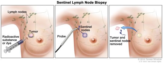
Let us assume the cancer is a train. The tumour is the origin station, so the sentinel lymph node will be the first station the cancer cells spread to. Now, just as the train reaches the first station, the cancer spreads to this sentinel lymph node, and we can check if it has spread by examining the sentinel lymph node. The dye is injected into the blood vessels to localise the sentinel lymph node, then schedule them for removal for testing. The pathologist checks for cancer infection in the sentinel lymph nodes and if it is positive, i.e., cancer cells of the original tumour are present, then the cancer has metastasised, and further treatment is decided. This test is very crucial in the treatment of cancer and is only possible because of the dyes.
DYES USED IN SLNB procedures:
Major dyes used in SLNB procedures are Evans Blue and Trypan Blue.
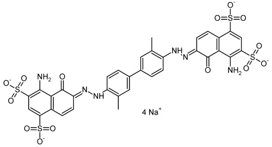
Evans blue:
Evans Blue is a bis-azo dye that has a good affinity for serum albumin (blood plasma found in the vertebrae). Because of this, it can be useful in detecting lymph nodes, the primary centres for blood plasma filtration. Since the lymph node contains this blood plasma, the Evans Blue in addition to Patent Blue, also known as Isosulfan Blue, stains the lymph node blue. Evans Blue fluoresces with excitation peaks at 470 and 540 nm and an emission peak at 680 nm. Evans Blue is also used to test the health of the Blood-brain barrier, a barrier that ensures that no plasma flows through the brain. If parts of the brain do become stained due to the dye, it means that the blood-brain barrier has been compromised. The dye was founded by an American Anatomist, Herbert McLean Evans. The dye was later marketed by The Eastman Kodak Company as Evans Blue, the name that got widely accepted globally.
Trypan blue:
Trypan blue is also an azo-based dye. In diagnostic medicine, it is a vital stain to colour the dead tissues or cells with compromised cell membranes blue. The Trypan Blue Dye cannot penetrate cell membranes, therefore only enters cells with abnormal or compromised membranes. Trypan Blue Dye is sensitive to some proteins like the *HER2 or **RAS family of genes, thus staining them blue upon contact.
*HER2: a special protein that is responsible for cancer growth and spread. Found in breast and ovarian cancer cells usually.
**RAS family of genes: responsible for the proteins in control of the cell communication pathways, cell death, and cell growth.
Chemical properties:
Trypan Blue and Evans Blue are closely related. They both have the same chemical formula, though their structures are different. Their uses, too, vary. While Evans Blue is used as lymph node tracer, Trypan Blue is used as a tracer for detecting compromised cell membranes or special cancer proteins.
Evans Blue

Trypan Blue
Both are non-toxic due to the small dosage administered.
Trypan Blue is slightly alkaline. This enables it to have an increased rate of staining. In the presence of another anionic dye with smaller molecules (like picric acid), trypan blue gets selective for collagen. Evan's blue also has comparable properties since they are closely related.
Downsides:
The major disadvantage of Evans blue and the Trypan blue dyes is that the spectral absorption peak lies in the visible region of the spectrum where reduced haemoglobin also has its maximum spectral absorption. To avoid interference between the dyes and haemoglobin, the patients must breathe oxygen.
Further research:
Researches are being conducted to make cancer detection as minimally invasive as possible.
Dr. Singhal, MD at Penn Medicine, took the initiative to exploit dyes for intraoperative uses. TumorGlowTM, a patented dye method, uses ICG, short for Indocyanine Green, as a dye. When the dye is pumped into the patient through an IV in high doses a day before surgery, the tumours and lymph nodes glow in green when IR rays are incident on them. This makes way for precision and clarity to remove tumours, in the minimally invasive approach, because it paints all the tumours in fluorescent green.
Dyes have been an extensive resource in the field of diagnostic medicine. More breakthroughs are likely on their way, making a diagnosis as minimally invasive as possible.
Bibliography
Image. (n.d.). Retrieved 7 7, 2021, from Wikipedia: The Free Encyclopedia: http://commons.wikimedia.org/wiki/File:TrypanBlueSalt.png
Image. (n.d.). Retrieved 7 7, 2021, from Wikipedia: The Free Encyclopedia: https://commons.wikimedia.org/wiki/File:Evans_blue.png
#breast cancer#slnb#dyes#biology#biomaterials#science#oncology#tumorglow#medicine#industry#molecular oncology
0 notes