#electron microscopy
Text
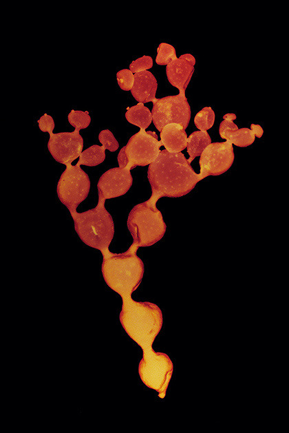


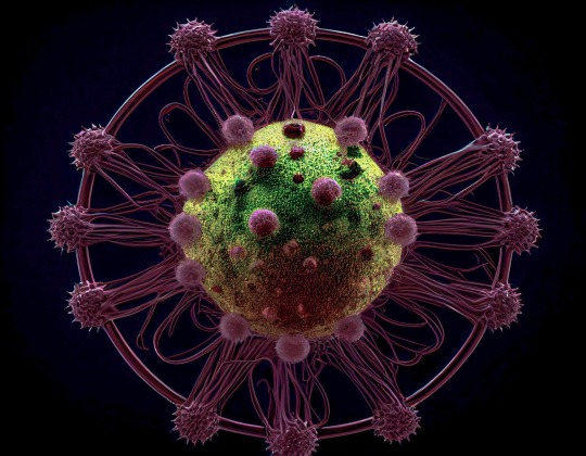
Stephanie Valentin
fathom
#Stephanie Valentin#macro#macrophotography#art#marine photography#plankton#photography#ocean#biotech#Stephanie Valentin fathom#electron microscopy#Australia
2K notes
·
View notes
Text
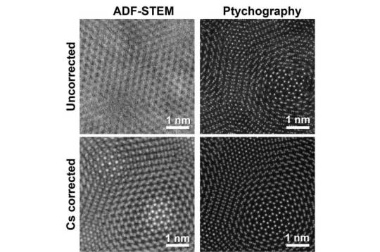
Reimagining electron microscopy: Bringing high-end resolution to lower-cost microscopes
Researchers at the University of Illinois at Urbana-Champaign have shown for the first time that expensive aberration-corrected microscopes are no longer required to achieve record-breaking microscopic resolution.
The field of microscopy is in the middle of a great revolution. Since the 1800s and the invention of the compound light microscope, there have only been a few major jumps in resolution to see different length scales: from bacteria and cells, to viruses and proteins, and even down to single atoms.
Generally, as resolution has been making these incredible jumps, so has the price of the microscopes used to achieve that resolution. Such hefty price tags severely limit the accessibility of these instruments. The current jump in resolution comes from a new technique called electron ptychography—a method that uses computation to boost the resolution of electron microscopes—which has taken the field by storm in the last 5-6 years.
Read more.
20 notes
·
View notes
Text
Neuronly Connect
'Flying' along neurites (projections that connect neurons to others) using an AI-based self-steering system called RoboEM in 3D electron microscopy data derived from mouse and human brain samples can replace human annotation input in complex connectome analyses
Read the published research article here
Video adapted from work by Martin Schmidt and colleagues
Department of Connectomics, Max Planck Institute for Brain Research, Frankfurt, Germany
Video originally published with a Creative Commons Attribution 4.0 International (CC BY 4.0)
Published in Nature Methods, March 2024
You can also follow BPoD on Instagram, Twitter and Facebook
10 notes
·
View notes
Text


/𝗶𝗺𝗮𝗴𝗶𝗻𝗲 𝗽𝗿𝗼𝗺𝗽𝘁:
A coloured scanning electron micrograph + head of a Jumping Spider (family Salticidae) + common housefly (Musca domestica) + cat flea (Ctenocephalides felis + head of a maggot or the larva of a bluebottle fly (Protophormia sp.) with tiny teeth-like fangs extending from its mouth + head of a soldier turtle ant (Cephalotes sp.) from the Amazonian rainforest + head of a honey bee
#nature#midjourney#midjourney art#midjourney ai#artificial intelligence#electron microscope#insects#electron microscopy
33 notes
·
View notes
Text


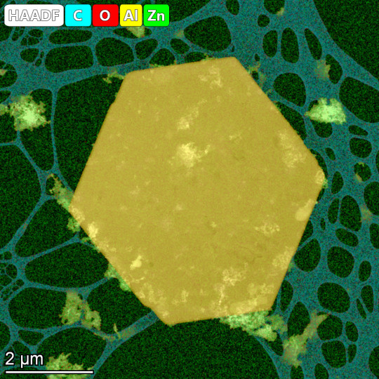
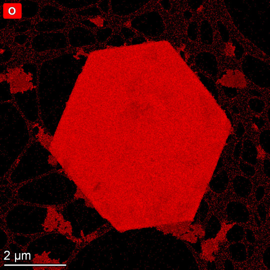
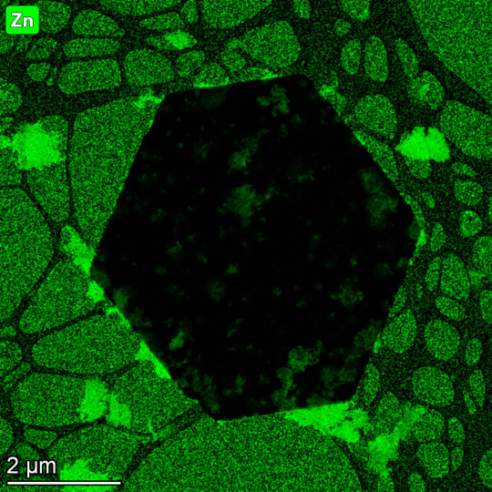
#electron microscopy#transmission electron microscopy#nanoparticles#rare earth hydroxides#zinc tungstate#silver on zinc oxide#zinc titanate#original content
6 notes
·
View notes
Photo

biotech.bae Researchers have discovered an enzyme that uses hydrogen in the atmosphere to create an electrical current.
The enzyme, Huc, was extracted from the bacterium Mycobacterium smegmatis and is able to consume hydrogen at levels as low as 0.00005% of the air we breathe.
The researchers used cutting-edge methods such as cryo-EM microscopy, electrochemistry, and molecular modelling to determine the enzyme's atomic structure and electrical pathways, as well as identify specific regions of the protein that allow hydrogen gas to enter the active site.
This discovery could pave the way for small air-powered devices as an alternative to solar-powered devices, but scaling up the production of the enzyme is still a key objective for future work.
DOI: doi.org/10.1038/s41586-023-05781-7
(Source: @biotech.bae Instagram page)
#science#Science and Technology#mad science#enzyme#enzymes#Molecular#molecular biology#cryo#electron microscopy#mycobacterium
9 notes
·
View notes

Text

electron micrograph of a green algae cell (Chlamydomanas reinhardtii) from Dartmouth College
0 notes
Text
Proton Therapy Treatment
It's a feasible alternative to traditional surgery for certain cancers when comprehensive surgical resection is not possible. The proton beam only penetrates tissue very little with most electrons simply missing cells entirely. This makes it an attractive option because there's less chance of damaging healthy tissues like organs around the tumor site like there would with standard treatment methods.
0 notes
Text
The Essential Role of Cylicins in Sperm Development and Fertility
Sperm development, known as spermiogenesis, is a complex process involving various stages and intricate structural changes. Recent research has shed light on the crucial role of Cylicins in spermiogenesis and male fertility in both mice and humans. Cylicins, specifically Cylicin 1 (Cylc1) and Cylicin 2 (Cylc2), are essential components of the perinuclear theca (PT), a critical part of the sperm…

View On WordPress
#Allele#Cas9#Chromatography#Codon#Conception#CRISPR#Cytoskeleton#DNA#DNA Extraction#Electron#Electron Microscopy#Electroporation#Exome Sequencing#Fertility#Gene#Genes#Genetic#Genetics#Genomic#Genomics#Genotyping#Infertility#Liquid Chromatography#Lysine#Mass Spectrometry#Membrane#Microscopy#Morphology#Nuclear Envelope#Palindromic Repeats
0 notes
Text
Electron Microscopy Market Analysis, Growth Opportunities by Forecast 2032
The global electron microscopy market size was valued at USD 3.94 billion in 2022 and it is projected to be worth around USD 8.67 billion by 2032, growing at a (CAGR) of 8.4% throughout the projection period 2023 to 2032.
The competitive analysis of the Electron Microscopy Market offers a comprehensive examination of key market players. It includes comprehensive company profiles, insights into revenue distribution, product advancements, regional market presence, strategic development plans, pricing strategies, selected target markets, and near-term industry leader activities. The information in this section will help readers better understand the forces that drive competition and what methods will help them stand out in pursuing new target markets.
Market projections and forecasts are underpinned by extensive primary research, further validated through precise secondary research specific to the Electron Microscopy Market. Our research analysts have worked very hard to compile important industry insights from significant industry players, such as Original Equipment Manufacturers (OEMs), top-tier suppliers, distributors, and pertinent government bodies.
Here are some competitive strategies for market research:
SWOT Analysis
Porter's Five Forces Analysis
Market Segmentation
Customer Surveys
Competitor Benchmarking
Market Trend Analysis
PESTEL Analysis
Consumer Behavior Research
Market Gap Analysis
Competitive Intelligence Gathering
Price Sensitivity Analysis
Product Positioning Analysis
Brand Perception Studies
Market Share Analysis
Distribution Channel Assessment
Product Life Cycle Analysis
Competitive Pricing Analysis
Social Media Listening
Customer Feedback Analysis
Technological Trends Research
Receive the FREE Sample Report of Electron Microscopy Market Research Insights @ https://stringentdatalytics.com/sample-request/electron-microscopy-market/2838/
Market Segmentations:
Global Electron Microscopy Market: By Company
• Bio-Rad Laboratories
• Bruker Corporation
• Carl Zeiss AG
• Danaher Corporation
• Danish Micro Engineering
• FEI Co.
• Hitachi High-Technologies Corporation
• Jeol Ltd
• Nikon Corporation
• Olympus Corporation
• Leica Microsystems GmbH
Global Electron Microscopy Market: By Type
• Transmission Electron Microscope
• Scanning Electron Microscope
• Others
Global Electron Microscopy Market: By Application
• Biology and Life Sciences
• Semiconductor and Data Storage
• Materials Research
• Industry
• Others
Regional Analysis of Global Electron Microscopy Market
All the regional segmentation has been studied based on recent and future trends, and the market is forecasted throughout the prediction period. The countries covered in the regional analysis of the Global Electron Microscopy market report are U.S., Canada, and Mexico in North America, Germany, France, U.K., Russia, Italy, Spain, Turkey, Netherlands, Switzerland, Belgium, and Rest of Europe in Europe, Singapore, Malaysia, Australia, Thailand, Indonesia, Philippines, China, Japan, India, South Korea, Rest of Asia-Pacific (APAC) in the Asia-Pacific (APAC), Saudi Arabia, U.A.E, South Africa, Egypt, Israel, Rest of Middle East and Africa (MEA) as a part of Middle East and Africa (MEA), and Argentina, Brazil, and Rest of South America as part of South America.
Click to Purchase Electron Microscopy Market Research Report @ https://stringentdatalytics.com/purchase/electron-microscopy-market/2838/
Here are a few main points you might find in such a Electron Microscopy report along with brief explanations:
Market Size and Growth:
Explanation: This section provides an overview of the current size of the market in terms of revenue or units sold and projects its growth over a specific period. It helps businesses understand the market's potential.
Market Segmentation:
Explanation: Segmentation divides the market into different categories based on factors like demographics, geography, behavior, and more. This information helps businesses target specific customer groups effectively.
Competitive Analysis:
Explanation: This section assesses the key players in the market, their market share, strategies, strengths, and weaknesses. It helps businesses identify their competition and opportunities for differentiation.
Trends and Drivers:
Explanation: It outlines the latest market trends, technological advancements, and factors driving consumer behavior. Understanding these trends is vital for adapting to market shifts and making informed decisions.
SWOT Analysis:
Explanation: A SWOT analysis evaluates the market's strengths, weaknesses, opportunities, and threats. It helps businesses identify areas for improvement and strategies to capitalize on.
Consumer Behavior and Preferences:
Explanation: This section delves into consumer attitudes, behaviors, and preferences, providing insight into what customers want and how they make purchasing decisions.
Regulatory Environment:
Explanation: It covers the regulations and policies that affect the market, which is particularly important in industries like healthcare, finance, and energy.
Market Entry Strategies:
Explanation: This section suggests strategies for entering or expanding in the market, such as partnerships, mergers, acquisitions, or product diversification.
Pricing Analysis:
Explanation: An analysis of pricing strategies used by competitors and the pricing structure within the market helps businesses set competitive and profitable prices.
Market Forecast:
Explanation: Based on historical data and trends, a market research report often provides forecasts for the market's future performance, including expected growth or decline.
Emerging Opportunities:
Explanation: Identifies new and emerging opportunities within the market that businesses can capitalize on, such as untapped niches or gaps in the market.
Challenges and Risks:
Explanation: Highlights potential obstacles or risks that could impact market growth, including economic downturns, supply chain disruptions, or regulatory changes.
Consumer Surveys and Feedback:
Explanation: Includes direct feedback from consumers through surveys or interviews, providing valuable insights into their needs and satisfaction with current products and services.
Case Studies and Success Stories:
Explanation: These showcase real-world examples of businesses that have successfully navigated the market, offering valuable lessons and inspiration for others.
Customization of the Report:
This report can be customized to meet the client’s requirements. Please connect with our sales team ([email protected]), who will ensure that you get a report that suits your needs. You can also get in touch with our executives on +1 346 666 6655 to share your research requirements.
Enquiry Before Buying @ https://stringentdatalytics.com/inquiry/electron-microscopy-market/2838/
Our More Reports:
1. Hot Swap Voltage Controllers Market
2. Surge Protectors Market
3. Camera Lenses Market
About Stringent Datalytics
Stringent Datalytics offers both custom and syndicated market research reports. Custom market research reports are tailored to a specific client's needs and requirements. These reports provide unique insights into a particular industry or market segment and can help businesses make informed decisions about their strategies and operations.
Syndicated market research reports, on the other hand, are pre-existing reports that are available for purchase by multiple clients. These reports are often produced on a regular basis, such as annually or quarterly, and cover a broad range of industries and market segments. Syndicated reports provide clients with insights into industry trends, market sizes, and competitive landscapes. By offering both custom and syndicated reports, Stringent Datalytics can provide clients with a range of market research solutions that can be customized to their specific needs.
Reach US
Stringent Datalytics
+1 346 666 6655
Social Channels:
Linkedin | Facebook | Twitter | YouTube

0 notes
Text
Small Scale Length Measurements
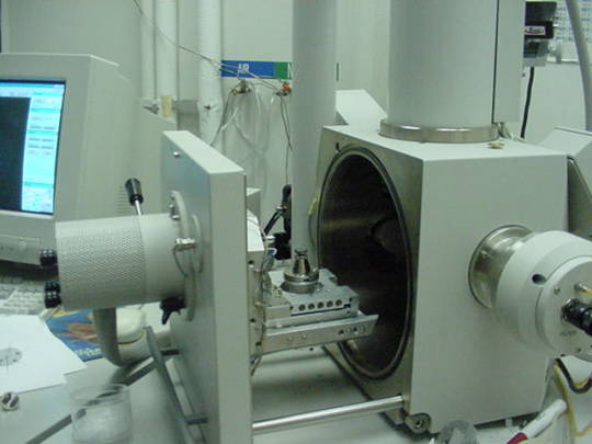
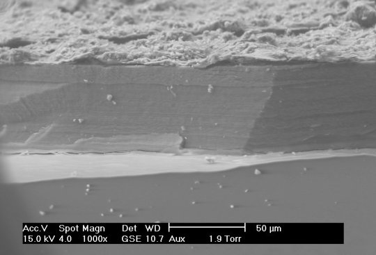
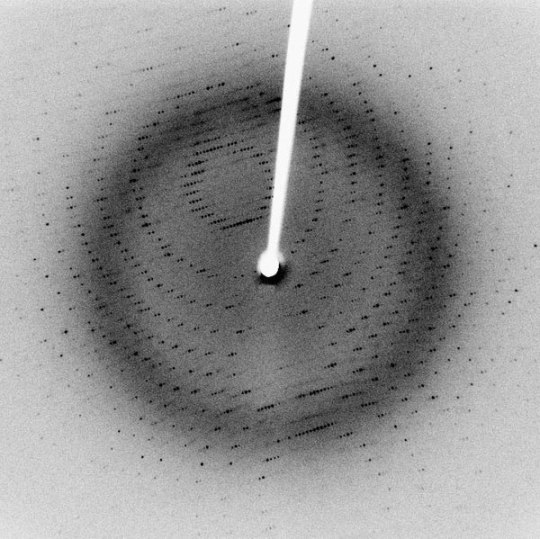
Many different techniques to measure length at small scales (typically microns to nanometers), including X-ray diffraction/crystallography, electron microscopy, and atomic force microscopy, among others. X-ray diffraction can be used to measure atomic spacing and lattice parameters (thereby determining crystal structure) using the wavelengths of the X-rays in question. Electron microscopy differs from many other techniques because they use, as the name implies, electrons to make their measurements, rather than any form of light. Though electrons are reflected back to a detector, the transit-time is not used to make measurements. Instead, Fourier transforms are used to generate an image.
Sources/Further Reading: (Images 1 and 4 - XRD Wikipedia) (Images 2 and 3 - SEM Wikipedia) (Measuring Length Wikipedia) (University of Wisconsin-Madison XRD) (University of Pennsylvania XRD) (University of Colorado XRD) (SEM Book Chapter)
#Materials Science#Science#Materials characterization#Electron microscopy#Diffraction#MeasurementMonday#2024Daily
12 notes
·
View notes
Text
Contact Details
Like the organs in our bodies, cells have their organelles each performing a specific function. Using 3D electron microscopy with high-speed molecular tracking, this study reveals nanoscale details of communication by molecular exchange at the contact site of two organelles – endoplasmic reticulum and mitochondria
Read the published research article here
Video from work by Christopher J. Obara and Jonathon Nixon-Abell, and colleagues
Janelia Research Campus, Howard Hughes Medical Institute, Ashburn, VA, USA
Video originally published with a Creative Commons Attribution 4.0 International (CC BY 4.0)
Published in Nature, January 2024
You can also follow BPoD on Instagram, Twitter and Facebook
6 notes
·
View notes
Text


/𝗶𝗺𝗮𝗴𝗶𝗻𝗲 𝗽𝗿𝗼𝗺𝗽𝘁:
Electron microscopy…
Inspired by the artwork of another AI artist and fascinated by the micro world of electron microscopy I decided to try and create some images for myself.
This ongoing exploration required an introduction to the terminology and methodology involved in imaging this hidden world. I barely scratched the surface with this set of images and though my prompts to the ai requested specific subject matter my novice knowledge and understanding can only marvel at the imagery.
14 notes
·
View notes
Photo
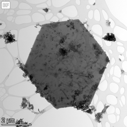
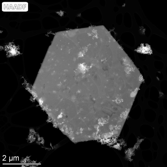
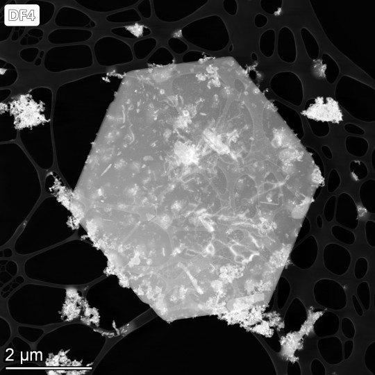


#electron microscopy#transmission electron microscopy#scanning transmission electron microscopy#lattice imaging#Fourier transform#energy dispersive x-ray spectroscopy#bright field image#dark field image#high angle annular dark field#original content
9 notes
·
View notes
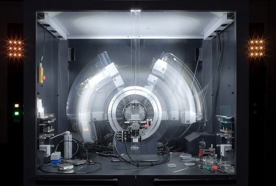
Photo

Nanoscale materials represent a class of substances that approach the size of individual atoms, about 0.1 to 10 nanometers, in at least one dimension. These materials exhibit properties that are unique from larger scale substances, and they are essential to advancing a wide array of technologies, from magnetic data-storage systems to superconducting quantum computers to biomaterials and biomedical applications.
To study and use these materials, tools are required to examine them at the atomic level while minimizing any disturbance to their structure. Electron microscopy, which uses electron beams to “see” atoms, is one of the fundamental means by which the structures of nanoscale materials can be studied.
Current designs of high-resolution transmission electron microscopes require a lens to focus an electron beam on the specimen surface, said University of Illinois Chicago physicist Robert Klie.
“A side effect of this design is that the sample material is inadvertently exposed to a high magnetic field, so turning the lens off to eliminate the magnetic field when studying magnetic or superconducting materials makes atomic-resolution analysis impossible,” he said.
UIC researchers will soon have a new tool built to overcome the side effect.
Through an $4 million National Science Foundation grant, UIC will be home to the world’s first analytical, aberration-corrected and monochromated transmission electron microscope with a magnetic field-free objective lens. UIC, through the Office of the Vice Chancellor for Research, will contribute the required cost-share of 30%, or $1.7 million, toward the total instrument cost. OVCR will also contribute nearly $4M to renovate space in the Center for Structural Biology not only to house this microscope, but to refresh some of their other shared research facilities.
“The instrument acquired through this grant has a new lens design, providing a magnetic-field-free sample region and, when combined with a nearly mono-energetic electron source, allows for atomic-resolution imaging as well as chemical analysis of these critical materials,” said Klie, UIC professor of physics and the project’s principal investigator.
The instrument will also be equipped with several sample or specimen stages that allow researchers to heat, cool or expose materials to liquids or a controlled magnetic field. The monochromated electron source will also allow for new spectroscopy measurements of materials that undergo changes in structure or properties, including magnetic ordering or superconducting transition, at atomic resolution.
Students and scientists at UIC, and across the Midwest and U.S., will also benefit from the microscope acquisition, according to the researchers. The only other microscope similar to this in the world is currently at the University of Tokyo. Read more.
0 notes
Last Seen Blogs

volos-wish
Kiki's Delibird Service

i-cum-on-ur-clit
Any and all things sexy!

nite0wl29
Removers In Texas news

krulu
Krulu

matchacowbee
need cuddles asap