#desmotubule
Text
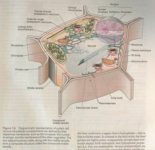
Plant cells are interconnected by cytoplasmic bridges called plasmodesmata (see Chapter 1), cylindrical pores 20 to 60 nm in diameter (Figure 6.18 and Figure 1.6).

"Plant Physiology and Development" int'l 6e - Taiz, L., Zeiger, E., Møller, I.M., Murphy, A.
#book quotes#plant physiology and development#nonfiction#textbook#plasmodesmata#lamella#plant cells#cell wall#tonoplast#cytoplasm#vacuole#desmotubule
0 notes
Text
FUNCTIONS OF ENDOPLASMIC RETICULUM
FUNCTIONS OF ENDOPLASMIC RETICULUM
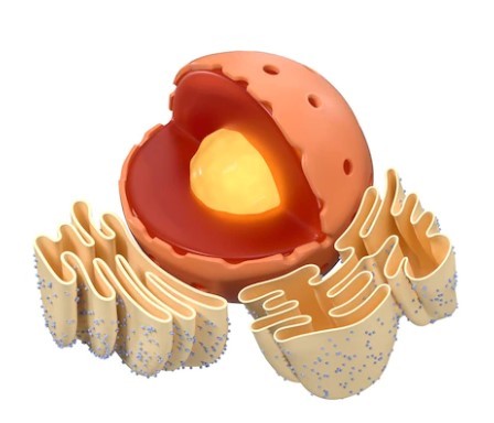
The ER enables the movement of materials from one area of the cell to another, constituting the circulatory system of the cell.
Desmotubule formation: Desmotubules are tubular extensions that stretch through plasmodesmata to connect the ER in two adjacent plant cells.
Support – The cytoskeleton, which also helps to preserve the cell’s shape, uses the ER as an…
View On WordPress
0 notes
Text
Plasmodesmata-digieduco
Formation of secondary wall materials on the primary wall does not take place uniformly, instead some thin areas are left out-those thin areas are called pit-fields. Thin and delicate strands or fibrils of cytoplasm, called plasmodesmata (singular : plasmodesma), pass through such pit-fields of the cell wall at intervals, thus connecting the living protoplasts of adjacent cells. Plasmodesmata usually occur in groups but they may be evenly distributed over the entire wall. When they are grouped they are localised In the primary pit-fields.
Most of the plasmodesmata are found to be restricted In thin areas (the primary pit-fields) of the young walls ; on the other hand, in mature walls with secondary layers, plasmodesmata sometimes occur in large groups only in the pit-membrane.
Two views regarding the structure of plasmodesmata
Secondary plasmodesmata may form between more mature cells which have not undergone division (e.g. in cells of carpels and other organs) or between the haustoria of some parasites and the cells of their host plants.
The distribution and number of plasmodesmata per unit area are determined during cell division. (adsbygoogle = window.adsbygoogle || []).push({}); Plasmodesmata may be quite numerous and meristematic cells may have plasmodesmata between 1000-100,000.
Diagram of a plasmodesmata as seen under electron microscope
Plasmodesmata have been also observed in the cell walls with the help ofdelectron microscope. The endoplasmic reticulum has been seen to contact with the plasmodesmata, thus forming a membrane system that can link nuclei of adjacent cells. Some workers think that tubules of endoplasmic reticulum extend through the plasmodesmata (Whaley et al 1960), although the connection between the elements of the reticulum through a plasmodesmata appears as solid structure. Plasmodesmata are cytoplasmic in nature-this can be proved by the fact that they are present only in living cells, they stain like that of cytoplasm, they exhibit a positive reaction for oxidases, on plasmolysis the protoplast withdraws from the wall in all portions except where plasmodesmata are present.
Plasmodesmata are found in red algae, mosses, liverworts, pteridophytes. gymnosperms and angiosperms. They may be seen throughout all living tissues including the meristematic. (adsbygoogle = window.adsbygoogle || []).push({}); Plasmodesmata, occurring in the outer walls of epidermal cells, are termed ectodesmata (Sievers, 1959 ; Franke, 1961).
Plasmodesmata can be observed readily traversing the thick cell walls of the endosperm of certain seeds e.g. Phoenix sp., Coffea arabica, Diospyros sp., Aesculus sp., Strychnos nux-vomica etc.
Practically, plasmodesmata are narrow channels through the cell wall, they are bounded by plasmalemma and containing cytoplasm and often a desmotubule. The desmotubule i.e. the central core, when present, is composed of protein sub-units and is a modified membranous structure which is continuous with the endoplasmic reticulum of the adjoining cells. Many workers think that the desmotubule is a derivative of the endoplasmic reticulum. Plasmodesmata are small, and upto 60 nm in diameter, normally organelles cannot pass through them (Gunning, 1976 : Robards, 1975-76).
Probable functions of Plasmodesmata :
(i) Plasmodesmata are mainly concerned with the translocation of food, specially in storage tissue like endosperm.
(ii) Plasmodesmata are concerned with the conduction of external and internal stimuli through plant tissues.
(iii) In some cases they establish union of isolated protoplasts of the plant body into a single protoplasmic structure.
(iv) Sometimes plasmodesmata are regarded as additonal layers for mechanical support.
(v) Plasmodesmata are regarded as channels for the movement of viruses from cell to cell (Esau, 1965).
(vi) It has been thought that most plant hormones move through the plasmodesmata (Carr, 1976).
Origin : Regarding the origin and development of plasmodesmata. (adsbygoogle = window.adsbygoogle || []).push({}); Foster (1949) writes that “additional secondary protoplasmic connections may arise during the development of elongating fibres and ramifying sclereids. in such cases, portions of the cell establish new cell contacts by “gliding” or"‘intrusive" growth between neighbouring elements. and during this phase new plasmodesmata may originate."
The origin and development of plasmodesmata has been studied by Frey-Wyssling (1959). (adsbygoogle = window.adsbygoogle || []).push({}); According to him the cell plate is partly protoplasmic in nature although its real nature is unknown. It is assumed that young growing walls are penetrated by the cytoplasm. With the accumulation of the cellulose microfibrils and pectic substances in the wall. the cytoplasmic connections become narrower gradually until they constitute thin threads i.e. the plasmodesmata. Some authors (Buvat and Pulssant, 1958) suggest that plasmodesmata are already present in the cell plate at the time of cell division. Generally with the increase of the wall surface the number of plasmodesmata also increases, this is probably brought about by the splitting of the original threads. When new areas of contact are formed between the cells during expansion of tissues, intrusive growth etc.. new plasmodesmata are formed in the maturing cell wall. According to Jones (1976), plasmodesmata are formed during cytokinesis, apparently at sites in the cell plate where strands of endoplasmic reticulum prevent fusion of vesicles.
via Blogger https://ift.tt/2G82w6c
0 notes
Text
However, plant cells, unlike animal cells, are further enclosed by a rigid, cellulosic cell wall (Figure 1.4). (...) Plant cells have two types of walls: primary and secondary (see Figure 1.4A).

The cytoplasm of neighboring cells is usually connected by means of plasmodesmata (singular plasmodesma), tubular channels 40 to 50 nm in diameter and formed by the connected plasma membranes of adjacent cells (see Figure 1.4A-D). (...) Secondary plasmodesmata form after cell division is completed, across primary or secondary cell walls (see Figure 1.4A), when small regions of the cell walls are digested by enzymes and plasma membranes of adjacent cells fuse to form the channel. The endoplasmic reticulum network of adjacent cells is also connected, forming the desmotubule (see Figure 1.4C and D) that runs through the center of the channel. Proteins line the outer surface of the desmotubule and the inner surface of the plasma membrane (see Figure 1.4D); the two surfaces are thought to be connected by filamentous proteins (spokes), which divide the cytoplasmic sleeve into microchannels. (...) The transport can be followed by studying the movement of fluorescently labeled proteins or dyes between cells (see Figure 1.4E-G). (...) Thus, single channels, referred to as simple plasmodesmata, can form branched plasmodesmata (see Figure 1.4A) when they connect with each other. (...) As a result, even virus-sized particles can readily move through the plasmodesmata to a neighboring cell (see Figure 1.4F and G). (...) The cell plate-spanning ER tubules establish the sites for the primary plasmodesmata (see Figure 1.4B-D). (...) Once made, guard cells remain cytoplasmically isolated from the rest of the leaf because during the last division that forms them, no plasmodesmata are made in the forming cell plate (note the absence of the green spots that indicate plasmodesmata in the guard cells in Figure 1.4E). (...) The prominent nuclear structure called the nucleolus (see Figure 1.4) consists of the rDNA of the NOR, the proteins that transcribe the rDNA and process the rRNA primary transcripts for assembly into ribosomes, and the immature ribosomes just being assembled. (...) The walls on the different sides of a cell may vary in thickness, in amount and type of impregnating substances, in sculpting, and in frequency of pitting and plasmodesmata – tiny membrane-lined channels that allow passive transport of small molecules and active transport of proteins and nucleic acids between the cytoplasm of adjacent cells (see Figure 1.4).
"Plant Physiology and Development" int'l 6e - Taiz, L., Zeiger, E., Møller, I.M., Murphy, A.
#book quotes#plant physiology and development#nonfiction#textbook#cells#plants#plasmodesmata#cell wall#rdna#rrna#transcription#proteins#transportation#active transport#passive transport#impregnating#nucleic acid#plant cells
0 notes
Text
Endoplasmic reticulum (ER)-digieduco
Previously the cytoplasm was considered to be structureless, but with the help of electron microscope an intricate membranous structure within the cytoplasm has been discovered. That structure is known as endoplasmic reticulum which consists of two lipoproteinaceous unit-membranes forming a canal-like anastomosing system. Generally three main types of endoplasmic reticulum are found e.g. long flattened units called cistemae, round units called vesicles and tube-like irregular units called tubules.
The endoplasmic reticulum may be smooth (non-granular) or rough (granular)-the latter type bears ribosomes . Sometime the ER occurs in the cytoplasmic strands. plasmodesmata traversing the cell wall of neighbouring cells. The portion of ER situated in tubular form in the centre of the plasmodesmata is called desmotubule. The main function of endoplasmic reticulum is to increase surface area within the cytoplasm for various metabolic activities, it also helps in translocation of metabolites and their storage within the vacuoles. It also plays an important role in cell wall formation, and also establishes the plane of cell division by the provision and transport of materials to be incorporated in the cell plate (Cutter, 1978). There are also views that the ER gives rise to membranes of Golgi bodies and microbodies.
Membranes of more or less similar structure are also found on the external surface of the cytoplasm i.e. ectoplasm and on the layer bordering the vacuoles Le. tonoplasm.
via Blogger http://bit.ly/2ViZTl8
0 notes
Last Seen Blogs

puxxx5135
Untitled
magicmayu
I'm back

aeschen
Blog, Poems and more

mi3ad
قلبي هنا

vaultflame94
Web Blog Ejaculação Precoce Tratamento Natural Exe