#rrna
Photo
Nucleic Acid MCQ 266 Link in Bio 👆 for more mcqs recommendation #pcr #covid #coronavirus #pixelcarracer #rrna #dna #laboratory #corona #pixelart #laboratorio #dopepixels #biotechnology #microbiology #medicina #biology #lab #pcrtest #policie #sequence #test #pixelcar #salud #science #virus #biologyteacher #enfermagem #genetics #pixelracer #pcgaming #genes (at Royal City Nanded) https://www.instagram.com/p/CpIF_EQPYAq/?igshid=NGJjMDIxMWI=
#pcr#covid#coronavirus#pixelcarracer#rrna#dna#laboratory#corona#pixelart#laboratorio#dopepixels#biotechnology#microbiology#medicina#biology#lab#pcrtest#policie#sequence#test#pixelcar#salud#science#virus#biologyteacher#enfermagem#genetics#pixelracer#pcgaming#genes
2 notes
·
View notes
Text
Abstract
Using morphological traits and molecular-genetic research methods, the authors have identified 24 species of naked amoeba from natural biotopes. The 18S rRNA gene sequences were obtained for the following species of naked amoeba: Amoeba proteus isolate AP07 (ON907618), Saccamoeba limax isolate SLU_22 (OP894078), Saccamoeba limax isolate SL_Uk19 (OQ520144), Saccamoeba sp. strain IDL777 (MZ079370), Thecamoeba striata isolate THS19 (OQ134482), Thecamoeba striata isolate THS20 (OQ134483), Thecamoeba similis isolate Prut river (OL604177), Thecamoeba similis isolate Baggersee Innsbruck (Baggersee Rossau) (OL604178), Thecamoeba quadrilineata isolate THQD2 (ON398269), Thecamoeba quadrilineata isolate THQA1 (ON398268), Thecamoeba sp. strain THS203 (MZ079371), Stenamoeba stenopodia isolate UKSS7 (OP375108), Stenamoeba stenopodia isolate POLSS7 (OP419588), Korotnevella stella isolate KSD2 (ON398267), Korotnevella stella isolate KSA1 (ON398266), Vexillifera bacillipedes isolate river Dnepr (OK649262), Vannella lata isolate Kamenka river (OL305063), Vannella lata isolate Varta river (OL305064), Vannella sp. strain VLS303 (MZ079372), Vannella simplex isolate Black Sea (OM403052), Vannella simplex isolate Mediterranean Sea (OM403053), Ripella sp. strain RPL100 (MZ079369), Mayorella vespertilioides isolate MV_7 (OP739500), Mayorella sp. isolate MY_7 (OP729930), Acanthamoeba sp. strain ATM123 (MZ079366), Acanthamoeba sp. isolate river Elbe (OK649261), Acanthamoeba polyphaga isolate AcPoly01 (ON908497), Acanthamoeba polyphaga isolate AcPoly15 (ON908496), Acanthamoeba griffini isolate Black sea (OM522832), Acanthamoeba griffini isolate Mediterranean Sea (OM522833), Cochliopodium actinophorum strain COP101 (MZ079367), Cochliopodium minus isolate river Stokhid (OK649264), Cochliopodium sp. strain COP102 (MZ079368), Vahlkampfia avara isolate VA7 (OP179657), Willaertia magna isolate river Teterev (OK649263). All of the naked amoebae species on the phylogenetic tree constructed based on the 18S rRNA gene are located within Amoebozoa and grouped with Tubulinea and Discosea. There are separate groups of freshwater, marine, and terrestrial biotopes; these groups are sister species relative to one another with low results of bootstrap analysis, which shows a low accuracy in the distances of particular amoeba species isolated from different natural biotopes.
0 notes
Text
However, plant cells, unlike animal cells, are further enclosed by a rigid, cellulosic cell wall (Figure 1.4). (...) Plant cells have two types of walls: primary and secondary (see Figure 1.4A).

The cytoplasm of neighboring cells is usually connected by means of plasmodesmata (singular plasmodesma), tubular channels 40 to 50 nm in diameter and formed by the connected plasma membranes of adjacent cells (see Figure 1.4A-D). (...) Secondary plasmodesmata form after cell division is completed, across primary or secondary cell walls (see Figure 1.4A), when small regions of the cell walls are digested by enzymes and plasma membranes of adjacent cells fuse to form the channel. The endoplasmic reticulum network of adjacent cells is also connected, forming the desmotubule (see Figure 1.4C and D) that runs through the center of the channel. Proteins line the outer surface of the desmotubule and the inner surface of the plasma membrane (see Figure 1.4D); the two surfaces are thought to be connected by filamentous proteins (spokes), which divide the cytoplasmic sleeve into microchannels. (...) The transport can be followed by studying the movement of fluorescently labeled proteins or dyes between cells (see Figure 1.4E-G). (...) Thus, single channels, referred to as simple plasmodesmata, can form branched plasmodesmata (see Figure 1.4A) when they connect with each other. (...) As a result, even virus-sized particles can readily move through the plasmodesmata to a neighboring cell (see Figure 1.4F and G). (...) The cell plate-spanning ER tubules establish the sites for the primary plasmodesmata (see Figure 1.4B-D). (...) Once made, guard cells remain cytoplasmically isolated from the rest of the leaf because during the last division that forms them, no plasmodesmata are made in the forming cell plate (note the absence of the green spots that indicate plasmodesmata in the guard cells in Figure 1.4E). (...) The prominent nuclear structure called the nucleolus (see Figure 1.4) consists of the rDNA of the NOR, the proteins that transcribe the rDNA and process the rRNA primary transcripts for assembly into ribosomes, and the immature ribosomes just being assembled. (...) The walls on the different sides of a cell may vary in thickness, in amount and type of impregnating substances, in sculpting, and in frequency of pitting and plasmodesmata – tiny membrane-lined channels that allow passive transport of small molecules and active transport of proteins and nucleic acids between the cytoplasm of adjacent cells (see Figure 1.4).
"Plant Physiology and Development" int'l 6e - Taiz, L., Zeiger, E., Møller, I.M., Murphy, A.
#book quotes#plant physiology and development#nonfiction#textbook#cells#plants#plasmodesmata#cell wall#rdna#rrna#transcription#proteins#transportation#active transport#passive transport#impregnating#nucleic acid#plant cells
0 notes
Text
The new generation of soil scientists barely even dig holes. What is the point!!
#like i am a lab girlie and will probably never have to profile again.#i kind of hate everything in my research rn and dont even want to think about soil#but at least I'm starting a soil microbio class which should be interesting#like it's so nice to go to class and hear words like ssu rRNA and plasmid and COCCUS and im like yeah!!! back in my natural environment!
0 notes
Photo

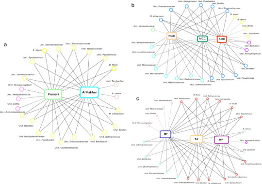
A new resource has been released that enables the comparison of microbial 16S rRNA and shotgun metagenomics data. The resource is a web-based tool that allows users to upload and compare two datasets, and to visualize the results as a heat map. The tool is designed to help researchers identify microorganisms that are present in different samples.
#Featured#News#OMICs#Topics#Translational Medicine#fault#microbial 16S rRNA#shotgun metagenomics#web-based tool#compare datasets#visualize results
0 notes
Text
Lobster shell microbes, epizootic shell disease, and climate change manuscript is published!
A collaborative paper on lobster shell bacteria has just been published in the journal iScience: “Water temperature and disease alters bacterial diversity and cultivability from American Lobster (Homarus americanus) shells.” This paper investigates what happens to bacterial communities on healthy and sick lobsters as they experience different water temperatures for a year.
You can read the paper…

View On WordPress
#16S rRNA#aquaculture#climate and microbes#climate change#epizootic shell disease#Lab Work#lobsters#maine#preprint#shell bacteria#UMaine#Updates
1 note
·
View note
Photo
Bacterial communities of hookah tobacco products are diverse and differ across brands and flavors. Malayil, et al., Applied microbiology and biotechnology doi: 10.1007/s00253-022-12079-7.
0 notes
Link
#allied market research#metagenomics sequencing market#16s rrna sequencing#de novo assembly#metatranscriptomics
0 notes
Text

Limestone jewel: A new colourful karst-dwelling pitviper (Serpentes: Viperidae: Trimeresurus) from the poorly explored borderlands of southern peninsular Thailand
Sabira S. Idiiatullina, Parinya Pawangkhanant, Tanapong Tawan, Thanawut Worranuch, Bunyarit Dechochai, Chatmongkon Suwannapoom, Tan Van Nguyen, Lawan Chanhome, Nikolay A. Poyarkov
Abstract
We describe a new species of pitvipers from Trang Province of Thailand, near the Thailand–Malaysian border, based on morphological and molecular (2427 bp from cyt b, ND4, and 16S rRNA mitochondrial DNA genes) lines of evidence...
The new species is currently known from a small karstic area in the Nakawan Range spanning the border of Thailand and Malaysia, in particular in limestone forests in Trang and Satun provinces (Thailand); it likely also occurs in the adjacent parts of Perlis State (Malaysia).
Our study also suggests that the taxonomy of T. kanburiensis species complex requires further studies; in particular our study suggests that the status of populations from Chumphon Province of Thailand and Pulau Langkawi Island of Malaysia should be re-assessed.
Read the paper here:
Limestone jewel: A new colourful karst-dwelling pitviper (Serpentes: Viperidae: Trimeresurus) from the poorly explored borderlands of southern peninsular Thailand (arphahub.com)
118 notes
·
View notes
Text
Published Feb 29, 2024
Highlights
SARS-CoV-2 Nsp1 localizes to the nucleolus, interacting with precursor rRNA
Nsp1 restrains nascent translation by disrupting rRNA processing and mature ribosomes
Repression of rRNA processing by Nsp1 is separable from mature ribosome inhibition
Summary
Severe acute respiratory syndrome coronavirus 2 (SARS-CoV-2) hinders host gene expression, curbing defenses and licensing viral protein synthesis and virulence. During SARS-CoV-2 infection, the virulence factor non-structural protein 1 (Nsp1) targets the mRNA entry channel of mature cytoplasmic ribosomes, limiting translation. We show that Nsp1 also restrains translation by targeting nucleolar ribosome biogenesis. SARS-CoV-2 infection disrupts 18S and 28S ribosomal RNA (rRNA) processing. Expression of Nsp1 recapitulates the processing defects. Nsp1 abrogates rRNA production without altering the expression of critical processing factors or nucleolar organization. Instead, Nsp1 localizes to the nucleolus, interacting with precursor-rRNA and hindering its maturation separately from the viral protein’s role in restricting mature ribosomes. Thus, SARS-CoV-2 Nsp1 limits translation by targeting ribosome biogenesis and mature ribosomes. These findings revise our understanding of how SARS-CoV-2 Nsp1 controls human protein synthesis, suggesting that efforts to counter Nsp1’s effect on translation should consider the protein’s impact from ribosome manufacturing to mature ribosomes.
#mask up#covid#covid 19#covid isn't over#pandemic#covid conscious#long covid#covid is airborne#wear a mask#coronavirus
24 notes
·
View notes
Photo

Carbohydrate MCQ 11 Link in bio 👆 for more mcqs recommendation #pcr #covid #coronavirus #pixelcarracer #rrna #dna #laboratory #corona #pixelart #laboratorio #dopepixels #biotechnology #microbiology #medicina #biology #lab #pcrtest #policie #sequence #test #pixelcar #salud #science #virus #biologyteacher #enfermagem #genetics #pixelracer #pcgaming #genes (at Delhi, India) https://www.instagram.com/p/CouKQEgvy5z/?igshid=NGJjMDIxMWI=
#pcr#covid#coronavirus#pixelcarracer#rrna#dna#laboratory#corona#pixelart#laboratorio#dopepixels#biotechnology#microbiology#medicina#biology#lab#pcrtest#policie#sequence#test#pixelcar#salud#science#virus#biologyteacher#enfermagem#genetics#pixelracer#pcgaming#genes
2 notes
·
View notes
Text
Exploring the Marvels of Biological Macromolecules: The Molecular Machinery of Life (Part 3)
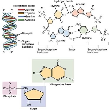
Nucleotide Structure: The Building Blocks
Nucleotides, the monomers of nucleic acids, consist of three fundamental components:
1. Phosphate Group (PO4): Provides a negatively charged backbone for the nucleic acid strand.
2. Pentose Sugar: In DNA, it's deoxyribose; in RNA, it's ribose. The sugar moiety forms the framework of the nucleotide.
3. Nitrogenous Base: Adenine (A), Guanine (G), Cytosine (C), Thymine (T) in DNA, and Uracil (U) in RNA. These bases are responsible for the genetic code.
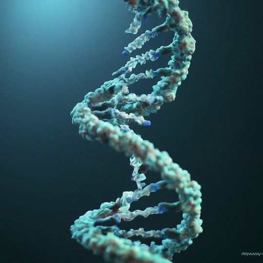
DNA (Deoxyribonucleic Acid): The Repository of Genes
DNA is a double-stranded helical molecule, with each strand composed of a linear sequence of nucleotides. It encodes the genetic information necessary for an organism's development, growth, and functioning. The Watson-Crick base pairing rules—A with T and C with G
DNA (Deoxyribonucleic Acid): The Repository of Genes
DNA is a double-stranded helical molecule, with each strand composed of a linear sequence of nucleotides. It encodes the genetic information necessary for an organism's development, growth, and functioning. The Watson-Crick base pairing rules—A with T and G with C—ensure DNA's complementary and faithful replication.
RNA (Ribonucleic Acid): From DNA's Blueprint to Protein Synthesis
RNA plays diverse roles in the cell, including serving as a messenger (mRNA) for protein synthesis, a structural component of ribosomes (rRNA), and an adapter molecule (tRNA) that brings amino acids to the ribosome during translation. Unlike DNA, RNA is often single-stranded and contains uracil (U) instead of thymine (T).
Genome Organization and Chromosomes
Genomic DNA is organized into chromosomes within the cell nucleus. These structures enable efficient storage, replication, and transmission of genetic information during cell division and reproduction.
Replication and Transcription
DNA replication ensures the faithful duplication of genetic material during cell division, while transcription converts DNA into RNA, providing a template for protein synthesis.
Translation
The cellular machinery, composed of ribosomes and tRNA, reads the mRNA code and assembles amino acids into polypeptides during translation, ultimately forming functional proteins.
Genetic Code
The genetic code, a triplet code of nucleotide sequences (codons), dictates a protein's sequence of amino acids. It is nearly universal, with only minor variations across species.
Epigenetics
Epigenetic modifications, such as DNA methylation and histone modifications, regulate gene expression without altering the underlying DNA sequence, pivotal in development and cell differentiation.
Macromolecular interactions are the essence of cellular life. Within the complex microcosm of a cell, countless molecules engage in precise and choreographed dances, forming intricate networks that govern every facet of biology. These interactions, governed by the principles of biochemistry, are the foundation upon which life's processes are built.
Amino Acids: The Building Blocks
Proteins are composed of amino acids organic molecules that contain an amino group (-NH2), a carboxyl group (-COOH), a hydrogen atom, and a distinctive side chain (R group). There are 20 different amino acids, each with a unique side chain that confers specific properties to the amino acid.
Primary Structure: Amino Acid Sequence
The primary structure of a protein refers to the linear sequence of amino acids in the polypeptide chain. The genetic information in DNA encodes the precise arrangement of amino acids.
Secondary Structure: Folding Patterns
Proteins don't remain linear; they fold into specific three-dimensional shapes. Secondary structures, such as α-helices and β-sheets, result from hydrogen bonding between nearby amino acids along the polypeptide chain.
Tertiary Structure: Spatial Arrangement
The tertiary structure is the overall three-dimensional shape of a protein, determined by interactions between amino acid side chains. These interactions include hydrogen bonds, disulfide bridges, ionic bonds, and hydrophobic interactions.
Quaternary Structure: Multiple Polypeptide Chains
Some proteins, known as quaternary structures, comprise multiple polypeptide chains. These subunits come together to form a functional protein complex. Hemoglobin, with its four subunits, is an example.
Protein Functions: Diverse and Essential
Proteins are involved in an astounding array of functions:
1. Enzymes: Proteins catalyze chemical reactions, increasing the speed at which reactions occur.
2. Structural Proteins: Proteins like collagen provide structural support to tissues and cells.
3. Transport Proteins: Hemoglobin transports oxygen in red blood cells, and membrane transport proteins move molecules across cell membranes.
4. Hormones: Hormonal proteins, such as insulin, regulate various physiological processes.
5. Immune Function: Antibodies are proteins that play a crucial role in the immune system's defense against pathogens.
6. Signaling: Proteins are critical in cell signaling pathways, transmitting information within cells.
Protein Denaturation and Folding
Protein Diversity: The vast diversity of proteins arises from the combinatorial possibilities of amino acid sequences, secondary structure arrangements, and three-dimensional conformations.
Nucleic acids, the remarkable macromolecules that govern all living organisms' genetic information, are life's quintessential molecules. These complex polymers of nucleotides play an unparalleled role in the storage, replication, and expression of genetic information, shaping the development, characteristics, and functions of every living entity on Earth. Let's embark on an exploration of the intricate world of nucleic acids.
Nucleotide Structure: The Building Blocks
Nucleotides, the monomers of nucleic acids, consist of three fundamental components:
1. Phosphate Group (PO4): Provides a negatively charged backbone for the nucleic acid strand.
2. Pentose Sugar: In DNA, it's deoxyribose; in RNA, it's ribose. The sugar moiety forms the framework of the nucleotide.
3. Nitrogenous Base: Adenine (A), Guanine (G), Cytosine (C), Thymine (T) in DNA, and Uracil (U) in RNA. These bases are responsible for the genetic code.
DNA (Deoxyribonucleic Acid): The Repository of Genes
DNA is a double-stranded helical molecule, with each strand composed of a linear sequence of nucleotides. It encodes the genetic information necessary for an organism's development, growth, and functioning. The Watson-Crick base pairing rules—A with T and G with C—ensure DNA's complementary and faithful replication.
RNA (Ribonucleic Acid): From DNA's Blueprint to Protein Synthesis
RNA plays diverse roles in the cell, including serving as a messenger (mRNA) for protein synthesis, a structural component of ribosomes (rRNA), and an adapter molecule (tRNA) that brings amino acids to the ribosome during translation. Unlike DNA, RNA is often single-stranded and contains uracil (U) instead of thymine (T).
Genome Organization and Chromosomes:
Replication and Transcription: DNA replication ensures the faithful duplication of genetic material during cell division, while transcription converts DNA into RNA, providing a template for protein synthesis.
Translation: The cellular machinery, composed of ribosomes and tRNA, reads the mRNA code and assembles amino acids into polypeptides during translation, ultimately forming functional proteins.
Genetic Code: The genetic code, a triplet code of nucleotide sequences (codons), dictates the sequence of amino acids in a protein. It is nearly universal, with only minor variations across species.
Epigenetics: Epigenetic modifications, such as DNA methylation and histone modifications, regulate gene expression without altering the underlying DNA sequence, pivotal in development and cell differentiation.
Macromolecular interactions are the essence of cellular life. Within the complex microcosm of a cell, countless molecules engage in precise and choreographed dances, forming intricate networks that govern every facet of biology. These interactions, governed by the principles of biochemistry, are the foundation upon which life's processes are built.
#science#biology#college#education#school#student#medicine#doctors#health#healthcare#genetics#genetic engineering#science nerds#dna activation#new dna
24 notes
·
View notes
Text
RNA: The Dynamic Molecule Driving Life's Diversity
DNA, the blueprint of life, often steals the spotlight when it comes to genetics. But lurking in its shadow is another crucial molecule, RNA (Ribonucleic Acid), playing a pivotal role in the symphony of life. More than just a passive messenger, RNA boasts a vibrant history and holds exciting potential for the future. Let's embark on a journey to unveil the world of RNA, exploring its captivating story and why it deserves your attention.
The story of RNA's discovery began in 1860 when Friedrich Miescher isolated a mysterious "nuclein" from white blood cells. However, it wasn't until the 1950s that James Watson and Francis Crick, alongside Rosalind Franklin (whose contributions were initially overlooked), unraveled the structure of DNA, relegating RNA to a supporting role as a mere messenger molecule. But the plot thickened in the 1960s when researchers like Howard Temin and David Baltimore stumbled upon reverse transcriptase, an enzyme that could convert RNA into DNA, challenging the long-held "central dogma" of DNA being the sole source of genetic information. This discovery opened the door to a whole new understanding of RNA's diverse capabilities.
The Many Faces of RNA
But RNA isn't just a protein puppet master. There are different types of RNA, each with unique jobs:
Messenger RNA (mRNA): Delivers the protein-making message.
Transfer RNA (tRNA): Brings the amino acids, the building blocks of proteins, to the party. Ribosomal RNA (rRNA): The foreman of the ribosome factory, making sure everything runs smoothly.
Non-coding RNA (ncRNA): A diverse bunch with various roles, from regulating genes to fighting viruses.
The true game-changer came in the early 2000s. Scientists stumbled upon a vast class of non-coding RNAs that don't code for proteins but have diverse and crucial functions. microRNAs (miRNAs), for example, regulate gene expression by silencing specific genes, while long non-coding RNAs (lncRNAs) control various cellular processes like development and disease. This discovery shattered the dogma that only protein-coding genes mattered, highlighting the crucial roles played by non-coding RNAs.
This newfound understanding of RNA's potential has ignited a revolution in medicine. Researchers are exploring RNA-based therapies for various diseases, from cancer and neurodegenerative disorders to viral infections. mRNA vaccines, like the ones used against COVID-19, harness the power of messenger RNA to deliver genetic instructions directly to cells, triggering immune responses. The future holds even more promise, with scientists exploring techniques like CRISPR-Cas9 to edit RNA and potentially treat genetic diseases.
New discoveries are constantly rewriting our understanding of this versatile molecule. Its adaptability and diverse roles make it a powerful tool for exploring the very essence of life, from evolution and development to disease and therapy. So, the next time you hear about genes, remember that RNA, the often-overlooked player, is just as crucial in shaping the tapestry of life. It's a story of constant evolution, unexpected discoveries, and immense potential, making RNA a molecule brimming with fascination and promise for the future.
#science sculpt#life science#science#molecular biology#biotechnology#biology#genetics#RNA#daily dose of science#dailyprompt#meaningful#scientific illustration#the glass scientists#microscopic world#microscopy#artists on tumblr#digital artist
11 notes
·
View notes
Text
Propaganda!


Ribosomes are macromolecular machines, found within all cells, that perform biological protein synthesis (mRNA translation). Ribosomes link amino acids together in the order specified by the codons of messenger RNA (mRNA) molecules to form polypeptide chains. Ribosomes consist of two major components: the small and large ribosomal subunits. Each subunit consists of one or more ribosomal RNA (rRNA) molecules and many ribosomal proteins (RPs or r-proteins). The ribosomes and associated molecules are also known as the translational apparatus.
Human vasopressin, also called antidiuretic hormone (ADH), arginine vasopressin (AVP) or argipressin, is a hormone synthesized from the AVP gene as a peptide prohormone in neurons in the hypothalamus, and is converted to AVP. It then travels down the axon terminating in the posterior pituitary, and is released from vesicles into the circulation in response to extracellular fluid hypertonicity (hyperosmolality).
19 notes
·
View notes
Text
Lobster shell microbes, epizootic shell disease, and climate change preprint manuscript is now online
Lobster shell microbes, epizootic shell disease, and climate change preprint manuscript is now online
It’s been a few years in the making, but our draft manuscript on lobster shell microbes, epizootic shell disease, and climate change is available online as a preprint (not yet peer reviewed)! You can read the preprint here, and the summary is below.
I joined this project back in the summer of 2020, when I was given a large 16S rRNA gene sequence dataset of bacterial communities from the shells…

View On WordPress
#16S rRNA#aquaculture#climate and microbes#climate change#epizootic shell disease#lobsters#maine#preprint#shell bacteria#UMaine#Updates
1 note
·
View note
Last Seen Blogs

strangeduckpaper
Holy Multiverse Batman!

stard308
Indian Gold Hair Care

lidaeli
redflagcore


randbhistorian
Untitled