#renal cyst
Text
Navigating Kidney Disease: An Understanding of Renal Dysfunction
The prevalence of kidney problems and renal cysts is a significant health concern that affects a large portion of the population. Although conventional medicine has many treatments available, an increasing amount of people are turning to alternative therapies such as homeopathy to treat renal cysts and kidney-related issues, including kidney infections, acute injury to the kidney, or kidney failure.

Understanding Renal Cysts
Renal cysts, fluid-filled sacs in the kidneys, can cause discomfort and complications when left untreated. homeopathic medicine for renal cyst is an alternative to conventional medicine, which typically recommends surgery or drainage.
Homeopathic medicine for renal cysts
The homeopathic practitioner will consider the health and constitution of each individual when treating renal cysts. The remedies are prescribed according to specific symptoms, such as pain, swelling, and urinary problems. Some remedies are prescribed for stinging, sharp pains, and swelling. Others may be recommended for burning sensations when urinating.
Homeopathic remedy for kidney infection:
It is essential to treat kidney infections quickly and effectively to avoid complications. Homeopathic remedy for kidney infection addresses the underlying cause of infection while supporting the immune response. Some of these are commonly used to treat kidney infections. Some are used for kidney infections.
Kidney Cyst Treatment by Homeopathy
The homeopathic kidney cyst treatment focuses on reducing the symptoms and preventing growth. Some remedies are recommended for slow-growing cysts, while others can encourage expulsion or cystic material. Some remedies are well-known for relieving pain caused by kidney cysts.
Homeopathy for Kidney Health
Homeopathy kidney treatment focuses on holistic health to maintain kidney function. Selecting constitutional remedies based on the unique characteristics of an individual can be crucial in preventing kidney issues. Here are some examples of constitutional remedies you may consider for kidney support.
Homeopathy for acute kidney injury:
Acute kidney damage (AKI) can be a severe and sudden condition that requires immediate attention. Homeopathic remedies complement conventional medicine's focus on supportive care by targeting the root causes. Acute kidney injury treatment remedies may be used to treat trauma-induced kidney damage, while others target inflammation and urinary symptoms.
Homeopathic medicine for kidney failure
The renals can no longer function properly. Homeopathic treatment for kidney failure aims to improve the quality of life, manage the condition and slow the progression. The remedies can be chosen based on each patient's symptoms and constitutional factors.
Conclusion:
Homeopathic medicine is a holistic, individualised treatment for renal cysts, acute kidney injury and kidney failure. Although more research is required to determine the effectiveness of homeopathic remedies for kidney-related conditions such as cysts, many people report positive results and improved quality of life through this alternative treatment. It is essential to consult a healthcare professional with expertise before using homeopathic medicine, particularly in cases of severe kidney disease. Homeopathic medicine for kidney failure can be a great alternative to or complement to conventional kidney treatments.
#homeopathy kidney treatment#kidney cyst treatment#homeopathic remedy#kidney infection#renal cyst#homeopathic medicine#Kidney treatment by homeopathy
0 notes

Text
#Renal Cysts#health care blog#healthy#healthcare#kauvery hospital#Renal Cysts Treatment#Best Urologist in chennai#healthcarenewsletter#newsletters#kidney diseases#kidneys#poly cystic kidney disease#Kidney transplant#kidney transplant hospital in chennai#kidney transplant hospital in india
0 notes
Text
Zinner Syndrome: A Rare Case of the Mesonephricduct Anomaly by Balagobi B in Journal of Clinical and Medical Images, Case Reports

Abstract
The present article reports 18-year-old boy came to the urology clinic with perineal discomfort and few episodes of incomplete emptying of the bowel for two months. He did not claim any symptoms related to his voiding or ejaculation. The abdominal and external genitalia examination was regular. He had average prostate size on digital rectal examination with a palpable painless cystic mass just above the prostate gland. Zinner's syndrome should be a differential diagnosis in young patients with urinary symptoms and unilateral renal agenesis. A detailed review of the relevant literature is also presented.
Keywords: Zinner's syndrome; congenital malformation; seminal vesicle cyst; renal agenesis; infertility.
Introduction
Zinner's syndrome is one of the rarest congenital malformations present with cysts in the seminal vesicle, ejaculatory duct obstruction, and ipsilateral renal agenesis [1]. Its origins in the development abnormality of the Wolffian duct at embryogenesis. Some of the patients with Zinner's syndrome may remain asymptomatic and discovered incidentally, while others present with clinical signs of bladder outlet obstruction, nonspecific pelvic pain, and symptoms related to ejaculatory dysfunction [2]. Herein, we present a case of Zinner's syndrome in which the patient presents with perineal pain and discomfort with a few episodes of incomplete defecation as an initial complaint.
Case presentation
An 18-year-old boy came to the urology clinic with perineal discomfort and few episodes of incomplete emptying of the bowel for two months. At the same time, he did not claim any symptoms related to his voiding or ejaculation. The abdominal and external genitalia examination was regular. He had average prostate size on digital rectal examination with a palpable painless cystic mass just above the prostate gland. The initial laboratory investigations did not reveal any abnormal values, including renal function tests and hormone analysis (LH, FSH, and testosterone). The ultrasound kidney, ureter, and bladder (KUB) showed the right side renal agenesis and homolateral seminal vesicle cyst (Figure 1). Subsequently, the urinary tract's magnetic resonance imaging (MRI) was requested, which diagnosed Zinner's syndrome by visualizing the seminal vesicle cyst on T1 and T2 weighted images as hypo intensity and hyper intensity signals, respectively (Figure 2). Furthermore, his sperm parameters were normal in limit and excluded the risk of future infertility. We offered simple nonopioid analgesics for the perineal pain and laxatives for the defecation issues. We did not do any invasive treatment for him as he is asymptomatic after initial treatment. Therefore, he is under surveillance at our urology clinic for more than one year.
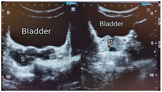
Figure 1: Ultrasound pelvis showing the multiple cysts within the right seminal vesicle(S) and dilated distal part of seminal vesicle within prostate gland (P).

Figure 2: (A) Coronal image of MRI abdomen showing absent right kidney with enlarged normal left side kidney. (B & C) Pelvic MRI. T1 and T2 – weighted axial images show the right side dilated cysts within the seminal vesicle, and it shows hyperintensity in T1 and hypointensity in T2-weighted images.
Discussion
Zinner's syndrome occurs due to abnormal growth of the Wolffian duct during embryogenesis, and it is marked by ejaculatory duct obstruction, cysts in seminal vesicles, and same-side renal agenesis [3]. In history, the seminal vesicle cysts were identified first by Smith in 1872, and later the association between unilateral renal agenesis and seminal vesicle cyst was first described by Zinner in 1914 [2]. Since that, more than 100 case reports in Zinner's syndrome reported in the English literature and this is the first reported case in Sri Lanka in this entity [4]. The ureteric bud originates from the distal part of the mesonephric duct and ascends cranially to meet the metanephric blastema, which will form the future kidney. Maldevelopment of the distal mesonephric duct occurs due to various insults during the first trimester that leads to ejaculatory duct atresia and abnormality of ureteral budding. Atresia of the ejaculatory duct causes obstruction and cystic dilatation of seminal vesicles and anomaly in the ureteral budding results in renal agenesis [5]. Patients with this congenital anomaly are usually asymptomatic until their second to the third decade of life [6]. The symptoms develop due to bladder irritation, bladder outlet obstruction, cyst distension, and obstruction of the ejaculatory duct. Therefore, the Patients may present with unspecific and various clinical manifestations, including voiding symptoms (frequency, dysuria, urgency, poor flow), haematuria, pelvic pain, perineal or scrotal pain or discomfort, urinary tract infection, painful ejaculation, and haematospermia [5].
The diagnostic evaluation of a Zinner's syndrome includes biochemical investigations, radiological imaging, and cystoscopic examination. Urinalysis and culture, blood analysis, renal function tests, and hormone profile (FSH, LH, Testosterone) is proper initial biochemical investigations. Transrectal ultrasonography is the most widely used tool for identifying and initial evaluating seminal vesicle cysts, and it reveals anechoic cystic pelvic lesions with a thick, irregular wall and calcifications [7]. Besides, the computed tomogram also can demonstrate the cysts in the seminal vesicle and renal agenesis, but it is not adequate to make the final diagnosis. Therefore, magnetic resonance imaging is the definitive diagnostic imaging of Zinner's syndrome without the need for additional invasive investigations. Typically, the cysts in seminal vesicles appear as hyperintensity lesions on T2- weighted images andhypointensity in T1- weighted images. Besides, an MRI scan reveals excellent soft-tissue anatomy between the cysts and surroundings in the pelvis, which is helpful for surgical management [3]. Urethrocystoscopy may show trigonal abnormality or bulge inside the bladder due to external compression [8].
The management of Zinner's syndrome should be planned according to the clinical presentation. The Conservative approach is helpful in mild symptomatic or asymptomatic cases with normal biochemical parameters [6]. Aspiration of the seminal vesicles and combined instillation of substances such as alcohol or minocycline and antibiotics are also tried in mild symptomatic group 3. Surgical treatment options should restrict to symptomatic cases or patients who failed conservative measures. Surgical treatment options can be a transurethral resection of the ejaculatory duct (TURED) or seminal vesiculectomy. TURED procedure includes the resection of the prostate at the level of verumontanum until the opening of the ejaculatory ducts. A study reported a remarkable improvement of semen volume, pH, and sperm count, increasing carnitine and fructose concentration after TURED in seven patients. Therefore, they recommended that TURED surgery be tried first to resumption natural fertility before trying other infertility treatments [9]. Open excision of the cyst was the most effective procedure for symptomatic patients until recently, and it was replaced by laparoscopic and robotic surgeries in the current era. Minimally invasive surgeries ensure retrovesical anatomy during surgery with minimal blood loss and minimal postoperative morbidity. Unfortunately, some of the patients with infertility fail to recover from poor sperm parameters despite all these efforts, and assisted reproduction techniques become the only hope for them [10].
Conclusion
Zinner's syndrome should be a differential diagnosis in young patients with urinary symptoms and unilateral renal agenesis. Even though transrectal ultrasonography is used as an initial screening tool, the MRI pelvis makes the final diagnosis, and surgical treatment options resolve the complaints in symptomatic patients except for the poor sperm parameters.
Acknowledgement
The authors wish to thank, S.Thiruvarangan, Research Assistant who assisted to this manuscript preparation and submission process in the final stage of this article.
For more details : https://jcmimagescasereports.org/author-guidelines/
#Zinner's syndrome#congenital malformation#seminal vesicle cyst#renal agenesis#infertility#LH#FSH#urology clinic#nonopioid analgesics#Balagobi B#JCMICR
0 notes
Link
We are all aware of the formation of cysts in different parts of the body, it is, however, very less heard of in the kidneys.
Renal Cyst Treatment in India
#symptoms of renal cyst#Cost of Renal Cyst Treatment in India#Hospitals for Renal Cyst Treatment in India'#renal cyst cause blood in the urine
0 notes
Text
Kidney Diseases, Types and Diagnosis
Kidney Diseases, Types and Diagnosis
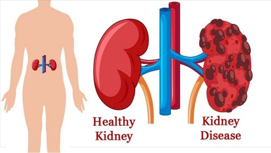
Kidney diseases can affect one or both kidneys, which are located on the left and right sides of the abdomen, in the middle of the back. They are bean-shaped and approximately the fist-sized. They are vital organs that remove toxins and extra water from the body.
What are kidney diseases?
A kidney disease does not occur overnight but develops over a period of time and in stages. Kidney disease…
View On WordPress
#ammonia#creatine#creatinine#end-stage renal disease#Kidney cysts#Kidney Disease#kidney failure#Polycystic kidney disease Polycystic kidney#Stones in the kidney#urea#uric acid#Waste products
0 notes
Text
click read more to see the pathology report on my kidney they took out
Surgical Pathology Report
SPECIMEN SOURCE:
A. LEFT KIDNEY
CLINICAL HISTORY:
Polycystic kidney disease
DIAGNOSIS:
A. LEFT KIDNEY:
Polycystic kidney disease with a small papillary adenoma (0.4
mm)
MD, PhD
Attending Pathologist
GROSS DESCRIPTION:
The specimen labeled "LEFT KIDNEY" is received fresh, now fixed in
formalin and consists of 1646 gram 28.0 x 15.0 x 13.0 cm
polycystic left nephrectomy specimen. The kidney alone measures
17.0 x 9.5 x 8.0 cm overall and is trabeculated and cystic. The
hilum displays vessels inclusive of: ureter (8.0 x 0.2 cm); renal
vein (0.6 cm in diameter) and renal artery (0.2 cm in diameter). A
fixed portion of Gerota's fascia is present measuring 24.0 x 11.0
cm. The renal vein is devoid of any lesion or thrombus. The
kidney is bivalved revealing brown liquid and polycystic kidney
with cysts ranging in size from 0.3 x 0.3 cm to 5.0 x 4.0 cm
overall. The cyst
walls tan-white and smooth measuring up to 0.2
cm in thickness. The urothelium of the renal pelvis is tan-white
and smooth surrounded by abundant yellow fatty tissue. The
calyces, medullary pyramids, and cortico-medullary junction, and
cortex are not grossly identified and totally obliterated by the
cysts. An adrenal gland is not present. A lesion is not gross
identified. The tissue is representatively submitted. Gross
pictures are taken.
SUMMARY OF SECTIONS:
A1 = Vascular margins, shaved, en face (ureter, renal vein and
renal artery) inked orange
A2=section of renal pelvis with cysts
A3=cyst with gerota's fascia
A4=cyst with hilar fat
A5=cyst with perinephric fat
A6=gerota's fascia and perinephric fat
A7-A9= representative sections of cysts
Total 9
7 notes
·
View notes
Text
Best Urologist In Kukatpally Hyderabad.
My health hospital is a leading surgery provider in the area of Kphb, Kukatpally, Hyderabad and is associated with one of the best urologists and andrologists in hyderabad who are trained in treatment of urological and andrological problems with 10+ years of experience . We have listed some of the most common problems & their treatments provided at My health hospital below. Book an appointment and meet your doctor today to know the best treatment option for the problem .
Urologist / Urology Specialist / Urology Doctor is a super specialist in the field of surgery dealing with problems related to Male and Female Urinary Tract which includes Kidney Stones treatment (laser), Prostate surgery (laser and conventional), treatment of urinary tract infections (simple and complex), urinary incontinence treatment (medical and surgical), circumcision(laser/stapler/conventional), treatment of testicular infections( Epidydimitis , orchitis, Epidydimo orchitis), surgical treatment of scrotal swellings(Hydrocele/Hernia), Renal cysts, kidney, bladder/prostate cancer, and treatment of lower urinary tract symptoms( Frequency/urgency/dysuria /blood in urine( Haematuria )/ Nocturia /poor stream) and performs necessary procedures for diagnosis (cystoscopy) and therepeutic purposes for the best treatment of patients having any of the above urological problems.
Andrologist / Male infertility specialist / ED (Ejaculatory dysfunction) doctor is a urologist trained in dealing with problems related to male reproductive system(penis/urethra/testis/seminal vesicles) which include problems related to unprotected sex (STD/STI- Sexually transmitted infections/sexually transmitted diseases) and problems with sexual intercourse( ED(Ejaculatory Dysfunction)/Premature ejaculation) and treatment of male infertility problems related to pregnancy(medical and surgical -varicocele and its treatment )
#best urologist in kukatpally hyderabad#urologist#best hospital in hyderabad#best kidney stone treatment in kukatpally#best hospital#hospitals near me
2 notes
·
View notes
Photo
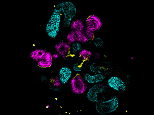
Mystery in Miniature
A rare genetic condition, tuberous sclerosis complex (TSC) causes the development of cysts and benign tumours in the kidneys and other organs, with potentially life-threatening consequences. While we know TSC is caused by mutations in the genes TSC1 or TSC2, the actual cellular processes affected have remained something of an enigma. In a recent breakthrough, researchers were able to investigate the effects of mutations in these genes using renal organoids, miniature self-organised clusters of cells that recapitulate the structures and functions of kidneys (pictured, with various kidney structures highlighted in different colours). When tracking the development of organoids made from cells lacking functional TSC1 or TSC2 they found disruptions to the developmental pathways of multiple cell types, contributing to the puzzling diversity of tumours associated with TSC. While there is currently no cure for TSC, these organoids could also prove a useful tool for testing potential drugs in the future.
Written by Emmanuelle Briolat
Image from work by Adam Pietrobon and colleagues
The Sprott Centre for Stem Cell Research, Regenerative Medicine Program, Ottawa Hospital Research Institute, Ottawa, Canada
Image originally published with a Creative Commons Attribution 4.0 International (CC BY-ND-NC 4.0)
Published in Cell Reports, July 2022
You can also follow BPoD on Instagram, Twitter and Facebook
#science#biomedicine#organoids#renal#kidney#gene mutation#genetic disorder#tuberous sclerosis#immunofluorescence
4 notes
·
View notes
Text

Kidney cysts result from genetic or nongenetic processes and occur in a variety of diseases in adults and children. (See 'Introduction' above.)
●To help diagnose and manage simple and complex kidney cysts, the Bosniak kidney cyst classification system was created. Based upon morphologic and enhancement characteristics with unenhanced and contrast-enhanced computed tomography (CT) scanning, cystic kidney masses are placed into one of five different categories (table 1). (See 'Bosniak classification of kidney cysts' above.)
●The evaluation of kidney cystic lesions is based upon initial classification according to the Bosniak system:
•Further evaluation of Bosniak category I and II lesions is not required. If the clinician is unable to distinguish between a category II and IIF cyst, follow-up imaging should be performed. (See 'Category I and II' above.)
•For category IIF lesions, we recommend making every attempt to obtain prior studies for purposes of comparison. If such studies are unavailable, we recommend an additional imaging study, such as a contrast-enhanced magnetic resonance imaging (MRI), for further characterization. If the radiologist is unable to clearly distinguish a category IIF from a III lesion, the lesion should be treated like a category III lesion.
•For category III and IV lesions, the approach varies among clinicians and depends upon patient factors. Options available include continued active surveillance with periodic imaging, fine-needle biopsy and possible ablation, or surgery with partial nephrectomy, if feasible. The decision between these options depends upon the appearance of the lesion and the comorbidities and life expectancy of the patient. In general, among patients who are good surgical candidates, surgery is preferred. For patients who are not good surgical candidates or who elect surveillance, we obtain a CT or MRI at six months, with follow-up imaging with either a CT or ultrasound yearly. (See 'Category III and IV' above.)
●Simple kidney cysts are observed frequently in normal kidneys. They are the most common kidney masses, accounting for roughly 65 to 70 percent of cases. The prevalence of simple kidney cysts varies with the population studied and the imaging modality utilized. These cysts most often occur in patients >50 years of age. (See 'Epidemiology' above.)
●Simple kidney cysts may be solitary or multiple and bilateral. They typically produce no symptoms or signs. Rarely, however, they can be associated with rupture (hemorrhage), hematuria, pain, abdominal mass, infection, and/or hypertension. (See 'Clinical features' above.)
●Simple kidney cysts have characteristic changes on ultrasonography. If basic ultrasonography criteria for a benign simple kidney cyst are met, further study is not required. A CT scan should be performed if the ultrasonogram is equivocal, if calcifications or septae are seen, or if multiple cysts are clustered in a pattern that could mask an underlying carcinoma. (See 'Diagnosis' above.)
●The major concern with simple kidney cysts is differentiating them from more serious disorders, such as polycystic kidney disease (PKD), complex cysts, and solid masses (such as a renal carcinoma or abscess). (See 'Differential diagnosis' above.)
●The vast majority of simple kidney cysts require no treatment. Therapy may rarely be required for symptoms, signs, and/or complications. (See 'Treatment' above.)
6 notes
·
View notes
Text
Them: Being a librarian must be so rewarding
Me: Today I had to look up renal cysts for a woman who also wanted pictures
3 notes
·
View notes
Text


June has been designated Worldwide LAM Awareness Month (WWLAM) by the Worldwide LAM Patient Coalition. WWLAM brings together the global community in a collaborative effort to educate the world about the signs and symptoms of LAM, raise funds to support women living with LAM and share our global achievements to inspire researchers and clinicians to optimize therapies and find a cure for LAM. Lymphangioleiomyomatosis (lim-FAN-je-o-LI-o-MI-o-ma-TO-sis), or LAM, is a rare and progressive lung disease that affects women almost exclusively and has no known cure. LAM is characterized by an abnormal growth of smooth muscle cells, especially in the lungs, lymphatic system, and kidneys. Abnormal growth of these cells can lead to loss of lung function, accumulation of lymph-rich fluid in the chest and abdomen, and growth of benign kidney tumours. When you breathe, oxygen passes through the airways to tiny air sacs (alveoli) in the lungs, where oxygen transfers into the blood through tiny blood vessels called capillaries. The LAM cells (abnormal smooth muscle cells) cause blockage of the small airways and damage the lung tissue, reducing airflow and oxygen absorption in the blood. Scientists are working to understand the mechanisms at work in the lung that cause the formation of cysts and lung destruction.
It is estimated that for every million women, three to seven will have LAM. However, a 2023 study of four European countries suggests the prevalence is at least 21 cases per million women– substantially higher than prior estimates. All races are affected, and women with LAM have been identified in more than sixty countries. The average age of women at the time of diagnosis is approximately 35 years old. Most women with LAM have had symptoms for several years before ultimately being diagnosed.
To learn more about LAM and to donate, visit The Lam Foundation. You can also help spread awareness by sharing this post. Thank you.
Source: The Lam Foundation.
Junio ha sido designado Mes Mundial de Concientización sobre LAM (WWLAM) por la Coalición Mundial de Pacientes de LAM. WWLAM reúne a la comunidad global en un esfuerzo colaborativo para educar al mundo sobre los signos y síntomas de LAM, recaudar fondos para apoyar a las mujeres que viven con LAM y compartir nuestros logros globales para inspirar a investigadores y médicos a optimizar las terapias y encontrar una cura para LAM. La linfangioleiomiomatosis, o LAM, es una enfermedad pulmonar rara y progresiva que afecta casi exclusivamente a las mujeres y no tiene cura conocida. LAM se caracteriza por un crecimiento anormal de células del músculo liso, especialmente en los pulmones, el sistema linfático y los riñones. El crecimiento anormal de estas células puede provocar la pérdida de la función pulmonar, la acumulación de líquido rico en linfa en el pecho y el abdomen y el crecimiento de tumores renales benignos. Cuando respiras, el oxígeno pasa a través de las vías respiratorias hasta pequeños sacos de aire (alvéolos) en los pulmones, donde el oxígeno se transfiere a la sangre a través de pequeños vasos sanguíneos llamados capilares. Las células LAM (células anormales de músculo liso) bloquean las vías respiratorias pequeñas y dañan el tejido pulmonar, lo que reduce el flujo de aire y la absorción de oxígeno en la sangre. Los científicos están trabajando para comprender los mecanismos que actúan en el pulmón y que causan la formación de quistes y la destrucción pulmonar.
Se estima que por cada millón de mujeres, entre tres y siete tendrán LAM. Sin embargo, un estudio de 2023 en cuatro países europeos sugiere que la prevalencia es de al menos 21 casos por millón de mujeres, sustancialmente más alta que las estimaciones anteriores. Todas las razas se ven afectadas y se han identificado mujeres con LAM en más de sesenta países. La edad promedio de las mujeres en el momento del diagnóstico es de aproximadamente 35 años. La mayoría de las mujeres con LAM han tenido síntomas durante varios años antes de ser diagnosticadas.
Para obtener más información sobre LAM y realizar una donación, visita The Lam Foundation. También puedes ayudar a crear conciencia compartiendo esta publicación. Gracias.
Fuente: The Lam Foundation.
#lam#lam disease#lung disease#rare disease#lam awareness month#rare disease awareness#the lam foundation#frenchygvart#frenchygv#drawing#painting#illustration#nature#typography#under the sea#sea garden#anchor#seagull#symbolism#hope#lammie#esperanza#sea shells#ocean#star fish#sea life#seaweed#algae#kelp#starfish
0 notes
Text
Kidney Cysts vs. Tumors: Understanding the Differences
Kidney health is a critical aspect of our overall well-being. Among the common concerns that can arise are kidney cysts and tumors. While they might seem similar, understanding their differences, symptoms, and treatment options is essential. This blog will provide clarity on these conditions and highlight the best urology care available in Lucknow.
What Are Kidney Cysts?
Kidney cysts are fluid-filled sacs that form on the kidneys. They can be simple or complex:
Simple Cysts: Usually harmless, these are filled with a watery fluid and rarely cause complications.
Complex Cysts: These may contain thicker fluid or solid material and could potentially be malignant.
Symptoms of kidney cysts can include pain in the side or back, fever, and frequent urination. However, many cysts are asymptomatic and found incidentally during imaging tests for other issues.
What Are Kidney Tumors?
Kidney tumors can be benign (non-cancerous) or malignant (cancerous). Common types of kidney tumors include:
Renal Cell Carcinoma (RCC): The most common type of kidney cancer in adults.
Wilms' Tumor: Typically occurs in children.
Oncocytomas and Angiomyolipomas: Benign tumors that usually don't spread.
Symptoms of kidney tumors may involve blood in the urine, persistent back pain, weight loss, and fatigue. Early detection through imaging and regular check-ups is crucial for effective treatment.
Diagnosis and Treatment
The diagnosis of kidney cysts or tumors often involves:
Ultrasound: To detect cysts and differentiate them from tumors.
CT Scan or MRI: For detailed imaging and assessment of the growths.
Biopsy: Sometimes needed to determine the nature of a complex cyst or tumor.
Treatment options vary based on the condition's severity and nature:
Monitoring: Simple cysts often require no treatment but periodic monitoring.
Surgery: Required for complex cysts causing symptoms or suspected to be cancerous, as well as for malignant tumors.
Minimally Invasive Procedures: Such as laparoscopic surgery or robotic-assisted surgery for kidney tumors.
Kidney Transplant: For severe cases where kidney function is compromised.
Seeking the Best Urological Care in Lucknow
For individuals in Lucknow, finding the right urological care is crucial. Dr. Aditya Sharma offers exceptional expertise in various urological fields, ensuring comprehensive care for all patients.
Best Endo Urology Care in Sector B Lucknow: Specializing in minimally invasive techniques for kidney and urinary tract conditions.
Best Uro Oncology Care Near Me: Expert care for urinary tract cancers, including kidney tumors.
Kidney Transplant Near Me: Providing advanced kidney transplant services with a focus on patient outcomes.
Best Female Urology Care: Tailored treatments for women's urological health concerns.
Best Pediatric Urology Care: Specialized care for children with urological conditions, including Wilms' tumor.
Best General Urology Care Near Me: Comprehensive urology services for all general concerns.
Male Infertility Care Near Me: Expertise in diagnosing and treating male infertility issues.
Urological Hospital Near Me: Equipped with state-of-the-art facilities for all urological needs.
Conclusion
Understanding the differences between kidney cysts and tumors is essential for timely and effective treatment. If you experience any symptoms or require specialized care, seek out the best urology services in Lucknow. With expert providers like Dr. Aditya Sharma, you can trust that you are in good hands, whether you need routine check-ups, advanced surgical procedures, or a kidney transplant. Prioritize your kidney health and consult with top urology experts near you.
#best endo urology care in sector b lucknow#best female urology care dr. aditya sharma#best general urology care near me#best pediatric urology care#best uro oncology care near me#kidney transplant near me#male infertility care near me
0 notes
Text
Nephrologist in Nelamangala, Bangalore: Comprehensive Guide
Introduction to Nephrology
Nephrology, the branch of medicine focused on kidney health, plays a crucial role in diagnosing and treating kidney diseases. With kidneys being essential for filtering blood, balancing fluids, and producing urine, maintaining their health is vital. Nephrologists are specialists dedicated to understanding and managing conditions related to kidney dysfunction, including chronic kidney disease (CKD), acute renal failure, and electrolyte imbalances. Nephrologist in Nelamangala Bangalore access to top-tier nephrology care is essential for those facing kidney-related health challenges.
Importance of Kidney Health
Kidneys perform several critical functions, including:
Filtering waste products and excess substances from the blood.
Regulating blood pressure through the renin-angiotensin system.
Maintaining electrolyte balance by managing levels of sodium, potassium, and calcium.
Producing erythropoietin, a hormone that stimulates red blood cell production.
Given these vital roles, any impairment in kidney function can lead to severe health complications, making the role of nephrologists indispensable.
Common Kidney Disorders and Their Management
Chronic Kidney Disease (CKD)
CKD is a progressive condition where kidney function deteriorates over time. It is often caused by diabetes, hypertension, and glomerulonephritis. Early detection and management are crucial to slow progression and prevent end-stage renal disease (ESRD).
Acute Kidney Injury (AKI)
AKI is a sudden loss of kidney function, often due to injury, severe infections, or medications. Timely intervention is necessary to restore function and prevent long-term damage.
Glomerulonephritis
This condition involves inflammation of the glomeruli, the tiny filtering units within the kidney. Causes include autoimmune diseases, infections, and certain medications. Treatment often involves managing the underlying cause and reducing inflammation.
Polycystic Kidney Disease (PKD)
PKD is a genetic disorder characterized by the growth of numerous cysts in the kidneys. Management focuses on controlling symptoms and slowing cyst growth through medications and lifestyle changes.
Advanced Diagnostic and Treatment Options
In Nelamangala, Bangalore, nephrologists employ state-of-the-art diagnostic tools and treatment modalities to manage kidney disorders effectively. These include:
Blood and Urine Tests
Routine blood and urine tests help monitor kidney function, detect abnormalities, and assess the severity of kidney disease.
Imaging Studies
Ultrasound, CT scans, and MRI are commonly used to visualize the kidneys and detect structural abnormalities, cysts, or tumors.
Kidney Biopsy
A kidney biopsy involves taking a small tissue sample for microscopic examination. It is essential for diagnosing specific kidney diseases and tailoring treatment plans.
Dialysis
For patients with severe kidney dysfunction, dialysis is a lifesaving procedure. It involves filtering waste products and excess fluids from the blood, mimicking the natural function of healthy kidneys. Two main types are hemodialysis and peritoneal dialysis.
Kidney Transplant
In cases of end-stage renal disease, a kidney transplant may be the best option. It involves replacing a diseased kidney with a healthy one from a donor. Post-transplant care is crucial to prevent rejection and ensure long-term success.
Leading Nephrology Centers in Nelamangala, Bangalore
Nelamangala, a rapidly growing suburb of Bangalore, is home to several renowned nephrology centers that offer comprehensive kidney care. These centers are equipped with advanced technology and staffed by experienced nephrologists dedicated to providing personalized care.
Nelamangala Nephrology Hospital
This facility is known for its state-of-the-art infrastructure and a team of highly skilled nephrologists. Services include advanced diagnostics, dialysis, and kidney transplant programs. The hospital’s patient-centric approach ensures high-quality care and better health outcomes.
Bangalore Kidney Care Center
Located in the heart of Nelamangala, this center specializes in treating a wide range of kidney disorders. The center offers a full spectrum of services, including preventive care, early diagnosis, and advanced treatment options such as dialysis and transplantation.
Global Health Nephrology Institute
This institute is a leading name in kidney care, known for its comprehensive services and cutting-edge technology. The institute’s multidisciplinary team approach ensures holistic care for patients, addressing both medical and lifestyle aspects of kidney health.
Preventive Measures for Kidney Health
Healthy Diet
A balanced diet rich in fruits, vegetables, whole grains, and low in sodium can significantly impact kidney health. Avoiding processed foods and controlling blood sugar levels are essential for preventing diabetes-related kidney damage.
Regular Exercise
Physical activity helps maintain a healthy weight, reduce blood pressure, and improve overall kidney function. Aim for at least 30 minutes of moderate exercise most days of the week.
Hydration
Staying well-hydrated supports kidney function by aiding in the elimination of toxins. However, individuals with kidney disease should follow their nephrologist’s advice on fluid intake.
Regular Check-ups
Routine medical check-ups and screenings for kidney function can detect issues early, allowing for timely intervention and better management of potential problems.
Avoiding Overuse of Medications
Non-prescription medications, especially pain relievers like NSAIDs, can harm the kidneys if used excessively. Always consult a healthcare provider before starting or continuing any medication regimen.
Conclusion
In Nelamangala, Bangalore, the availability of top-notch nephrology services ensures that residents have access to the best possible kidney care. From advanced diagnostics to cutting-edge treatment options, nephrologists in this region are well-equipped to handle the complexities of kidney disorders. By prioritizing kidney health through preventive measures and regular check-ups, individuals can significantly reduce the risk of developing severe kidney conditions.
For more information on maintaining kidney health and finding the right nephrologist in Nelamangala, contact your local healthcare provider today.
Read More : https://www.amruthahospital.com/services/nephrology/
0 notes
Text
13th May 2024
Yale: Introduction to Radiology - Ultrasound
youtube
Ultrasound - Learning Objectives:
Distinguish solid vs cystic masses on ultrasound.
Historical Context/Basic Principles:
Conventional radiography and CT Scans use X-rays.
Ultrasounds use Sound Waves.
Tranducer:
Tranducers are specific to exam type. E.g. deeper/superficial tissue.
Transducers contain a special structure made of piezoelectrical crystals. Piezoelectric crystals change shape with electrical impulse, vibrate and generate soundwaves,
Sound Waves: 2 - 20 Mhz
(Audible Sound = 20 - 20,000 Hz)
Sound travels differently in different structures.
At any interface, sound may be:
Transmitted
Reflected
Something in Between
Whatever signal goes back to the transducer deforms the piezoelectrical crystal again, resulting in an electrical impulse that gets recorded by a computer.
The differences in the speed of sound in different tissues results in:
Transmission - Anechoic (Black)
Reflection - Hyperechoic (White)
Something In Between = Shade of Grey
Ultrasound Image - Orientation:
The different planes that Radiologists use are axial (divides the body into top and bottom halves), coronal (perpendicular), and sagittal (midline of the body). Radiologists call images that are axial or coronal view differently as they reverse left and right.
Axial Plane
Sagittal Plane
Right/Cephalad
Left/Caudal
Definitions:
Axial (Horizontal) / Transverse Plane - divides the body into cranial and caudal (head and tail) portions
Sagittal Plane / Lateral Plane (Logitudinal, Anteroposterior) divides the body into left and right
Cephalad - toward the head, or anterior part of the body
Caudal - at or near the tail or the posterior part of the body
The top of an ultrasound image is always the most superficial aspect (skin), with the bottom of the image being the deeper tissues.
Probe Marker - marked by small letter "p" on ultrasound, also found on the tranducer itself as a small bump.
Sound Waves Interaction with Tissues:
Transmitted > Fluid > No Signal > Anechoic = Dark/Black
Between > Soft Tissues/Muscles/Fat > Iso/Hypo/Hyperechoic = Shades of Grey
Reflected > Bones, Air > Lots of Signal > Hyperechoic = Bright
Ultrasound - Simple Cyst Characteristics:
Anechoic (Black)
Smooth walls, well circumbrised
Posterior acoustic enhancement
Sharp posterior wall
Definitions:
Anechoic - free from echo
Renal Mass - abnormal growth in kidney
Circumscribed - to draw a line around, encircle
Posterior Acoustic Enhancement - the increased echoes deep to structures that transmit sound exceptionally well. This is characteristic of fluid-filled structures such as cysts, the urinary bladder and the gallbladder
0 notes
Last Seen Blogs

paladinwife
Queen of the Afterlife

jeremieoster
OSTER Jérémie

stripepocket14-blog-blog-blog
LOL - Lover of Life

iocredonellafantasia
Io credo nella fantasia..