#endoderm
Photo
1 note
·
View note
Text
just saw a tiktok clip that said that every dino boy needs a shark boy. so true. whos gonna be my dino boy.
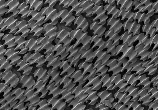
#guys i love sharks did u know that#do i talk abt it enough skjdfhjskdf#like!!! thresher sharks r so cool!!!#theres three species that we know of but a lot of professionals think theyre might be four??? thats sick????#ALSO ALSO ALSO theyre endoderms!!!! thresher sharks have special thermoregulation so they produce heat internally which like helps with-#-their metabolism and muscle shivering!!!#shoutout to the bigeye thresher btw their eyes r in keyhole shaped socket things so their eyes can rotate up its so sick#they have their bigger eyes so they can hunt in the dark!!!!#they also have like a. whats it called.#DIET VERTICAL MIGRATION#so they stay deeper underwater during the day then at night they come up closer to the surface to feed!!!#since its a thresher it still has thermoregulation too OH YEAH I FORGOT TO MENTION threshers are warm blooded!!! so their body temp is-#-higher than the water temp that they usually stay in#anyway. those r my shark facts for the day. did not mean to just ramble there JKGHSKJGHKJS#mlm#mlm yearning#mlm post#mlm love#gay mlm#trans mlm#t4t#mlm blog#mlm thoughts#t4t yearning
27 notes
·
View notes
Photo

Gut Geometry
The human gut is approximately 20-feet long, yet it squeezes into our bodies. During development, the gut tube forms a reproducible looped pattern in order to fit into that space, suggesting that it’s controlled by our genes. How cells influence changes in the shape of whole organs isn’t easy to track, especially for internal organs like the gut! But researchers have developed a new imaging technique to analyse just that. They visualised the folding and elongation of the midgut of flies and generated time-lapse movies that show how the layers of muscle cells (yellow) and endodermal cells (blue) which will form the inner lining of the gut, interact to mould it into four chambers before starting to coil. With further analysis, they found that muscle constrictions that induce shape changes in the endodermal cells to form the chambers are triggered by signalling molecules controlled by Hox genes – known for controlling tissue pattern development.
Written by Sophie Arthur
Image from work by Noah P Mitchell and colleagues
Department of Physics, University of California, Santa Barbara, Santa Barbara, CA, USA
Image originally published with a Creative Commons Attribution 4.0 International (CC BY 4.0)
Published in eLife, May 2022
You can also follow BPoD on Instagram, Twitter and Facebook
#science#biomedicine#gut#intestine#organ development#endodermal cells#drosophila#fruit flies#hox genes#tissue patterning#developmental biology#embryo development#immunofluorescence
7 notes
·
View notes
Text
In most plant roots, a medial longitudinal cross section reveals long cell files that converge in the subapical region of the root (Figure 17.21A). (...) In Arabidopsis, four distinct sets of initials, all of of the which are adjacent to the QC, can be defined in terms of their position and the tissues they produce (Figure 17.21B):
Columella initials. Located directly below (distal to) the QC, these initials give rise to the central portion (columella) of the root cap.
Epidermal-lateral root cap initials. Located to the side QC, these initials first divide anticlinally to set off daughter cells, which divide periclinally to form two files of cells that mature into the lateral root cap and epidermis.
Cortical-endodermal initials. Located and adjacent to the epidermal-lateral root cap initials, the cortical-endodermal initials divide anticlinally to set off daughter cells, which then divide periclinally to form the cortical and endodermal cell layers.
Stele initials. Located directly above (proximal to) the QC, these initials give rise to the vascular system, including the pericycle.

Ablation of the QC led to another abnormal division and precocious differentiation of the adjacent initials (see Figure 17.21B), suggesting that the QC produces a mobile signal that acts on the adjacent cells to prevent their differentiation and thus maintain their ability to divide.
"Plant Physiology and Development" int'l 6e - Taiz, L., Zeiger, E., Møller, I.M., Murphy, A.
#book quotes#plant physiology and development#nonfiction#textbook#plant roots#arabidopsis#qc#quiescence center#columella#root cap#daughter cells#epidermal lateral root cap#cortical endodermal#cell division#cortex#endodermis#plant cells#stele#pericycle#cell differentiation
1 note
·
View note
Note
Why do broish circles of the internet use the term "endomorph"? What's it mean? Why the prefix "endo-"? I only ever find blogarticles whenever I try to search the term.
I've seen the term before so I'm not completely surprised by it.
In fact the height comparison website I found for fandom shenanigans uses the system:
So after some basic searching I've found it's eugenics. Why is it always eugenics?
Tldr its a system of body categorization created by William Herbert Sheldon.
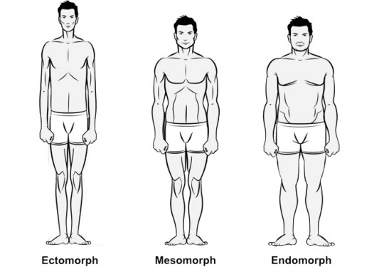
His system was based on the bodies of ivy league men, from his Wikipedia article, a quote from Ron Rosenbaum about it:
"He believed that every individual harbored within him different degrees of each of the three character components. By using body measurements and ratios derived from nude photographs, Sheldon believed he could assign every individual a three-digit number representing the three components, components that Sheldon believed were inborn -- genetic -- and remained unwavering determinants of character regardless of transitory weight change. In other words, physique equals destiny."
Heres the Wikipedia about the system and the creator.
ecto- meso- endo- are references to layers of development "He created these terms borrowing from the three germ layers of embryonic development: The endoderm, (which develops into the digestive tract), the mesoderm, (which becomes muscle, heart, and blood vessels) and the ectoderm (which forms the skin and nervous system)." In short it's inner, middle, outer. morph means "form or structure".
Now to the point: broish alpha male believer types are eugenists with a fresh coat of paint. Not surprised they are frothing over ANOTHER chance to say they are genetically destined to be great or to be a loser. They look for excuses for not working on their personalities or manners and eugenics is easy. They want easy excuses and think using outdated crap "science" makes them look smart.
As for why other groups of people would use the term (such as the team behind that height comparison website) I can assume the worse or I can assume they needed a system for sorting and thought the terms sounded "professional".
-mod squirrel
39 notes
·
View notes
Note
Can I have a weird animal fact plz
Wanna hear about SHARKS and EARLY EMBRYONIC DEVELOPMENT and COMMON CULTURAL MISCONCEPTIONS?
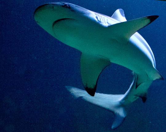
And no, it's not that some species will eat their siblings before birth, though that does happen. If you're not an only child, be glad you're not a sand tiger shark.
Anyway, we're mostly talking about teeth. Now teeth, despite common cultural mindsets, aren't actually bones.
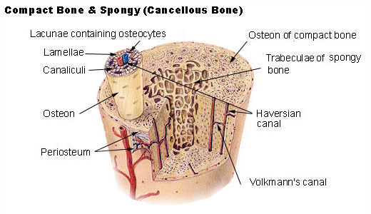
Bone is made of, well, bone: Most of your long bones have a rigid, dense layer outside (Cortical bone) and a spongy, lighter layer inside (cancellous bone), and a space inside for marrow and other tissues. Your bones aren't hollow, why waste that space when you could be making blood cells in there?
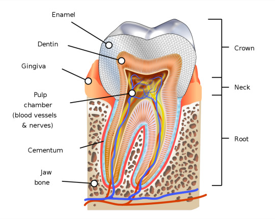
Teeth, on the other hand, don't have any actual bone inside. The outer layer, enamel, is the hardest thing in our bodies, which is one reason teeth are so easily fossilized. If our teeth were actually bones, then we'd be gumming our food by the time we're twenty.
I know some of you are asking: But, but, how do we know teeth didn't start out as bones and just got super specialized over time? I'd say, great question! We know they didn't for two main reasons.
Teeth come from a different embryonic layer than bone. Embryogenesis gets pretty complicated, but I'll try to explain it simply.
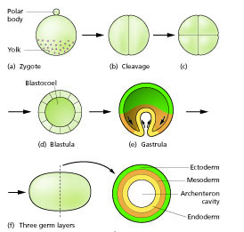
Back when you (and every animal) is first conceived, you start life as a single cell called a zygote, then that divides into a solid ball of cells. That ball of cells keeps dividing and becomes hollow inside. Then at some point, like sticking your thumb into the clay to make a little bowl in elementary school, a hole forms and the hollow ball of cells folds inside of itself to create some layers. Now look at you! You've got three layers of cells! You've got an outside, or ectoderm. You've got an inside, or endoderm. And you've got the cells between your inside and outside, or mesoderm.
Each layer of cells gives rise to different parts of you. The endoderm mostly makes the inner lining of your digestive tract. The mesoderm makes most of your insides, including your bones. And the ectoderm makes your skin and eyes and outsides, including your teeth.
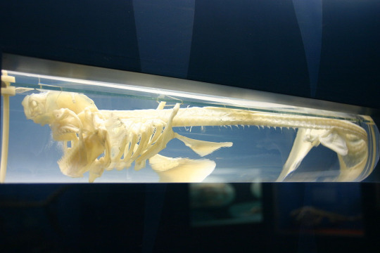
We also know teeth aren't bones because teeth evolved before true bones. Sharks and their kin have well-developed teeth with enamel, but they don't have any mineralized bones in their bodies. Even their jaws aren't true bone, but an evolutionary precursor. It takes a whole different leap of evolution to add full-body mineralized bone to the vertebrate repertoire!
Now, let's bring some things together. Sharks, though we love to sing their smooth praises, have skin that has been used throughout history as sandpaper or protective coverings. This is due to a layer of dermal denticles covering the skin- note that I didn't say scales! Scales evolved from dermal bone, or thin plates of bone arising from the ectoderm layer. These structures aren't bony at all, they're made of a different material: enamel. Yep, the same stuff in teeth. Yep, arising from the same embryonic layer as teeth.
This entire post has been a very roundabout way of saying that teeth aren't bones, and shark skin is actually covered in teeth.
(While in good fun, this is an educational scientific post. I ask that you do not continue the smooth sharks joke here.)
818 notes
·
View notes
Text
// cpsm novel spoilers (chp 592/593)
"Just cut the crown prince into four pieces."
... lloyd. lloyd you can't just say it like that-
"My lord is not a planaria."
"Planaria? Hold it. "I think there's some misunderstanding."
"What misunderstanding do you mean?"
"I mean dividing your master into four parts? "How did you understand how we were going to share it?"
"Isn't it divided into four parts: head, torso, limbs, and soul?"
"Are you dreaming of becoming a butcher?"
"... ... no?"
"I'm not going to divide it like that. The human body is not like Legos. "It seems like we should just divide it into endomorph, ectoderm, mesomorph, and soul."
"Endoderm, ectoderm...... "What is it?"
"There is such a thing. anyway-"
Archangel Lloyd scratched the back of his neck.
"The quartering I mentioned is very different from what you think."
well no shxt it's different lloyd!! YOU CAN'T JUST SAY LET'S CUT HIM INTO FOUR PARTS AND NOT EXPECT THEM TO ALL THINK YOU'RE GOING TO FCKING QUARTER HIM INTO PIECES LIKE IN THE MEDIEVAL AGES??
#ying talks about cpsm#crown prince sells medicine#cpsm#cpsm liveblog#cpsm spoilers#chp 592#chp 593#why are you like this#also yea he continues to say there's such a thing#mysterious bastard.
16 notes
·
View notes
Text
Health: Adrenal Glands
TCM: Jing, Kidneys
GNM: Off Track (cortex), Unbearable Stress (medulla)
Greek: Sanguine (cortex), Choleric (medulla)
Chakra: Root
Astrology: Mars, Aries-Libra; ex. managing adrenal health can be especially important for natal placements like Mars in Libra/7th, Mars in Pisces/12th, South Node conjunct Mars, etc.
The adrenal glands, which sit atop the kidneys, play a central role in the body's response to stress, fatigue, immune challenges, and several key physiological and metabolic functions. Issues related to the adrenal glands is very common yet highly under-diagnosed. They consist of two regions:
Adrenal Cortex: Derived from the mesodermal base substance cholesterol, the adrenal cortex produces hormones such as cortisol, corticosterone, aldosterone, and male sex hormones. These hormones play vital roles in physiological functions. Cortisol and corticosterone, known as stress hormones, contribute to the regulation of metabolism, inflammation, and blood sugar, and assist in long-term stress response by enriching the blood with minerals and glucose. Their anabolic effects also aid in healing and regeneration. Aldosterone helps maintain blood pressure by regulating the balance of salt and water in the body. The adrenal cortex's production of male sex hormones can influence the reproductive system. If unresolved, chronic stress may lead to excessive cortisol production, causing problems like weight gain and high blood sugar.
Adrenal Medulla: The endodermal adrenal medulla manages emotional and physical stress by producing the hormones noradrenaline, dopamine, and adrenaline. These hormones are pivotal in activating the "fight, fright, flight" response, a physiological reaction that occurs in response to a perceived harmful event or threat. This acute stress response increases heart rate, blood sugar, and mental alertness, along with other physiological changes. Chronic anger or emotional upheaval can strain the adrenal medulla, leading to an energetic drain.
Stress
The adrenal glands are highly sensitive to stress, and chronic stress can lead to various disorders:
Psychiatric Disorders: Neuroses, post-traumatic stress disorder (PTSD), depression, anxiety disorders, bipolar disorder.
Neurological Disorders: Migraines, peripheral neuropathy, dizziness, tremors.
Glandular Disorders: Issues related to other glands including the thyroid which is responsible for cell growth; for instance, uterine cancer, polyps, hypothyroidism, hyperthyroidism.
Cardiovascular Disorders: Coronary artery disease, stroke, heart attack, hypertension, arrhythmias.
Respiratory Disorders: Asthma, chronic obstructive pulmonary disease (COPD), difficulty in breathing.
Immunological Disorders: Possible tumor promotion, reduced resistance to infection, autoimmune disorders, chronic inflammation.
Metabolic Disorders: Diabetes, obesity, metabolic syndrome, difficulty in regulating blood sugar.
Gastrointestinal Disorders: Ulcers, irritable bowel syndrome (IBS), chronic indigestion, malabsorption.
Genitourinary Disorders: Impotence, incontinence, menstrual problems, urinary tract infections, kidney dysfunction.
Musculoskeletal Disorders: Muscle weakness, chronic fatigue, fibromyalgia, joint pain.
Skin Disorders: Acne, eczema, psoriasis, skin thinning.
Cysts and Cancer
Adrenal Weakness: If the adrenal glands don't produce enough adrenaline, the heart may pump slower, leading to fluid accumulation and cysts in the prostate, ovaries, and breasts. These cysts can harden and potentially lead to cancer.
Blood Flow: Increasing blood flow can help resolve cysts if addressed quickly.
Signs of Imbalance
Frequent sickness, fatigue, low libido, low backache, chronic health issues, dark circles under the eyes, hair loss, early greying, frequent urination at night, cold hands and feet, brain fog, pain and weakness in the lower back, loins, thighs, knees and lower body, urinary weakness and debility, polyuria and nocturia, impotence and male sexual dysfunction, moodiness and irritability, depression, muscle or bone loss, autoimmune conditions, chronic fatigue, hormone imbalance, body aches, unexplained weight loss, lightheadedness, skin discoloration (hyperpigmentation), weakened stress response, insulin resistance, sleep problems, weight gain, sweet and salty food cravings, difficulty getting up in the morning, increased PMS or menopausal symptoms, inability to handle stress, increased allergies, frequent sighing, cravings for salty foods, higher energy levels in the evenings, overuse of stimulants like caffeine.
Traditional Chinese Medicine (TCM)
Jing (essence): The statement by the Chinese that the kidneys harbor our Jing refers to the adrenals having the highest concentration of neural crest cell derivatives in the entire body. Strong Jing corresponds to robust characteristics like strong teeth, which are made by neural crest cells, while weak Jing relates to signs of aging like grey hair and deafness, also linked to neural crest cells. Jing's manifestations include the progression from youth to adulthood, reflected in functions controlled by the pituitary (aided by neural crest cells), and can be seen in the structure of the face and jaw. Neural crest cells also create the heart's connective tissue, affecting lifespan. Jing as a concept is not the same as neural crest cells but they represent the body's inherent organizational strength. Weak organizational energy leads to neural crest cells that don't form properly or function well, resulting in genetic disorders affecting facial development. The Chinese recognized these markers as indicators of weak Jing.
Kidneys & Urinary Tract: The adrenal glands are connected to the kidneys not just through the renal fascia, but also by way of the renal artery, draining into the renal vein, and receiving nerve connections from the renal plexus. The kidneys regulates the body's water content and are essential for maintaining healthy bones, as well as producing healthy bone marrow and blood. It determines the level of adrenaline and dopamine in the body, affecting our energy and rest. In addition, it forms a relationship with the heart through various hormones. During puberty, the adrenal cortex starts to produce sex hormones like testosterone and oestrogen, and this production continues throughout adulthood. Caffeine depletes kidney qi, yang, yin, and essence, contributing to liver and adrenal issues, and long-term exhaustion. Regular coffee drinkers, especially those who don’t feel its effects, may be nearing adrenal exhaustion. Adrenal fatigue is often considered a kidney yang deficiency. If left untreated, it can progress to a kidney yin deficiency. These deficiencies are often marked by a light low groaning tone to the voice, a darkish pallor under the eyes, negative attitudes of insufficiency or inadequacy, needing to sit and not being able to stand for long, and worrying about money. Additionally, those who experience traumatic shock or long term stress often have their hair turn gray or fall out.
Kidney Yang (medulla): Linked to the reactive sympathetic nervous system and the hormones adrenaline, dopamine, and norepinephrine, produced in the adrenals. A deficiency in kidney yang can lead to symptoms like cold hands and feet, edema, night urination, and low libido. This deficiency parallels disorders like adrenal fatigue, hypothyroidism, and sexual dysfunction. This condition is worsened by the use of marijuana, diminishing our natural drive, motivation, and willpower. Adrenaline is used to treat conditions like asthma, anaphylaxis, and slow heart rates. It works on the cell membrane's outside, which is known as the yang aspect of the cell. It never enters the cell but attaches to a receptor on the outside, initiating a cascade of chemicals that open or close gates on the cell's exterior. In the heart and muscles, adrenaline prompts more calcium to enter, which amplifies the force of contraction; in the lungs, it leads to the relaxation of the muscles, permitting more air to flow in; in the brain, it initiates the emotional reactions connected with fear.
Kidney Yin (cortex): Associated with the parasympathetic nervous system and the hormone cortisol, produced in the adrenals. A kidney yin deficiency may manifest as sore back, leg weakness, insomnia, and anxiety. It may parallel disorders like diabetes, high blood pressure, and hyperthyroidism. Although necessary for daily function and stress response, excessive cortisol can lead to fluid retention, osteoporosis, muscle wasting, depression, and diabetes. If our bodies stopped producing cortisol, it would result in significant illness. While adrenaline binds to the exterior of the cell (yang), cortisol is absorbed into the very core (yin). Contrary to the effects of adrenaline which are more immediate, the effects of cortisol can take years.
Greek Medicine
Hot Temperament: The adrenals are generally represented as stimulating and energizing various functions to adapt to stress.
Adrenal Cortex: Sanguine, nutritive, and anabolic, enriching the blood and decreasing swelling.
Adrenal Medulla: Choleric, energetic, and catabolic, stimulating acute stress response.
Faculty Support: Adrenal glands produce supplementary amounts of sexual hormones, bridging a connection between vital and generative faculties; supporting heart and lungs (vital), kidneys and pancreas in blood sugar regulation (natural), enhancing mental alertness and stimulating the sympathetic nervous system functioning (psychic), supporting male sexual function and response (generative).
Root Center: The adrenals form the basic energetic support for the entire organism, associated with the Root Chakra.
Kidneys & Urinary Tract: Weak adrenals may adversely affect the vitality and functioning of the kidneys and urinary tract, which are interlinked with the adrenal glands'. When the kidneys are not effectively eliminating fluids, it can lead to the accumulation of phlegm and moisture. Additionally, the kidneys have a connection to the soles of the feet which may be affected by cold conditions. Conditions of melancholy or devitalization of the adrenals, kidneys, and urinary tract often correlate with chronic fatigue.
Adrenal Exhaustion: Chronic stress and irregular habits can lead to fatigue, irritability, pain, and impotence in men usually caused by flare-ups of Choleric anger or any experiences which feel like a roller coaster. Sexual overindulgence also exhaust the adrenals.
Male Sexual Function: The adrenal glands provide energetic support for male sexual function. Issues with adrenal energy may result in sexual dysfunction, including impotence or premature ejaculation.
Blood Sugar Regulation: In instances of dangerously low blood sugar, the adrenal glands jump into action, raising levels through adrenaline. Chronic instability may involve adrenocortical hormones, aggravating factors in type II diabetes.
German New Medicine (GNM)
Adrenal Cortex (Off Track)
Conflict: Feeling like you've made a wrong choice or gone down the wrong path.
Under-Functioning: Waterhouse-Friedrichsen syndrome, adrenal gland insufficiency, Addison's disease. Reduced cortisol production, feeling stressed and tired.
Over-Functioning: Excess cortisol (Cushing's disease) with high blood pressure, round face, obesity, muscle atrophy or aldosterone (Conn's syndrome) with high blood pressure, low potassium, weak muscles, thirst, frequent urination.
Adrenal Medulla (Unbearable Stress)
Conflict: Extreme tension from stress, feeling overwhelmed. Something is beyond reach.
Diagnosis: Pheochromocytoma, neuroblastoma. High blood pressure, racing heart, increased blood sugar, sweating.
Astrology
Mars: This planet governs adrenaline, testosterone, male sexual function, playing a role in the catabolic metabolism where fuel is burned. It also oversees the release of toxins, the regulation of red blood cells, iron, and muscle tissue, including tendons and ligaments. It extends to the entire muscular system, embodying vitality and physical strength. The energy of Mars is hot and dry so it's temperament is Choleric. Mars co-rules the adrenals (with Aries/Libra) and the male genitalia (Scorpio).
Aries: Aries is a Choleric sign, and its will to action makes it prone to anger and stress, which deplete and weaken the adrenal glands. The sign is energetic, sharp-eyed, cheerful, and alert, but may become irritable and have a restless tendency to keep going until burnout. This can lead to eyestrain and poor vision as the health of the eyes is dependent on the strength of the adrenals. The preference for stimulants like chili pepper, caffeine, and their attraction to the Sun can overstimulate the adrenal glands, leading them to seek cooling substances like icy drinks, fruits, and seafood.
Libra: Libra, the Sanguine Air sign that rules the kidneys and lower back, is a counterbalance to Aries. The kidneys and genitourinary tract are only as strong and healthy as the adrenal glands, which are their energetic support. Underlying adrenal weakness and exhaustion weakens the kidneys and genitourinary system, leading to urinary debility, urinary tract infections, inflammation or irritation, uremia, and gout. Since male sexual function is also dependent on the strength and health of the adrenal glands, problems may arise in this area.
Aries-Libra Dynamic: Kidneys, adrenals, eyes, head and cranium, low back, lumbar spine, bladder, genitourinary system (especially male), hips and loins. Aries represents personal power, autonomy, honesty, and decisiveness, often leaning towards individualism and assertiveness. Libra, on the other hand, emphasizes cooperation, diplomacy, compromise, and social harmony. While Aries is direct and often acts alone, Libra seeks mutual choice and operates through consensus. This represents a struggle between competition and cooperation, selfhood and partnership, war and peace, doing and being. Finding balance is needed for acid/alkaline balance, as Aries tends to run acidic, and for hormonal balance, where Aries tends toward excess testosterone. The tension between these two forces may cause a lot of frustration and aggravation, particularly in males, manifesting as irritation or inflammation of the genitourinary tract.
Healing and Relief
Diet: Paleo; reduce caffeine, sugars and sweeteners, gluten, starch, white flour, processed foods, and hydrogenated oils.
Nutritional Supplements: Ionic Magnesium, B Vitamins, Omega-3 Fatty Acids, Vitamin C, Zinc, Probiotics, L-Theanine
Herbal Adaptogens: Ashwagandha, Rhodiola Rosea, Holy Basil, Licorice Root, Siberian Ginseng, He Shou Wu
Lifestyle Considerations: Address societal pressure to perform, lack of self-care, and the conflict in real life. Regular exercise of varied intensity. Practice meditation, mindfulness, Tai Chi, Qigong. Express creatively and spend time in nature. Align sleep schedule with circadian rhythm to nourish yin. Hydrate more and add salt to water. Avoid various toxins like dioxin or insecticides which contribute to adrenal malfunction. Spend more time in the sun each day. Don't sit for too long. Try wearing a haramaki, stretch cotton (for summer) or stretch wool (for winter).
These are research notes from the work of Daniel Keown, Michel Odoul, Michael Tierra, Lesley Tierra, Johan Boshwinkel, David Osborn, Judith Hill, Eileen Nauman, Björn Eybl, Caroline Markolin, Deb Shapiro, and Jody Smith.
#adrenaline#kidneys#urinary tract#adrenal glands#adrenal fatigue#somatic#medicine#mind body connection#naturopathy#astrology#holistic#chakras#gnm#tcm#chinese medicine#healing#aries#libra#psychosomatic#health
40 notes
·
View notes
Text
Quick Roll Table for Lancer NPCs
Starting off with IPS-N, for when you need to populate a rickety O'Neil cylinder with salty cosmonauts.
1. IPS-N Marine: Nearly two and a half meters tall and loaded with myolace, endodermal NxRA integuments, and titanium-carbide osteoplate. Putting her in a hardsuit only makes her more of a terror in close quarters. She treats her squadmates like a mother bear treats her cubs and takes no shit when it comes to their safety, whether that be from the enemy or IPS-N suits. With any luck, she’ll include you in her pack too.
2. IPS-N Subline Jockey: They’re twitchy as all hell and just can’t seem to sit still. Quite short despite a lifetime in space, and always wears their truncated environmental filter system. They’ve got an impeccable record sitting in the casque of a Walleye; if you need a flak screen penetrated or a patrol cutter outran, they’re the one to call. Willing to fly anyone or anything anywhere for a surprisingly generous rate, no questions asked.
3. IPS-N Suit: Totally unflappable and results driven, station deckhands have taken to calling them ‘Rocky’ for their stone-cold demeanor. One wouldn’t expect someone as relatively unassuming as them to hold the levers of military and civilian procurement in the palm of their hand. Despite the strict IPS-N regs on employing freelancers, they’re willing to take all comers for work. Be absolutely sure you don’t disappoint.
4. IPS-N Mechanic: As salty as any cosmonaut comes, and has little patience for those who don’t know what they’re doing or waste his time. Carries an entire toolbox, fusion torch and all, inside an oversized and very rusty mechanical arm. Some say that he can put a NLS drive back together with his eyes closed. Does repair work on the cheap, but will make you stay and watch him. Take notes, he’ll quiz you once he’s done.
5. IPS-N Miner: A persistent ray of sunshine despite spending all of her time working in deep-core nickel-iron asteroid mines. She lost all of her limbs and an eye doing her job. IPS-N set her up with some second-rate cybernetics, clunky, poorly neuro-tuned, and constantly belching steam. They still expect her to work and pay them back. She quietly wishes she could hurt the company very badly for putting her in this situation.
6. IPS-N Captain: Older than most everyone on the station, doesn't look a day over 65 thanks to the vagaries of nearlight travel, stasis-sleep, and a healthy diet of Ispahsalari cigars and Ras Shamran whiskey. Rough around the edges, with a low brow sense of humor befitting of an old salt cosmonaut. If you can get past both those, you'll find that he's well worth his weight in O2, knows the best deck-teks and merchants on the station on a first name basis

22 notes
·
View notes
Text
Life-Span Development John Santrock Chapter 3 Notes
-gernminal period- is the period of prenatal development that takes place during the first two weeks after conception. It includes the creation of the fertilized egg, or a zygote; cell division; and the attachment of the zygote to the uterine wall
-differentiation- specialization of cells to perform various tasks
-blastocyst- consists of an inner mass of cells that will eventually develop into the embryo
-trophoblast- an outer layer of cells that later provides nutrition and support for the embryo
-implantation- the attachment of the zygote to the uterine wall
-embyronic period- the period of prenatal development that occurs from two to eight weeks after conception. During this period, the rate of cell differentiation intensifies, support systems for cells form, and organs appear.
-embryo- the blastocyst attaches to the uterine wall and the mass of cells is now called an embryo
-endoderm- the inner layer of cells, which will develop into the digestive and respiratory systems
-mesoderm- the middle layer, which will become the circulatory system, bones, muscles, excretory system, and reproductive system
-ectoderm- the outermost layer, which will become the nervous system and brain, sensory receptors (ear, nose, and eyes, for example), and skin parts (hair and nails, for example)
-the endoderm primarily produces internal body parts, the mesoderm primarily produces parts that surround the internal areas, and the ectoderm primarily produces surface parts
-amnion- a sac (bag or envelope) that contains a clear fluid in which the developing embryo floats
-umbilical cord- contains two arteries and one vein, and connects the baby to the placenta
-placenta- consists of a disk-shaped group of tissues in which small blood vessels from the mother and the offspring intertwine but do not join
-organogenesis- the name give to the process of organ formation during the first two months of prenatal development
-fetal period- lasting about seven months, is the prenatal period between two months after conception and birth in typical pregnancies. Growth and development continue their dramatic course during this time.
-neurons- or nerve cells, which handle information processing at the cellular level in the brain
-Four important phases of the brain's development during the prenatal period involve: (1) the neural tube, (2) neurogenesis, (3) neuronal migration, and (4) neural connectivity.
-As the human embryo develops inside it's mother's womb, the nervous system begins forming as a long, hollow tube located on the embryo's back. This pear-shaped neural tube, which forms at about 18 to 24 days after conception, develops out of the ectoderm. The tube closes at the top and bottom ends at about 27 days after conception.
-A massive proliferation of new immature neurons begins to take place at about the fifth prenatal week and continues throughout the remainder of the prenatal period. The generation of new neurons is called neurogenesis, a process that continues through the remainder of the prenatal period but is largely complete by the end of the fifth month after conception. At the peak of neurogenesis, it is estimated that as many as 200,000 neurons are generated every minute.
-neuronal migration- involves cells moving outward from their point of origin to their appropriate locations and creating the different levels, structures, and regions of the brain
-teratogen- is any agent that can potentially cause a birth defect or negatively alter cognitive and behavioral outcomes.
-The field of study that investigates the causes of birth defects is called teratology.
-The does, genetic susceptibility, and the time of exposure to a particular teratogen influence both the severity of the damage to an embryo or fetus and the type of defect:
-Dose- The dose effect is rather obvious--the greater the dose of an agent, such as drug, the greater the effect.
-Genetic susceptibility- The type or severity of abnormalities caused by a teratogen is linked to the genotype of the pregnant woman and the genotype of the embryo or fetus. For example, how a mother metabolizes a particular drug can influence the degree to which the drug's effects are transmitted to the embryo or fetus. Also, for unknown reasons, male fetuses are far more likely to be affected by teratogens than female fetuses.
-Time of exposure- Exposure to teratogens does more damage when it occurs at some points of development than at others. Damage during the germinal period may even prevent implantation. In general, the embryonic period is more vulnerable than the fetal period.
-embryonic period- when organs are being formed
-a critical period is a fixed time period very early in development during which certain experiences or events can have a long-lasting effect on development.
-psychoactive drugs- act on the nervous system to alter states of consciousness, modify perceptions, and change moods
-fetal alcohol spectrum disorders (FASD)- are a cluster of abnormalities and problems that appear in the offspring of mothers who drink alcohol heavily during pregnancy
-afterbirth- is the third stage, at which time the placenta, umbilical cord, and other membranes are detached and expelled. This final stage is the shortest of the three birth stages, lasting only minutes.
-midwifery- is a profession that provides health care to women during pregnancy, birth, and the postpartum period
-doula- is a Greek word that means "a woman who helps." A doula is a caregiver who provides continuous physical, emotional, and educational support for the mother before, during, and after childbirth
-analgesia- is used to relieve pain. Analgesics include tranquilizers, barbiturates, and narcotics.
-anesthesia- is used in late first-stage labor and during delivery to block sensation in an area of the body or to block consciousness
-oxytocin- is a hormone that promotes uterine contractions; a synthetic form called Pitocin is widely used to decrease the duration of the first stage of labor
-natural childbirth- is the method that aims to reduce the mother's pain by decreasing her fear by providing information about childbirth and teaching her and her partner to use breathing methods and relaxation techniques during delivery.
-Bradley Method- a type of natural childbirth that is used today which involves husbands as coaches, relaxation for easier birth, and prenatal nutrition and exercise
-prepared childbirth- or the Lamaze method. It includes a special breathing technique to control pushing in the final stages of labor, as well as more detailed education about anatomy and physiology
-cesarean delivery- the baby is removed from the mother's uterus through an incision made in her abdomen. Cesarean deliveries are performed if the baby is lying crosswise in the uterus, if the baby's head is too large to pass through the mother's pelvis, if the baby develops complications, or if the mother is bleeding vaginally.
-breech position- the baby's buttocks are the first part to emerge from the vagina
-Apgar Scale- is widely used to assess the health of newborns at one and five minutes after birth. The Apgar Scale evaluates an infant's heart rate, respiratory effort, muscle tone, body color, and reflex irritability.
-Brazelton Neonatal Behaviroal Assessment (NBAS)- named after American pediatrician T. Berry Brazelton (1918-2018). The NBAS typically is performed 24 to 36 hours after birth. It is also used as a sensistive index of neurological competence up to one month after birth for typical infants and as a measure in many studies of infant development. The NBAS assesses the newborn's neurological development, reflexes, and reactions to people and objects.
-Neonatal Intensive Care Unit Network Neuro behavorial scale (NNNS)- provides another assessment of the newborn's behavior, neurological and stress responses, and regulatory capacities
-low birth weight infants- weigh less than 5 pounds 8 ounces at birth. Very low birth weight newborns weigh less than 3 pounds 4 ounces, and extremely low birth weight newborns weigh less than 2 pounds 3 ounces.
-preterm infants- are those born three weeks or more before the pregnancy has reached its full term- in other words, 35 or fewer weeks after conception
-small for date infants- are those whose birth weight is below normal when the length of pregnancy is considered. They weigh less than 90 percent of all babies of the same gestational age. Small for date infants may be preterm or full term.
-extremely preterm infants- are those born at less than 28 weeks gestation
-very preterm infants- are those born at less than 33 weeks of gestational age
-kangaroo care- involves skin-to-skin contact in which the baby, wearing only a diaper, is held upright against the parent's bare chest. Kangaroo care is typically practiced for two to three hours per day, skin-to-skin over an extended time in early infancy.
-postpartum period- the period after childbirth or delivery that lasts for about 6 weeks or until the mother's body has completed its adjustment and has returned to a nearly prepregnant state. It is a time when the woman adjusts, both physically and psychologically, to the process of childbearing
-postpartum depression- involves a major depressive episode that typically occurs about four weeks after delivery. Women with postpartum depression have such strong feelings of sadness, anxiety, or despair that for at least a two-week period they have trouble coping with their daily tasks
-bonding- the formation of a connection, especially a physical bond between parents and the newborn in the period shortly after birth
All From The Life-Span Development by John W. Santrock Chapter 3
#developmental psychology#life span development#psychology#college#college life#student#textbook notes
5 notes
·
View notes
Text

Você sabia que a Daucus carota sativus é o nome científico da cenoura, uma planta da família Apiaceae? A cenoura é alimento muito cultivado e conhecido em todo o mundo por suas raízes tuberosas comestíveis.
Que as cenouras são ricas em fontes de nutrientes, fibras, vitaminas e minerais, provavelmente você já sabe. Mas você sabe como é composta as partes de uma cenoura? Vem comigo que vou te falar sobre algumas.
Raiz: possui raiz tuberosa, com epiderme (composto por células justapostas, e por pelos absorventes que facilitam a absorção de água de nutrientes), córtex (composto por células parenquimatosas) , endoderme (conhecida por ser uma camada que delimita o córtex e o cilindro vascular), periciclo (camada localizada entre o cilindro vascular e a endoderme), zona de crescimento (região que fica próxima as extremidades da raíz, possuindo células meristemáticas).
Cilindro Vascular: esta parte da cenoura é responsável pelo transporte de água, composto também por xilema (com traqueídes e elementos de vasos que são responsáveis pelo transporte de nutrientes e água que são absorvidos pela raiz; além de uma parede celular espessa), floema (com células crivadas), câmbio vascular ( com células meristemáticas), periciclo.
Folhas: possui uma epiderme (com células epidérmicas e estômatos); cutícula (ajuda na redução de perda de água), mesofilo (com parênquima paliçádico e parênquima lacunoso), xilema e floema ( responsáveis pelo transporte de água, nutrientes e produtos fotossintéticos).
Caule: epiderme (com células epidérmicas que fornecem proteção do mesmo); córtex com parênquima cortical (se localiza abaixo da epiderme e é responsável pelo armazenamento de nutrientes e água); feixes vasculares (xilema e floema); câmbio vascular (com células meristemáticas, contribuindo para o crescimento secundário do mesmo); medula.
Estas são algumas curiosidades sobre a cenoura! Gostou? Então não deixe de curtir e compartilhar com seus amigos sobre o assunto! Nos mande também seus conhecimentos e dúvidas sobre, que responderemos vocês!! No próximo post, traremos então algo novo para acrescentar seus conhecimentos sobre anatomia vegetal no seu dia a dia.
2 notes
·
View notes
Text
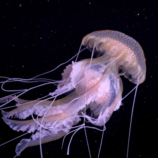
🪼How much water are jellyfish made of?
Have you ever thought about what jellyfish are made of? Maybe you thought of jelly? Jellyfish are elementary organisms that can range in different colors and sizes, but what is fascinating is that 95% of a jellyfish's body is water. Who knew that some jellyfish could be so dangerous, yet they are proactively just water? Jellyfish are also composed of three layers: the epidermis, the mesoglea, which is the slimy jelly-like substance you often observe, and then the endoderm gastrodermis. These beautiful animals play a vital role in our ecosystem, mainly as a good food source for turtles, but if you ever see a jellyfish, feel free to report your sightings to help scientists further their studies. To find all kinds of information about jellyfish, please click the link below:)
2 notes
·
View notes
Photo

Feeling the Squeeze
A critical process in early development, gastrulation defines the transition from a single layer of cells, known as the epiblast, to a more complex structure formed of three germ layers – the endoderm, mesoderm and ectoderm – which later give rise to different structures in the body. A key process in gastrulation is the epithelial-to-mesenchymal transition (EMT), in which epiblast cells undergo major changes in shape and gene expression to detach from their neighbours and move away. By labelling cell membranes with fluorescent proteins and using time-lapse microscopy, researchers were able to film cells undergoing the EMT (pictured in green) in developing mouse embryos, revealing how cells extract themselves by incrementally contracting their outer, or apical, surfaces (in red). EMTs are also a feature of cell movements later in life, from wound healing to cancer cell migration in metastasis, so studying these transitions could inspire new ways to prevent cancers from spreading.
Written by Emmanuelle Briolat
Image from work by Alexandre Francou, Kathryn V Anderson and Anna-Katerina Hadjantonakis
Developmental Biology Program, Sloan Kettering Institute, Memorial Sloan Kettering Cancer Center, New York, NY, USA
Image copyright held by the original authors
Research published in eLife, May 2023
You can also follow BPoD on Instagram, Twitter and Facebook
#science#biomedicine#embryonic development#developmental biology#epithelial to mesenchymal#metastasis#immunofluorescence#cancer
7 notes
·
View notes
Text

الرد على شبهة وجود خطأ علمي في الآية الكريمة (فكسونا العظام لحماً)
بقلم : عبدالله محمد (طبيب أسنان)
نص الشبهة؛
يقول الملحد لقد أخطأ قرآنكم في ذكر تخلق العضلات بعد العظام لان كليهما ينشآن من طبقة الميزوديرم (mesoderm) وبالتالي يتكونا في نفس الوقت ، فلا تسبق العظام تكون العضلات (اللحم)
الرد:
ملحوظة قبل البداية : اللحم هو العضلات وليس الجلد فحين تشتري لحما من الجزار يعطيك من العضلات وليس من الجلد
أولا سنتكلم عن تكوين الجنين :
تشارك ثلاث طبقات في تكوينه و هي : الاكتوديرم (ectoderm)و الميزوديرم (mesoderm) و الاندوديرم (endoderm)، و ما يهمنا في هذا الموضوع هي طبقة الميزوديرم التي تتكون منها العظام و العضلات.
ادعى الملحدون أن العظام و العضلات يتكونان في نفس الوقت بحجة أنهما يتكونان من نفس الطبقة، و لكنه لم يدرك أننا لو تتبعنا نمو أنسجة الجسم سنجد : القلب و الغدد التناسلية و الغضاريف و العظام و العضلات وغيرها وغيرها من الاعضاء (1)، و كل ذلك لا يتكون في نفس الوقت.
و نسأله : ما رأيك الآن؟! هل القلب يتكون مع الاعضاء التناسلية؟ هل الغدد تتكون مع العظام؟! هل الغضاريف تتكون مع العظام؟!
كل هذه الأنسجة تتكون من نفس طبقة الميزوديرم (mesoderm)، ولا تتكون في نفس الوقت فهذه حجة سخيفة تدل سطحية تفكيرك و معلوماتك ، فأنت تضع استنتاجا زائفا و أنت لست أهلا للاستنتاج اصلا.
على سبيل المثال، الغضاريف تتكون في منتصف الأسبوع الخامس (2) بينما العظام يبدأ تكوينها عند السادس، و العضلات ما بين منتصف السادس حتى الثامن. و هذا يعني أن الغضاريف تتكون من نفس الطبقة و لكن لا تتكون في نفس الوقت.
و إذا ما طلبت منهم مرجعا علميا لإثبات كلامهم هذا لا تجد إلا قولهم "يخرجان من نفس الطبقة، إذاً يخرجان في نفس الوقت!!!" وقد اثبتنا بطلان هذه الحجة
ثم لو سلمنا لكلامهم فهذا لا صلة له بمعنى الآية، فالآية تقول : فكسونا العظام لحما، "فكسونا" و بالتالي فالآية تتكلم عن مرحلة الكسو، اي مرحلة ارتباط العضلات بالعظام، و ليس مرحلة تكون العضلات نفسها.
لأن العضلات تتكون منفصلة وكذلك العظام منفصلة ثم بعد تكونهمها تقوم الأربطة بربط العضلات بالعظام فالآية تتحدث عن عميلة الربط هذه وليس عن تكون العضلات نفسها
ولاحظ أن الله عز و جل ((لم)) يقل : ثم كسونا العظام لحماً !!، بل قال : فكسونا، و الفاء في اللغة تدل على السرعة في الفعل الذي يليها.
و لكن سنتنزل و نفترض أن الآية تتكلم عن تكوين العضلات.
هذا المرجع الواسع المتخصص في علم الأجنة (3) يذكر ان بداية تكون العظام يحدث عند بداية الاسبوع السادس فيقول حرفيا "at sixth week : mesenchymal condensation for appendicular skeletal bone" فنلاحظ هنا انه يتحدث عن عظم حقيقي "bone" بينما في الاسبوع السادس ايضا يحدث فقط بداية تكون الخلايا التي تشكل العضلات وليس العضلات نفسها فيقول حرفيا "segmental elaboration of somite myotome occurs at sixth week" بينما تشكل العضلات يظهر عند الاسبوع الثامن فيقول حرفيا "at eighth week : individual muscles develop" فهنا يتحدث عن عضلات حقيقية "muscles". في الأسبوع الثامن . فاذهبوا وحاكموا العالم صاحب هذا المرجع الطبي !! او لا تصدعوا رؤوسنا ، و على الرغم أن هذا ليس معنى الآية، و لكن لو اعتبرنا أنه معناها فنحن أيضا نمتلك الأدلة على أن خلايا العظام أسبق من حيث التمييز.
و إذا ما عدنا للمعنى الصحيح للآية نجدها تتكلم عن مرحلة الكسو أو مرحلة ارتباط العضلات بالعظام.
في موقع EHD , أحد المواقع المتخصصة في علم الأجنة، سنجده يقول أن تكون العظام يحدث ما بين الأسبوع السادس و الأسبوع السابع فيقول حرفيا "Bone formation begins between 6 and 7 weeks, starting with the clavicle..."(4)، يعني قبل السادس لا يحدث تكامل للعظام. و لنرَ هذا الحديث الموافق لهذه المعلومة الطبية، يقول رسول الله ﷺ : "إذا مر بالنطفة اثنتان و أربعون ليلة بعث الله إليها ملكا، فصورها و خلق سمعها بصرها و جلدها و لحمها و عظمها" (5)، و هذه الأعضاء كلها لا يكتمل نموها إلا بعد الأسبوع السادس، أي ��عد الاثنتين و أربعين ليلة، و قبل ذلك لا يكتمل نموها. لدرجة أن الجلد يستمر في النمو إلى ما بعد الأسبوع الحادي عشر و لا يكتمل تكوينه قبل الأسبوع السادس أبداً ، ملحوظة: حرف الواو في الحديث السابق لا يفيد و لا يستلزم الترتيب، و إنما تدل على العطف فقط ، وهذا الحديث دليل على إختلاف الجلد عن اللحم وانهما ليسا شيئاً واحداً
ملحوظة : قد تجد اختلاف في موعد تكون الأنسجة من مرجع لمرجع آخر وذلك لإختلاف العينات المفحوصة لدى صاحب كل مرجع لذلك فقد تختلف في بعض الايام التقريبية .
و بخصوص ارتباط العضلات بالعظام، نفس موقع يقول أنه بحلول منتصف الأسبوع السابع تقوم الأربطة بربط العضلات بالعظام فيقول حرفيا "From 7 to 7½ weeks, tendons attach leg muscles to bones...."(6).
و بهذا يتبين لنا أن اللفظ القرآني دقيق جدا حين قال فكسونا، و لم يقل ثم كسونا.
"فكسونا" تدل على السرعة، و بالفعل كل الفرق بينهما أقل من نصف أسبوع. و هذا كله مثبت بأدلة علمية من مواقع طبية متخصصة في علم الأجنة ((كما وضحنا)).
من ناحية أخرى، نجد أن العضلة لها مبدأ و منتهى، مبدأ العضلة يُسمى المبدأ العظمي (origin)، أي الطرف المتصل بالعظام. فكيف يكون للعضلة مبدأ و منتهى (أطراف) (insertion) قبل تكون العظمة التي من المفترض أنها ركيزة صلبة تتصل بها العضلة لكي تؤدي وظيفتها(7).
و هذا إثبات بديهي سهل من ناحية أخرى، فلا يمكن أن تتصل العضلة بالعظم إن كانت العظام لم تتكون بعد.
وبذلك يتبين جهل وتدليس من يزعم بوجود خطأ علمي في هذه الآية ويتبين الإعجاز والسبق العلمي في القرآن الكريم وهو ما يؤكد وحيه من خالق هذا الانسان
والسلام عليكم ورحمة الله وبركاته
المراجع :
(1) https://www.researchgate.net/.../Schematic-overview-of...
(2) https://www.ehd.org/dev_article_unit6.php
(3) Netter's Atlas of human Embryology ch:8 (musculoskeletal system)
(4) https://www.ehd.org/dev_article_unit7.php
صحيح مسلم 2645 (5)
(6) https://www.ehd.org/dev_article_unit8.php
(7) https://www.visiblebody.com/learn/muscular/muscle-movements
2 notes
·
View notes
Text
Tree of Life 6: Metazoa (animals)
Brace yourselves, this one’s long.
[Disclaimer: taxonomy is a complex, ever-changing field, and this overview is certainly not going to be exhaustive, especially concerning extinct groups]
← Part 5 (Unikonta: amoebae, fungi, and such)
Part 6.5 (Porifera, Cnidaria, Plathyhelminthes, Annelida) →
We finally come to Animalia, or Metazoa “animals beyond” (as opposed to Protozoa, which are no longer considered animals at all). Perhaps you won’t find it easy to think of what sponges, chickens, oysters, starfish, and millipedes have in common. Here are the distinctive features that zoologists have identified for Metazoa:
First of all, all animals are heterotrophs, i.e. they cannot produce their own organic molecules, though very few species have become partially autotrophic thanks to symbiosis.
All animals have a diplont life cycle (see part 3), which goes as follows. It starts with two gametes, a female egg cell and a male sperm cell, which has a single flagellum (or, rarely, none), and fuses into the egg cell thanks to an anterior organ called acrosome. Each gamete is haploid, meaning it carries one copy of each gene; the fusion of the gametes produces a diploid zygote, which has two copies of each gene. The zygote multiplies by mitosis to form a multicellular body in which each cell is also diploid (unlike fungi and plants). Animals are always multicellular; while some fungi, namely yeasts, have reverted to unicellularity (see part 5), no animal has done so. Finally, each individual produces new haploid gametes, male and/or female, by meiosis, which discards half the genome in those cells.
(All multicellular organisms -- not just animals, but plants and fungi too -- go through a unicellular stage at some point in their life cycle. Why is it so, given that many species can regenerate their body from small fragments and reproduce by division? Probably to keep cancerous mutations in check: if you start as a single cell, either that cell will be healthy, and mutant lineages will have to start all over again; or it will be mutant, and so will be all its descendants, with no healthy cells to exploit, so that the mutation will destroy itself.)
During the development from zygote to multicellular body, the embryo folds to form an inner cavity, and the cells organize themselves in two or three germ layers: an internal endoderm, an external ectoderm, and in most cases a mesoderm which lies in between. Generally speaking, the endoderm forms the lining of the digestive tube with annex glands (e.g. the liver), as well as our lungs; the ectoderm forms the outer coating of the body, with skin, armor, and sense organs, as well as the nervous system; the mesoderm forms other organs (e.g. kidneys, sex glands), bones, and muscles.
Animals have at least two types of tissues: epithelial, which cover surfaces, and connective, which fill up volumes. Epithelial tissues, such as skin and mucous membranes, are made out of one or more layers of tightly packed cells growing over a basal lamina which supplies food. The cells are linked to each other by junctions such as desmosomes, which sew cell membranes to each other making the whole tissue more resistant. In connective tissues, cells are spaced wide apart, sunk in an extracellular matrix. Usually the matrix is a mix of gelatin and elastic fibers made out of proteins, most importantly collagen; but sometimes it may be liquid, as in blood, or built out of hard mineral, as in bone. To these two basic kinds, most animals add two more: muscles and nerves.
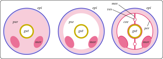
Cross-sections of three generic animals. Left to right, acoelomate, pseudocoelomate, eucoelomate. The endoderm (in yellow) forms the gut; the ectoderm (in blue) forms the outer surface; the mesoderm (in pink) fills the space in between.
The body of an acoelomate, e.g. a tapeworm, is filled with a mass of mesodermal cells called parenchym (par), in which organs (such as kidneys or gonads, mor) and the gut are immersed. The body is surrounded by an ectodermal epidermis (epi). Simple enough, but the organs are vulnerable to squishing.
In a pseudocoelomate, e.g. a hookworm, the other organs are immersed in a fluid-filled cavity called pseudocoelom (pse). Since water is not compressible, it gives the body a fixed volume and some mechanical resistance, as well as distributing substances, and storing products. Unfortunately a puncture wound is easily fatal.
In an eucoelomate, e.g. an earthworm, the mesoderm forms a lining or peritoneum (per) that wraps around the coelom (coe) and divides it in sections, as well as mesenteries (mes) that keep organs in place. The separate chambers of the coelom help against liquid loss and allow new kinds of movement, such as burrowing in sand. However, blood vessels (ves) are now required to distribute materials.
This is not a strict evolutionary sequence: animal phyla change from one to the other depending on their niche and conditions.
The animal kingdom is -- no wonder -- vast and complex. The most important high-level categories are known as phyla, commonly used in studies of animal diversity and large-scale evolution; here, I’ve written the names of phyla in all caps. The phyla Mollusca, Arthropoda, and the above-phylum group Deuterostomia will all get their own posts; the phyla Porifera, Cnidaria, Platyhelminthes, and Annelida will be described together in part 6.5.
?? 0. † Vendobionta: Let’s start with the strangest, shan’t we? Vendobionta is almost certainly not a clade, but a convenience term for all the weird macroscopic organisms that flourished in the Ediacaran period (once known as Vendian), between 580 and 540 Ma. It’s not clear even that they were all animals, let alone where they might fit in the tree: some have been described as algae, fungi, colonies of protists, or even representatives of a completely lost kingdom of life. In general, they didn’t have visible mouths or sense organs, nor any way to move; they probably (?) absorbed nutrients from water, grazed on mats of bacterial slime, and/or were photosynthetic. They suddenly disappeared at the beginning of the Cambrian, presumably because new organisms with eyes, jaws, and muscles predated them into extinction and/or consumed all their food.
† Trilobozoa “three-lobed animals”: These Vendobionts are radially symmetrical: their body, circular as a whole, is composed of three segments rotated around the central axis. This is quite unique: no living animal has a rotational or a three-fold symmetry (although some Cnidarians have a non-rotational, six-fold one). Branching grooves (a ciliated organ to collect food, maybe?) converge at the center of the upper surface. The most famous representative is † Tribrachidium, found in Russia and Australia.
† Medusoidea “jellyfish-like”: These too are flattened and radially symmetrical, often with concentric rings. The largest, like † Aspidella and † Cyclomedusa, could grow several cm wide. † Arkarua adami has a peculiar five-fold symmetry, like Echinoderms, though it’s unilkely to be related. Sometimes included among the † Medusoidea is † Eoandromeda octobrachiata, a strange galaxy-like being with eight spiral arm coiling out from the center.
† Petalonamae “petals from Namibia”: A set of two groups with plant-like shapes. † Rangeomorpha, most common in Newfoundland, had frond-like fractal structures -- possibly an adaptation to absorb as much food and oxygen as possible from the water. (Maybe sunlight too, but it seems some lived too deep for photosythesis.) Two, three, or six rows of branching fronds emerged from a central axis. Some, like † Charnia, stood upright in the water; others, like † Fractofusus, lay on the seabed. † Erniettomorpha such as † Pteridinium, found in Namibia, had lateral "vanes” formed by rows of sacs.
† Proarticulata “before articulated ones”: The most familiar, maybe. They had flattened bodies and bilateral (which is to say, left-right) symmetry. They also seemed to have internal branching canals that might be remains of a digestive system. † Dickinsonia grew as a thin segmented disk up to a meter long; it might actually be a Ctenophoran (see below), showing both righ-left and front-back symmetry. † Spriggina, from the very end of the Ediacaran, head a sort of “head” shield and rows of segments that might not have been exactly symmetrical, but somewhat offset.

A selection of Vendobionta. Far left: † Charnia masoni († Rangeomorpha, England). (Museo delle Scienze di Trento, via Wikipedia) Upper row, from left: † Spriggina floundersi († Proarticulata, Australia) (Wikipedia); † Ernietta plateauensis († Erniettomorpha, Namibia) (Ivantsov & Zagrevskaya 2021). Lower row, from left: †Tribrachidium heraldicum († Trilobozoa, Australia) (I. & Z. 2021); † Dickinsonia costata († Proarticulata, Australia) (Verisimilus, Wikipedia); † Mawsonites spriggi († Medusoidea) (Nordelch, Wikipedia)
1. PORIFERA “hole-carriers”. The (possibly paraphyletic) phylum of sponges. In general, they have a system of internal ciliated canals collecting food from the water that streams in through lateral pores, and out through a channel at the top. The whole structure is kept up by a basket of various composition. See part 6.5 for more.
? 2. PLACOZOA “plate animals” Honestly, it’s a bit embarassing to call this thing an animal. Until a few years ago, phylum Placozoa counted a grand total of one species, Trichoplax adhaerens from tropical seas (now, the count is three). Its body is simply a single layer of epithelial tissue surrounding a thin syncitium (i.e. a network of fused cells that retain separate nuclei). On the lower side, the epithelium has cilia and digestive glands; on the upper, it contains little crystal spheres that might serve as defense. Trichoplax reproduces by splitting, and feeds by crawling over bacterial mats; it secretes digestive enzymes to break food down, but each epithelial cell takes it up on its own. Recent studies (e.g. Laumer & al 2019) suggest that Placozoa might belong next to Cnidaria.

Trichoplax adharens, top view and cross-section. The whole thing is about 0.5 mm across and 0.025 mm thick. (Oliver Voigt, Wikipedia; Hickman & al 2008)
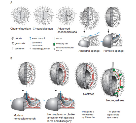
A model of the origin of animals, starting from a colony of Choanoflagellates (see part 5), passing through the ancestors of sponges, and ending with the likely common ancestor of Eumetazoans, which gave rise to both us and jellyfish. (Nielsen 2012)
3. Eumetazoa “animals well beyond”: All animals more complex than that pass through a process called gastrulation: when the embryo is still just a little ball of cells, it folds inwards producing a hollow or archenteron, the future gut. It communicates with the exterior through an opening called blastopore, the first prototype of a mouth. The two layers of cells produced by the folding, inner and outer, become the endoderm and ectoderm. Thanks to this hollow, Eumetazoans practice extracellular digestion: the uptake of food is not a responsibility of individual cells, like in sponges and Placozoans, but rather food is degraded in the internal cavity by digestive enzymes, and the resulting simple molecules are distributed to all tissues. In Eumetazoans we also see the first appearance of the nervous system, which allows coordinated action of the whole body.
In recent classifications, it’s being phased out in favor of Parahoxozoa, the animals defined by the presence of Hox genes, which specify different body regions and ensure that each anatomical structure forms in the correct part of the body; this would include Cnidaria and perhaps Placozoa too, but would exclude Ctenophora.
3a. CNIDARIA “nettles”: The phylum of corals, sea anemones, jellyfish, man-o’-wars, and curious parasites that have long been believed to be protists. They have radial symmetry (4-, 6-, or 8-fold), a single opening where the radia meet in the center, and specialized stinging cells for defense and hunting. See part 6.5 for more detail.
?? 3b. CTENOPHORA “comb-carriers”: What a strange situation! Ctenophorans have distinct tissues, appendages, a nervous system, and even have all three germ layers, unlike Porifera, Placozoa, and Cnidaria which lack the mesoderm. This would put them next to Bilateria (see below); except that recent molecular studies (e.g. Laumer &al 2019) insist on placing them in a basal position, further away from us than even sponges! An artifact of unusually fast evolution rates, perhaps?
Most Ctenophorans (e.g. Mertensia or Pleurobrachia) have bag-shaped bodies with the mouth at one pole (serving both to eat and to expel waste) and a branching, blind-end gut. Eight rows of combs, each of which is a band of cilia, run along the sides: hence the common name of “comb jellies”. In fact, they are the largest organisms on Earth that swim with cilia instead of muscles. Two tentacles may be retracted into pouches or extended to catch preys thanks to colloblasts, specialized glue-producing cells. Others, like Beroë, lost their tentacles; Coeloplana flattened its body to crawl on the seabed; the “Venus’ girdle” (Cestum veneris) extended it sideways to swim like a lopsided worm. Class † Scleroctenophora, endowed with armor plates, is known from the Cambrian but now extinct.
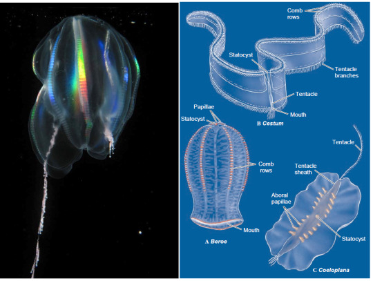
A variety of comb jellies. Left: Mertensia ovum, with one tentacle retracted. The rainbow shimmer of the comb rows is a distinctive feature of Ctenophora. Right: Beroë, Cestum, and Coeloplana. The mouth of Coeloplana is on the bottom, its lining forming the whole lower surface. Note that the body of Cestum is not long and narrow -- it’s short and very broad! (Kevin Raskoff, Wikipedia; Hickman & al 2008)
3c. Bilateria “two-sided”: The name says it all: animals in this clade have bilateral symmetry, with a left and a right side that are (in general) mirror images of each other, while keeping a distinction (thanks to signalling gradients during embryo development) between a front and back end. This allows them to concentrate their feeding and sensorial apparatus in the front, when the body first encounters new stuff, while the expulsion of waste and gametes moves to the back: a good plan if you’re an organism that moves consistently in one direction. In general, Bilaterians have a linear gut with an anterior mouth and a posterior anus, where food always flows from front to back.
3c1. Acoelomorpha: A group of marine worms, with thin bodies a few mm long. The simplest sort of Bilaterians: unlike almost all others, they don’t have a distinct mouth and anus, but a single opening at the center of their ventral surface, like jellyfish. That opening, which serves both as mouth and anus, does not lead into a gut (not even a jellyfish-style cavity), but onto a simple mass of endodermal cells that break down food. The body is covered by a thin ciliated epidermis; between these two layers, there are mesodemal muscles, nerves, ovaries, and testicles (they’re all hermaphroditic). That’s... about it. It’s not clear whether this simplicity is primitive, or a secondary loss of traits. They comprise three phyla, ACOELA, NEMERTODERMATIDA, and also XENOTURBELLIDA, whose 2 species were until recently classified next to Echinoderms (e.g. starfish).
3c2. Protostomia “mouth first”: This group is defined by a number of features, but few of its members possess them all. In theory, when the embryo folds to create a proto-gut, the resulting opening, or blastopore, should give rise to the mouth, with the anus appearing later; in Deuterostomes the opposite is true. In practice, this is only true in some cases. The nervous system is formed by a number of ganglia strewn along paired ventral nerve cords; these cords form a loop around the mouth, which in some cases (e.g. in spiders and octopodes) becomes a brain. Many groups of Protostomes develop from planktonic larvae that swim and collect food with bands of cilia.
3c2a. ? Mesozoa “middle animals” So called because they were once thought to lie between the simpler sponges and the more complex Eumetazoa. All there is to their body is a single layer of ciliated cells surrounding a mass of maturing eggs. In fact, they are descendants of relatively complex Protostomes that shed most of their body structures as a consequence of living as parasites: ORTHONECTIDA live in the body cavities of various marine invertebrates, RHOMBOZOA only in the kidneys of squids and octopodes.
Left: a generic Acoela, possibly the model from which all Bilaterian animals from snails to pigeons and from wasps to starfish developed. Right: the Mesozoan Rhopalura (Nemertodermatida; female above, male below).
3c2b. Gnathifera “jaw-carriers”: A diverse clade of invertebrates. Most are microscopic, and form a pretty good part of marine animal plankton. They have some variety of jaws made out of chitin, a nitrogen-rich sugar that we already saw in the cell wall of Fungi (see part 5).
? 3c2b1. CHAETOGNATHA “hairy jaws”: They go by the common name of “arrow worms”. Unlike most worms, they swim in open water, with undulations of their flattened body which extends sideways into fish-like fins. They even have, like vertebrates, a post-anal tail that doesn’t contain the intestine. The head has a large hood that conceals rows of chitinous grasping spines.
3c2b2. GNATHOSTOMULIDA “little jawed mouths”: Delicate mm-sized worms that live between grains of sand on beaches and seafloors. They have rasping jaws to scrape bacteria and fungi off sand grains and into a blind-end gut.
3c2b3. MICROGNATHOZOA “animals with tiny jaws”: A phylum with only one species, Limnognathia maerski on Greenland. A tiny wrinkled thing only 0.15 mm long, but with bizarrely complex retractile jaws, made out of 15 different elements. All other structures are very simple; it uses cilia to move and feed. Only females have ever been found.
3c2b4. ROTIFERA “wheel-carriers” or Syndermata “fused skins”: Another of the big ones. They are named after the wheel-like ciliate crown (corona) which, beating, creates the water current they use to swim and feed. Almost always microscopic, they have a great variety of body shapes, sac-like, worm-like, or spiny, sometimes with a thick segmented armor. They have little jaws and usually a foot with adhesive glands to stick to a substrate, which can fold onto itself like a spyglass. Under the cuticle, their skin is a syncytium, that is, fused cells that share their cytoplasm. Rotiferan class Bdelloidea is famous for having only females, which reproduce by parthenogenesis, their males having gone extinct some 25 million years ago. Until recently considered a separate phylum, Acanthocephala (“spiny heads”) is a group of Rotifers that specialized for life as parasites, losing jaws and gut, and developing an eversible spiny proboscis to attach to the intestine of vertebrates.
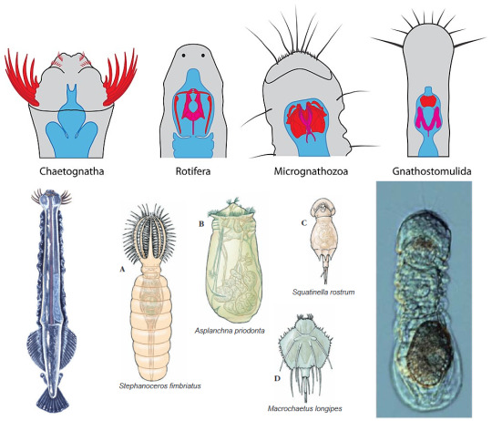
Variety of Gnathifera. On top: the chitinous jaws of Gnathiferan phyla, in red (Nicolas Bekkouche, Wikipedia). Below, from left: the arrowworm Pterosagitta draco (Chaetognatha), with its grasping spines extruded (Animal Diversity Web); some examples of Rotifera (Hickman & al 2008); Limnognathia maerski, only species of Micrognathozoa, carrying an enormous, highly decorated egg (Brusca 2016).
3c2c. Rouphozoa “sucking animals”
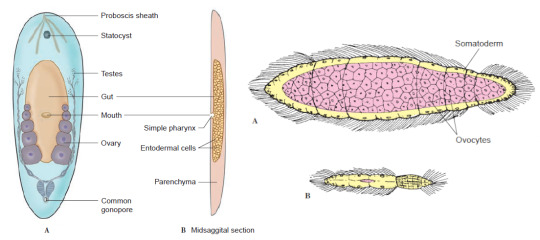
3c2c1. PLATYHELMINTHES “flat worms” The phylum of, well, flatworms; which includes such fascinating beings as flukes and tapeworms. They have lost their anus, and so have only a mouth leading into a branching gut; but often they are parasitic, and can absorb nutrients from the host through their skin. See part 6.5.
3c2c2. GASTROTRICHA “hairy bellies”: Millimeter-sized aquatic worms with a rounded back and a flattened belly, all covered in scales, spines, and bristles. Unlike flatworms, they have distinct mouth and anus, but for the rest their body organization is similar. They have a distinct brain that might also contain simple eyespots, and adhesive glands to attach to a substrate.
3c2d. Lophotrochozoa “animals with crest or wheel” or Spiralia: This group has several common traits but none that truly universal. The former name refers to the trochophora, a particular type of larva that swims with several ciliated bands, and has a sensorial apical organ connected to nerve ganglia. This larva was probably found in the earliest Protostomes but lost in most of their other branches. The latter name refers to a feature of embryonal development called spiral cleavage: the cells on top of the early embryo shift around so they’re no longer aligned with the ones below.
3c2d1. MOLLUSCA “soft ones” The phylum of chitons, tusk shells, clams, oysters, scallops, slugs, snails, squids, octopodes, and cuttlefish. The defining features are a muscular foot used for locomotion (which divides into the tentacles of squids), a tongue-like radula used to scratch at food, and a mantle that surrounds the organ mass and generally secretes a calcareous shell. We shall see more of them in part 7.
3c2d2. ANNELIDA “ringed” The phylum of segmented worms, such as ragworms, tubeworms, earthworms and leeches. They have a body divided in compartments with a true coelom, and blood vessels along its length; each segment contains its own excretory apparatus and usually locomorory appendages or bristles. See part 6.5.
3c2d3. NEMERTEA (named after the sea nymph Nemertes, “the unerring”) or Rhynchocoela “hollow muzzle”: Known as “ribbon worms”, they are mostly marine, though there are a few terrestrial species. They have a sharp-tipped muscular proboscis, normally tucked in a pocket in their head, which can shoot out like the tongue of a chameleon to catch preys. They are the simplest animals that have closed blood vessels, like us, rather than have blood soaking their whole body. For the rest, they are rather similar to Turbellarian flatworms, which we’ll see in part 6.5.
3c2d4. CYCLIOPHORA “wheel carriers”: A micro-phylum with no more than three species, all in genus Symbion, first discovered in 1995. All of them live exclusively on the mouthparts of cold-water lobsters, stealing a tiny fraction of their meals. They are sac-shaped, with an adhesive foot and a ciliate mouth at the top, only 0.3 mm long, and reproduce by budding.
Left: minute Lepidodermella squamatum (Gastrotricha), next-of-kin of flatworms (Giuseppe Vago, Wikipedia). Right: the ribbon worm Gorgonorhynchus repens (Nemertea), extruding its uniquely branched proboscis in response to a threat (Rachel Koning, Wikipedia)
3c2d5. Lophophorata “crest-carriers”: Distinguished by having a lophophore, a hydraulic ridge covered in ciliated tentacles that catch microscopic food particles when exposed to a water current. The gut is U-shaped, with the mouth opening at the center of the lophophore, and the anus usually just outside. Usually unable to move much -- they are very ‘coral-like’ in lifestyle.
3c2d5a. † HYOLITHA “Y-shaped stone”: A completely extinct phylum from the Paleozoic era, some 500-to-250 million years ago. They had conical shells with a cap to close them, and coiling structures that perhaps served to prop them up on the seafloor. These structures are called helens, after the daughter of the paleontologist who described them.
3c2d5b. BRYOZOA “moss-animals” or Ectoprocta “anus outside” or Polyzoa “many animals”: Very coral-like indeed: colonial animals with tiny polyps that grow on hard surfaces, forming a chitinous or calcareous communal skeleton. Alcyonidium forms gelatinous finger-like columns; Membranipora forms delicate lace-like webs; Plumatella forms rigid branches. Food particles are carefully selected by the tentacles and can be rejected. The guts of the polyps are connected internally, so they don’t all need to catch food on their own; some are free to specialize for different functions, such as whip-like vibracula and beak-shaped avicularia, which defend the whole colony.
3c2d5c. ENTOPROCTA “anus inside”: Basically the same as Bryozoans, except the anus opens inside the lophophore, too. Can be colonial or solitary.
3c2d5d. PHORONIDA (named after the Phoronis or Io, a lover of Zeus): A very small phylum with animals that resemble Bryozoa but are not colonial. They have tube-shaped body several cm long, anchored to rock or buried in sand, into which their flower-like lophophores can be retracted.
3c2d5e. BRACHIOPODA “arm-feet”: A sad story: between 500 and 350 million years ago, Brachiopods or “lampshells” were absolutely everywhere in Earth’s seas, but they were hit badly by the Devonian and Permian mass extinctions, and now only a few hundred species are left, their niches mostly taken over by Bivalve mollusks (e.g. clams). They are indeed quite clam-like, but whereas Bivalves have symmetrical left- and right-valves, Brachiopods have a larger upper valve and a smaller lower one. Enclosed between these valves is a pair of spiral-shaped lophophores which deliver food particles to the mouth.

Top left: two Symbion pandora (Cycliiophora) with larvae growing from their side (Nielsen 2012). Bottom left: Frenulina sanguinolenta (Brachiopoda) from the tropical Pacific, with the half-open valves revealing the curled lophophores inside (Brusca 2016). Right: the freshwater colonial Bryozoan Cristatella mucedo, with dozens of lophophorate polyps producing a common gelatinous matrix to creep about like a slug (Ernst Haeckel).
3c2e. Ecdysozoa “molting animals”: The distinctive feature of this clade is covering their whole body with a cuticle made out of organic molecules. The cuticle is strong and water-proof, but since it’s not made of cells it cannot grow: as the animal develops to adult size, it must shed or molt periodically its old cuticle and secrete a new one from its bare skin. They have few or no cilia (even their sperm cells often lack flagella!), and may have hard “teeth” or scales around the mouth.
3c2e1. Scalidophora “platelet-carriers”: A group of phyla whose cuticle is moade out of chitin, forming various kinds of plates, scales, and spines. They all live in sand or gravel.
3c2e1a. LORICIFERA “armor-carriers”: A small phylum discovered only in the 1980s. They have an egg-shaped armor or lorica and a retractile, cone-shaped head surrounded by long spines. Less than a mm long, they live in gravelly seafloors, eating bacteria. Three species discovered in the deep Mediterranean in 2014 are the only multicellular organisms known to survive in perpetual lack of oxygen.
3c2e1b. KINORHYNCHA “mobile muzzle”: Sometimes known as “mud dragons”, these tiny mud-dwelling worms have an 11-segmented trunk covered in back-pointing spines, and a further ring of spines surrounding their mouth. They cannot swim, but only crawl or burrow with their hydraulic extensile head: the spines prevent them from sliding backward.
3c2e1c. PRIAPULIDA “little Priapus”. These seafloor-dwelling worms have evertible hydraulic heads they use to burrow in the sand, which make them look like... well, there’s a reason they are named after the Greek god of penises. Size’s about the same, too. The cylindrical body is mostly filled with liquid and by very large gonads, and ends posteriorly with mysterious hollow appendages probably used to breathe.
The strange and unfairly obscure Scalidophora. Left: Pliciloricus enigmatus, the first Loriciferan ever discovered (Carolyn Gast). Top right: the “mud dragon” Echinoderes (Kinorhyncha), with its spiny head fully protracted (Brusca 2016). Bottom right: Priapulus caudatus (Priapulida), lying on a Norwegian beach, with its burrowing head inflated (Hickman & al 2008).
3c2e2. Nematoidea “thread-like”: A clade of two rather similar phyla of worms.
3c2e2a. NEMATODA “threads”: One of the big ones: there are tens of thousands of named species. Their body is smooth and cylindrical (hence the name of “roundworms”); a pseudocoelom filled with liquid under pressure, contained by a tough collagen cuticle, gives them shape; they have lost any trace of cilia or flagella. They have a linear gut that starts with a large muscular pharynx to suck microscopic preys. Muscles run only along the body length, and are used to swim or burrow in a whip-like motion. Most, living in water and soil, are under 1 mm long, but parasite species can be larger; for example the cm-sized Ascaris lumbricoides, commonly found in the gut of people who ingest dirt. Placentonema, which lives in the uterus of sperm whales, can grow to 8 meters long! Other parasites are whipworms (Trichuris), hookworms (e.g. Ancylostoma), dog heartworms (Dirofilaria), and the terrible Wuchereria which causes elephantiasis. Most species however are harmless eaters of bacteria. The microscopic Caenorhabditis elegans is famous among biologists as one of the most extensively studied organisms in the world, since it’s so easy to raise and lives only a few days. A peculiarity of Nematodes is eutely, which means their development is so strictly organized that each species has a fixed number of cells: an adult hermaphrodite C. elegans, for instance, has exactly 959 cells; here’s the full list.
3c2e2b. NEMATOMORPHA “thread-shaped”: A much smaller phylum than Nematoda, and exclusively parasitic. Perhaps you’ll have seen haunting videos of huge tangles of worms sliding out of an insect’s abdomen upon being immersed in water: those are Nematomorphs, also known as “horsehair worms”. They are fairly similar to Nematodes, but their gut is fused into a solid mass of cells; most of their food they absorb as larvae, living in the “blood” of arthropods.
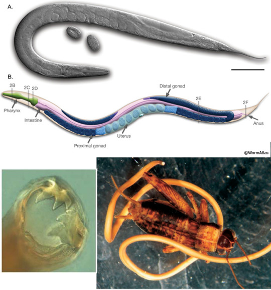
Top: a hermaphrodite adult of Caenorhabditis elegans (Nematoda), about 1 mm long; this species has males and hermaphrodites, but no specialized females (WormAtlas). Bottom left: the friendly smile of the hookworm Ancylostoma caninum (Nematoda), which uses those hooks to attach to the intestine wall of dogs (Brusca 2016). Bottom right: the horsehair worm Paragordius tricuspidatus (Nematomorpha) leaving the husk of its host, a cricket (Hickman & al 2008). As they need to hatch in water, Nematomorphs of that species alter the behavior of their host to induce it to drown itself in the closest pond.
3c2e3. Panarthropoda “all arthropods”: Or rather, one should say, Arthropods plus their closest relations, which usually have some kind of paired limbs. Loosely synonymous with Lobopodia “lobed feet”. Their coelom is very reduced, and replaced by a haemocoel filled with blood (more properly hemolymph) in which organs are directly soaking. A muscular heart ensures blood circulations, but other than it there are no blood vessels.
?3c2e3a. † DINOCARIDIDA “fearsome shrimps”: Wonderfully weird quasi-Arthropods from the seas of the Cambrian. They were the top predators of their environment, with well-developed eyes, relatively large brains, and massive raptorial appendages. Just take † Anomalocaris, with its pair of articulated mouth tentacles, looming with its half meter of length over a mostly cm-sized fauna, or † Opabinia, with its five (!) eyes and mouth at the end of a flexible proboscis. The later † Tamisiocaris even took to whale-style filter-feeding with comb-like appendages. They didn’t have real limbs, but lateral flaps that probably beat up and down, “rowing” in the water.
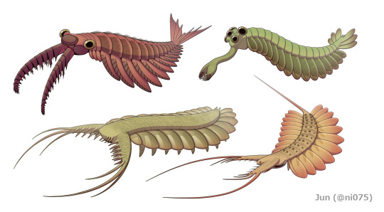
A beautiful selection of † Dinocaridida by Junnn11 (Wikipedia). Top left: † Anomalocaris canadensis, the largest predator of Burgess Shale. Top right: † Opabinia regalis. Bottom left: † Pambdelurion whittingtoni. Bottom right: † Kerygmachaela kierkegaardi. All are from the Cambrian; top row from Burgess Shale (Canada, about 510 million years ago), bottom row from Sirius Passet (Greenland, about 520 million years ago).
3c2e3b. TARDIGRADA “slow walkers”: You know these: “water bears”, those adorable microscopic eight-pawed creatures famous for being incredibly resistant to environmental stresses and dangers by dehydrating and stopping their own metabolism. (Boiling water! Liquid nitrogen! Pure alcohol! Hard vacuum! In reality, no species is resistant to all.) They have, guess what, a muscular pharynx with retractile stylets to pierce and suck fluids from animal or plant cells. They are so small that their muscles only have a handful of cells each, and they can expel eggs or waste only when molting their cuticle.
3c2e3c. ONYCHOPHORA “claw-carriers”: While all the other phyla are partly, mostly, or even exclusively aquatic (and often just marine), Onychophorans are the only animal phylum that is exclusively terrestrial. To be sure, its first representatives in the Cambrian, like Burgess Shale’s Aysheaia, lived in shallow seas; but their descendants are found only in the litter of tropical forests. Known as “velvet worms” because of their soft chitinous cuticle, they have cylindrical bodies walking on up-to-40 pairs of stubby clawed legs. The mouth is flanked by antennae and by massive slime glands that can shoot strings of gluey mucus to entangle preys and predators. Like insects, they breathe through lateral spiracles that allow air directly into the body. They are usually live-bearing, and may even have a placenta.
Left: the velvet worm Peripatoides novaezelandiae (Onychophora) (Uwe Schneehagen, Wikipedia). Elsewhere: a variety of Tardigrades: Hypsibius dujardini (top), Milnesium tardigradum (bottom left), and Echiniscoides sigismundi (bottom right) (Animal Diversity Web).
3c2e3d. ARTHROPODA “articulated feet”. Nothing much. Just like, you know, >90% of the whole Animal kingdom by species count. Spiders, scorpions, mites, crustaceans, millipedes, and all sorts of insects are here -- see parts 8 and 9 on that!
3c3. Deuterostomia “mouth second”: The other major part of Bilateria, distinguished from Protostomia by features of embryonal development, chiefly the fact that the blastopore develops into the anus and not the mouth. The two main phyla of Deuterostomes are Echinodermata (starfish, sea urchins, and such) and Chordata (vertebrates and our kin). See part 10 to continue our journey in that direction.
...
Special secret bonus phylum! MONOBLASTOZOA “single-layer animal”. Only one species known, Salinella salve, which was observed exactly once in 1892 in a salt pan in Argentina and then was never seen again, so that today most zoologists doubt it ever even existed. They supposedly have only one kind of cells, forming a single ciliated layer than encloses a gut with a mouth and an anus.
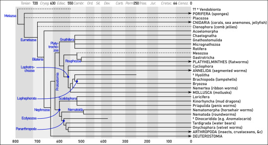
Summary. Dates (mostly from Edgecombe & al 2011 and Parfrey & al 2011) are in millions of years ago. Note how most of the radiation occurs between the Ediacaran (”Ediac.”) and Cambrian (“Cambr.”) periods -- roughly between 600 and 500 million years ago.
Sources
Adl & al (2019), “Revisions to the Classification, Nomenclature, and Diversity of Eukaryotes” (link)
Brusca (2016), Invertebrates (3rd edition), Sinauer
Dunn &al (2018), “Ediacaran developmental biology” (link)
Edgecombe & al (2011), “Higher-level metazoan relationships: recent progress
and remaining questions” (link)
Fernández & Gabaldón (2020), “Gene gain and loss across the metazoan tree of life” (link)
Giribet (2016), “Genomics and the animal tree of life: conflicts and future prospects” (link)
Hickman & al (2008), Integrated Principles of Zoology (14th edition), McGraw-Hill
Ivantsov & Zagrevskaya (2021), “Symmetry of Vendobionta (Late Precambrian Metazoa” (link)
Laumer & al 2019, “Revisiting metazoan phylogeny with genomic sampling of all phyla” (link)
Nielsen (2012), Animal Evolution: Interrelationships of the Living Phyla
Parfrey & al (2011), “Estimating the timing of early eukaryotic diversification with multigene molecular clocks“ (link)
4 notes
·
View notes
Note
12
Okay so. The captions are in spanish for obvious reasons and I hadn't thought on that until I tried to explain it?? Anyways.
This is a trilaminar embryo. Yes I know it looks like this emoji 🤯. Which I'm gonna use at my favor multiple times in the text below. But its only 15-17 days old so leave the embryo alone

The 🤯 it's surrender by three cell layers that are called (from the outside to the inside) syncytiotrophoblast (the amorphous mass, and this multinucleated) cytotrophoblast and chorionic mesoderm, and on the whole they form the chorion. They are in charge of the implantation process.
Then we have the 🤯. *sighs* it's called trilaminar embryo because it technically has three layers too: ectoderm, mesoderm and endoderm.
It's... Complicated to explain it here (on tumblr, via a pencil draw and not my first language) but I'm gonna do my best.
So.
Originally the embryoblast had two layers: epiblast and hypoblast, and was called embryoblast. From the hypoblast came the yolk sac and two extraembryonic mesoderms: the somatic mesoderm (that originally were where the corial mesoderm is now) and the splanchnic mesoderm (that covered the 🤯), but they migrated at some point to cover the yolk sac (that's the inferior part of the 🤯) and the amniotic sac (the superior part of the embryo).
Now, from the epiblast came The Three Layers we are talking about now. It's cells start to migrate in the direction of the hypoblast (displacing also cells of this) and these will transform into the Endoblast. Then more cells migrate and these are the Intraembryonic Mesoderm (there's more about this ñwnzlak but I'm gonna explain it later) and the cells that do not migrated turn into the Ectoderm.
Now. The Intraembryonic Mesoderm.
It's the embryo's middle layer, and it's divided in Paraxial mesoderm (or somitic mesoderm) and intermediate mesoderm (then the extraembryonic mesoderms migrate too and transform in lateral Intraembryonic mesoderm but that's irrelevant rn).
From the paraxial mesoderm are born the somites, that have an important roll in the creation of muscle, bone and skin.
And theres the notochord. By this point, the embryo already went thru a process called neurulation, aka the embryo's nervous system creation is already in motion. The notochord comes from ectoderm cells.
And that's all I can think about without having a stroke?? This of explaining science in english without knowing scientific english it's way harder than I thought ngl
5 notes
·
View notes
Last Seen Blogs

foxknow54
Untitled

bytheriversedgefic
Meet me by the river's edge

blibliblue
Kawa-ii

awesomearime
Anime, Art, Ships and Stuff
blibliblue
Kawa-ii