#mESC cells
Text

embryonic development rate varies among species & is correlated w lifespan, body size, life history etc
human embryonic development is 2-3 times slower than mouse
this this paper: in vitro model of developmental rate in hESC and mESC cells
hESC cells differentiate slower (and divide slower)
mass-specific CCM metabolic ratesin hESC cells are slower than mESC cells
this is because human cells are larger and contain more stuff lol
inhibit ETC slows development rate: not related to ATP production but related to NAD+/NADH ratios
increased NAD+ leads to faster embryonic development by driving faster protein translation rates
Nature Jan 2023, PMID: 36599986
#developmental metabolism#embryonic development#species-specific developmental rates#hESC cells#mESC cells#ETC#NAD to NADH ratio#protein translation
0 notes
Text
[DELAYED Prolonged BREAST Enhancement INFECTION WITH MYCOBACTERIUM FORTUITUM].
A free selenium atom certain with cysteine regarding catalytic dyad may be uncovered inside crystallographic constructions involving Mpro along with ebselen along with MR6-31-2 suggesting hydrolysis with the molecule sure organoselenium covalent adduct and also formation of an phenolic by-product, validated simply by muscle size spectrometry. The mark wedding together with selenation mechanism associated with self-consciousness recommends wider beneficial applying these materials against SARS-CoV-2 along with other zoonotic beta-corona trojans.Scouting around for nonresident life's hard since we don't know very well what signatures are special one's. All of us display precisely why intricate molecules seen in substantial large quantity are general biosignatures along with demonstrate the initial intrinsic experimentally tractable way of molecular intricacy, referred to as molecular construction directory (MA). To get this done many of us compute the complexity of varied million compounds and also verify that the intricacy can be experimentally determined by mass spectrometry. This approach allows us identify molecular biosignatures from the pair of varied biological materials from around the globe, outer space, as well as the laboratory, showing it is possible to create a life diagnosis try things out based on Mother that is stationed to be able to extraterrestrial areas, and also utilized as a complexity level to quantify limitations needed to immediate prebiotically plausible procedures inside the lab. This tactic is important for finding lifestyle somewhere else in the whole world or making de-novo living from the laboratory.The impact as well as splash of liquid lowers about reliable substrates are all-pervasive in lots of critical fields. Even so, past studies have mainly centered on spherical drops as the non-spherical situations, including raindrops, charged falls, oscillating drops, and declines impacted by electromagnetic field, continue to be mostly untouched. Utilizing ferrofluid, can certainly various decline styles along with illustrate the basic role regarding form inside affect and also sprinkle. Findings reveal that various fall styles produce significant different versions within distributing characteristics, splash oncoming tno155 inhibitor , and also splash amount. Nonetheless, main every one of these variants we discover widespread mechanisms over different drop designs the outcome dynamics will be governed by the actual superellipse model, the actual splash over beginning is brought on from the Kelvin-Helmholtz lack of stability, along with the amount of sprinkle is dependent upon the power dissipation before liquid heading out. Our study generalizes the actual drop influence research beyond the circular geometry, and divulges the opportunity of using fall contour around control impact along with sprinkle.Decoding the particular mechanisms that will management the pluripotent terrain express is vital with regard to comprehension embryonic development. Nevertheless, the particular epigenetic regulating ground-state mouse embryonic originate cells (mESCs) isn't entirely comprehended. The following, all of us identify the epigenetic proteins MPP8 as being essential for ground-state pluripotency. Its lacking contributes to cellular never-ending cycle criminal arrest and also impulsive distinction.
#inhibitor#small molecules#chemicals#kinase inhibitors#tyrosine kinase inhibitors#enzyme inhibitors#protein inhibitors#proteins kinase inhibitors#phosphatase inhibitors#compound library#activators#modulators#agonists#antagonists
1 note
·
View note
Text
Identification of anisotropy in chromosome dynamics by principal component analysis using integrated spatial genomics
Eukaryotic interphase chromosomes maintain a three-dimensional structure within the nucleus and undergo fluctuations. It has been reported that such dynamics are involved in transcription, replication, and DNA repair. However, the analysis of chromosomal dynamics has been limited to high-throughput chromosome conformation capture data, which records the contact frequencies between chromosomal regions and lack direct information about the dynamic. Herein, we investigated chromosome fluctuations as polymers based on experimental data from sequential fluorescence in situ hybridization (seqFISH)+ using a multiomics methodology. To describe the principal modes of chromosome fluctuations, we applied principal component analysis to the three-dimensional structure information of single chromosomes in 446 mouse embryonic stem cells (mESCs) obtained from seqFISH+ data analysis for spatial genomics and nuclear factors (histone marks, repeat DNAs, and nuclear compartments). We found that chromosome fluctuations exhibit both isotropic and anisotropic modes. The properties of anisotropy in chromosome fluctuation vary among chromosomes and appear to depend on the interaction between repeat DNAs on the chromosomes and nuclear factors. Furthermore, our principal component analysis revealed anisotropic chromosome fluctuations before and after the mitotic phase, specifically when chromosomes adopt a spindle-like shape. This result suggests the potential involvement of anisotropic chromosomal fluctuations in the transition of nuclear organization during the cell cycle. Our results represent the first study to elucidate the dynamics of chromosomes as polymers based on real multiomics data. http://dlvr.it/T27PZv
0 notes
Text
Human Amnion-derived Mesenchymal Stem Cells Improved The Reproductive Operate Of Age-related Diminished Ovarian Reserve In Mice Through Ampk Foxo3a Signaling Pathway Full Text
Tan et al. observed that hAESCs resulted in important modifications of T-cells, macrophages, dendritic cells, and monocyte/macrophage infiltration along with the elevation of lipoxin-A4 [126]. Importantly, the regenerative potential of the Amnio-M just isn't limited to simple utilization as a protection bandage but extends to include its high content material of regenerative key components. Advanced technologies supported the transformation of the Amnio-M into 3D scaffold that can fit appropriately into a defect. They have additionally helped its reintegration with other natural and artificial biomaterials as proven in Table Table3.3. For instance, the gel type of the Amnio-M could be advantageous over powder or cryopreserved Amnio-M in wound therapeutic as it'll provide a hydrating wound barrier, overcome the wound contractility, and management the speed of release of therapeutic components [154]. Moreover, anti-microbial agents within the AF corresponding to beta-lysin, bactericidin, lysozyme, and transferrin might be concerned in mounting that effect [92].
In these ways, tradition conditions have been discovered to affect the sort of pluripotency displayed by ESCs 4. In this manner, optimization of growth situations is relevant to any utility by which enhanced, long-term, or secure pluripotency is necessary. The derivation of hESC traces has trailed developments within the research of mouse embryonic stem cells (mESCs), which were first established as cell traces in ,7. Although hESCs and mESCs are each derived from blastocyst-stage embryos, they have very completely different organic properties with respect to underlying mechanisms of self-renewal eight.
On the internal face of the amnion of ruminants, particularly close to and on the umbilicus, are numerous raised, rough, discrete, spherical foci referred to as amniotic plaques or pearls. They are wealthy in glycogen however of unknown perform and normally disappear after 6 months of gestation. Towards the tip of being pregnant, clean, discoid, rubber-like lots might float in the allantoic fluid. They originate from invaginations of the allantochorion round accumulations of compacted histiotroph between the chorion and endometrium. They are known as ‘hippomanes’ (in cows more hardly ever ‘boomanes’) and have no practical significance (Rűsse & Grunert 1993).
Live cells had been gated to exclude lifeless cells primarily based on propidium iodide (Sigma) or LIVE/DEAD fixable dead cell stains (Thermo Fisher Scientific). Monoclonal antibodies towards human CD3, CD4, CD8, CD25, FOXP3, IFN-γ, TNF-α, and PD-1 had been used. For intracellular staining, 2 μM monensin (Fujifilm) was added for four h at 37 °C and cells permeabilized utilizing the FOXP3/Transcription Factor Staining Buffer Set (Thermo Fisher Scientific) in accordance with the manufacturer’s instructions.
placenta banking benefits
The supernatant was discarded, and the pellet was resuspended and replenished till it reached P2 or was cryopreserved. Some animals presented important neurological improvement, such because the restoration of nociception and skill to stay on station. Despite the need additional research, until the current moment, cell therapy has been possible and has no dangerous effects on animals. After three weeks of tradition in osteogenic induction medium, feline AECs distinctly changed their morphology and were surrounded by calcium deposits positive to von Kossa staining. In controls, cells did not change in morphology and did not stain positively to von Kossa.
And the expression of FSHR positioned within the granulosa cell floor showed the identical development. These outcomes indicated that the hAMSC transplantation may improve the expression of protein concerned in reproductive hormone signaling in ovarian cells to improve the ovarian operate (Figure S3). The mice (36-week-old) on the day 7 of the final hAMSC transplantation have been injected intraperitoneally with anesthetic until their limb muscles turned weak. The obtained blood was left at room temperature for 30 min, then serum was collected following centrifugation at 3000 rpm for 15 min and stored at − eighty °C earlier than hormone analysis.
At present, many strategies for large-scale manufacturing of exosomes had been out there [43]. With recent advances in the study of exosome-based therapies with MSCs, it could be very important assess their localization, monitoring, and monitoring after transplantation in vivo [93]. One method is magnetic resonance imaging (MRI), which permits the localization of exosomes labeled with distinction agents (such as ultra-small superparamagnetic iron oxide nanoparticles) [94]. Compared the relative expression of cytokines in culture supnantant of hAMSCs and hUMSCs. In all of the animals that underwent the decompression procedure, a metal-compatible picture artefact was observed. This picture artefact presumably resulted from filings derived from the wear and tear of the surgical milling cutter in touch with the free stripper, used to prevent and defend constructions throughout bone put on.
Several international locations, corresponding to the united states or China, launched their specific cellular and gene therapy guidance and established regenerative drugs products’ rules [160,161]. Each European Union member state at present has its personal particular rules targeted on embryonic stem cells and perinatal tissues research. Perinatal stem cell clinical trials should be permitted by the local bioethical commissions and be performed in line with the EU clinical trials registration regulation [162,163]. Cells of fetal origin, also identified as perinatal stem cells, are derived from extraembryonic buildings, such because the placenta, umbilical cord, and amniotic fluid [164,165]. Perinatal tissues are additionally extensively used as a reservoir of hematopoietic progenitor cells obtained from the umbilical cord blood [166]. Furthermore, it's believed that the perinatal stem cells obtained at the very early phases of pregnancy (first trimester) possess the next regenerative efficiency in comparability with cells isolated from full-term or near-term feto-maternal tissues.
For chromosome evaluation, hESCs within the exponential progress stage had been treated for two h with colcemid (0.1 mg/mL). After colcemid therapy, digested single cells had been karyotyped using the G-band technique. In summary, this paper describes the successful institution of 4 hESC traces utilizing hAECs as a feeder layer.
In this study, we generated a high-resolution developmental roadmap of postimplantation NHP embryos and recognized the amnion as a key signaling structure important for mesoderm formation in primates. Embryos with lack of perform of ISL1 in most of the cells in the amnion fail to kind mesoderm as a outcome of a reduction in BMP4 signaling and aren't able to giving rise to viable offspring (Fig. 7). In these conditions, ISL1-null cells were able to self-organizing into embryonic-like sacs and broke symmetry similar to the wild type but did not develop further.
#placenta banking#placenta position#amnion#types of placenta#placenta tissue banking#placenta banking benefits#placenta cord blood banking#placenta stem cell banking#cord blood and placenta banking#placental cells#what are placental cells#amnion and placenta#amnion stem cells#placenta amnion#placenta and amnion
1 note
·
View note
Text
The impact of SWI/SNF and NuRD inactivation on gene expression is tightly coupled with levels of #RNA polymerase II occupancy at promoters [RESEARCH]
SWI/SNF and NuRD are protein complexes that antagonistically regulate DNA accessibility. However, repression of their activities often leads to unanticipated changes in target gene expression (paradoxical), highlighting our incomplete understanding of their activities. Here we show that SWI/SNF and NuRD are in a tug-of-war to regulate PRC2 occupancy at lowly expressed and bivalent genes in mouse embryonic stem cells (mESCs). In contrast, at promoters of average or highly expressed genes, SWI/SNF and NuRD antagonistically modulate #RNA polymerase II (Pol II) release kinetics, arguably owing to accompanying alterations in H3.3 and H2A.Z levels at promoter-flanking nucleosomes, leading to paradoxical changes in gene expression. Owing to this mechanism, the relative activities of the two remodelers potentiate gene promoters toward Pol II–dependent open or PRC2-dependent closed chromatin states. Our results highlight #RNA Pol II occupancy as the key parameter in determining the direction of gene expression changes in response to SWI/SNF and NuRD inactivation at gene promoters in mESCs. http://genome.cshlp.org/cgi/content/short/33/3/332?rss=1&utm_source=dlvr.it&utm_medium=tumblr
0 notes
Text
Organoids: from traditional 2D cell Culture to 3D culture models
In recent years, organoid culture technology is developing rapidly. Organoids has been awarded 'Method of the Year 2017' by Nature Methods, for their immense potential as tools to study human biology in health and disease.
Previously, the term ‘organoid’ has been used to encompass all 3D organotypic cultures derived from primary tissues, pluripotent stem cells (PSCs), including embryonic stem cells (ESCs) and induced pluripotent stem cells (iPSCs), established cell lines, as well as whole or segmented organs such as organ explants consisting of multiple tissue types. The ‘organoid’ was defined by Fatehullah et al. As an in vitro 3D cellular cluster derived exclusively from primary tissue, ESCs or iPSCs, capable of self-renewal and self-organization, and exhibiting similar organ function as the tissue of origin[1-3].
The development of organoids
Organoid technology is built upon the foundation of stem cell technologies, classical developmental biology and cell-mixing experiments. In the early 20th century, Wilson (1907) demonstrated that dissociated sponge cells can self-organize to regenerate a whole organism. Stem cell research began to thrive when murine ESCs (mESCs) were first isolated and established in 1981[4-6]. In 2009, Hans Clevers lab established a long-term primary culture to generate the intestinal organoid culture system. It was an outstanding technological leap for the stem cell field[5][7].
Lately, organoids have been successfully generated for an increasing variety of organs, including but not limited to gut, stomach, lung, kidney, liver, pancreas, mammary glands, prostate, thyroid, retina, inner ear, taste bud and brain[8].
Organoid model is a major technological breakthrough that acts as a valuable model for the study of tissue development, disease modeling, drug screening, personalized medicine and cell therapy[1].、

Figure 1. Diverse applications of organoid technology
[5]
.
Recently, organoids have been widely used in many areas, including developmental biology, disease modeling, precision medicine, regenerative medicine, toxicology, drug discovery studies, host-microbiome interactions, gene editing, multi-omics, and phylogenetic studies[5].
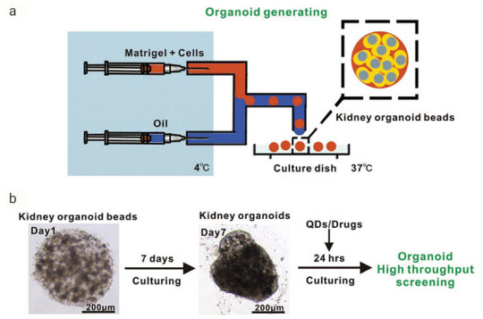
Figure 2. Generation of Reproducible Kidney Organoids
[9]
.Mouse kidney cells are suspended in Matrigel liquid precursor and fabricated organoid beads by microfluid machine and 3D printer. Organoid beads are cultured supplement with
Noggin
,
R-spondin 1
,
FGF-4
,
FGF-basic
,
SB-431542
,
CHIR-99021
. The size, shape, and composition of the kidney organoids are highly reproducible.Advantages of organoid model
Organoids represent an important bridge between 2D cultures and in vivo mouse/human models. They are more physiologically relevant than monolayer culture models and are far more amenable to manipulation of niche components, signaling pathways and genome editing than in vivo models [1][10]. Some of the advantages of the organoid models are;
1) Compared to traditional two-dimensional (2D) cell culture, organoids are similar to primary tissue in both their composition and architecture, harboring small populations of genomically stable, self-renewing stem cells that give rise to fully differentiated progeny comprising all major cell lineages at frequencies similar to those in living tissues[11].
2) Organoids can be expanded enormously, cryopreserved as biobanks, and easily manipulated using techniques similar to those established for 2D monolayer culture[11].
3) Primary-tissue-derived organoids lack mesenchyme/stroma that provides a separate system for studying a specific tissue of interest without being influenced by the local microenvironment[1].
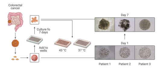
Figure 3. Comparison of Organoid Cultures with Two-Dimensional Cell Cultures and Studies in Animals
[10]
.
Compared with the traditional patient-derived cancer cell line (PDC) and patient-derived xenograft (PDX) model, the PDO model has unparalleled advantages. In the screening of drugs for tumor therapy, tumor organoid models derived from patient tumors have higher sensitivity, heterogeneity, and stability and can restore the genuine attributes of tumors more effectively. In addition, tumor organoids can be preserved, resuscitated, passed infinitely, and mechanically cultured on a chip for drug screening[13]. Therefore, organoid technology exerts enormous potential in evaluation of efficacy and toxicity of drugs, regenerative medicine, and precision medicine. Organoids have been established successfully for multiple cancer types, including but not limited to stomach cancer, colorectal cancer, liver cancer, pancreatic cancer[14].
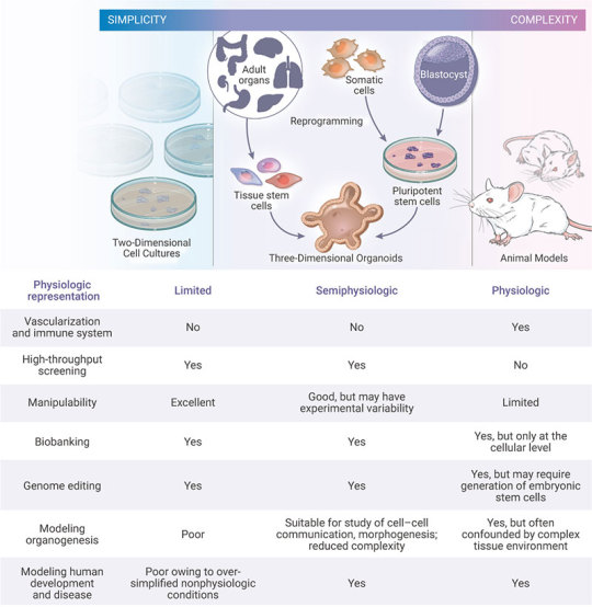
Figure 4. Establishment of patient-derived organoids as in vitro tumor models for colorectal cancer
[15]
.
Many pathogenic viruses that infect humans display species specificity and animal models can’t be used to study those viral infections. Hence, studying viral biology and identifying potential treatments benefits by developing in vitro cell systems (organoids) that closely mimic human physiology. In the current COVID-19 pandemic, organoids have emerged as powerful tools for SARS-CoV-2 research, bridging the gap between cell lines and in vivo animal models[16-17].
The "Magic" in organoid culture medium
Organoids can be generated from tissue-resident adult stem cells (ASCs) or from PSCs. Under appropriate conditions, supplementation of proper culture medium, growth factors and small molecules, stem cells embedded in Matrigel can undergo continuous self-renewal and differentiation, and self-organize into 3D-structures. The methods of culturing different organoids are similar[17].
1) Multiple sources:
1.1). ASCs-derived organoids: Primary tissue that is dissociated into functional sub-tissue units containing stem cells. These functional units are further digested into single cells and FACS-sorted to enrich for stem cells.
1.2). ESCs/iPSCs-derived organoids: stem cells undergo directed differentiation towards the desired germ lineage, eventually generating floating spheroids that are subsequently embedded in extracellular matrix (ECM) to initiate organoid culture[1].
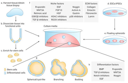
Figure 5. Organoid generation and culture from primary tissue and ESCs/iPSCs
[2]
.
2) Manipulability of niche components:
Organoids are typically cultured in an ECM surrounded by culture media supplemented with specific niche factors (different from air-liquid interface (ALI) method which is introduced recently)[18]. Organoids can either differentiate spontaneously or be induced to differentiate towards desired lineages or cell types by adding suitable differentiation factors and/or withdrawing factors that promote stemness. Common niche and ECM factors include R-spondin, EGF, Noggin, Activin A, and Collagen. Specific small molecules are added such as TGF-β inhibitor A-83-01, GSK3β inhibitor CHIR99021, and ROCK inhibitor Y27632[1].
Stem cells are maintained and perpetuated in organoids, continually giving rise to differentiated progeny. In addition, organoids can be dissociated and plated onto membrane supports coated with Matrigel or Collagen to form 2D monolayer organoid models[17].
MedChemExpress offers a variety of high-quality recombinant proteins and small molecules for organoid culture.
Related products
Cytokines
Human EGF
A well-known growth factor for epithelial tissues; binding to EGF receptors, induces hyperplasic changes.
EGF can be used for the generation of Gastrointestinal tract, liver, thyroid, brain organoids.
Human FGF-2/4/9/10
FGFs play crucial roles in a wide variety of cellular functions, including cell proliferation, survival, metabolism, morphogenesis, and differentiation,
as well as in tissue repair and regeneration. In a 3D extracellular matrix, FGF-2, FGF-7, FGF-9, and FGF-10 promote lung organoid formation.
Human HGF
HGF is a known hepatocyte mitogen that can be used for the liver organoid culture.
Human Wnt3a
Wnt is a master regulator in regulation of cell development, proliferation, differentiation, adhesion, and polarity.
Wnt3a is an essential niche component for maintaining the proliferation of Lgr5-positive stem cells in various organoids such as the small intestine,
large intestine, stomach, pancreas and liver.
Human BMP-4
BMPs play crucial roles in embryogenesis and development, and also in maintenance of adult tissue homeostasis.
BMP-2 and BMP-4 are widely used in in vitro protocols of generation of hepatic cells from induced pluripotent stem cells (iPS) and from embryonic stem cells (ESC).
Human Noggin
Noggin is an inhibitor of bone morphogenetic proteins that modulates cellular differentiation, proliferation, and apoptosis.
Noggin is one of the most important components of organoid media are growth factors.
Human DKK-1
DKK-1 is a canonical WNT inhibitor that can induce retinal progenitors to self-organize.
Small-molecule Inhibitor
Y-27632 dihydrochloride
Y-27632 is a Rho Kinase (ROCK) inhibitor; Has been used to increase the proliferation and reduce apoptosis of progenitor cells grown in vitro.
A 83-01
A 83-01 is an inhibitor of TGF-β type I receptor ALK5, the Activin/Nodal receptor ALK4, and the nodal receptor ALK7.
References:
[1] Aliya Fatehullah, Si Hui Tan, Nick Barker, et al. Organoids as an in vitro model of human development and disease. Nat Cell Biol. 2016 Mar;18(3):246-54.
[2] Marina Simian, Mina J Bissell. Organoids: A historical perspective of thinking in three dimensions. J Cell Biol. 2017 Jan 2;216(1):31-40.
[3] HansClevers. Modeling Development and Disease with Organoids. Cell. 2016 Jun 16;165(7):1586-1597.
[4] Madeline A Lancaster, Juergen A. Knoblich. Organogenesis in a dish: modeling development and disease using organoid technologies. Science. 2014 Jul 18;345(6194):1247125.
[5] Claudia Corrò, Vivian S.W. Li, et al. A brief history of organoids. Am J Physiol Cell Physiol. 2020 Jul 1;319(1):C151-C165.
[6] G R Martin. Isolation of a pluripotent cell line from early mouse embryos cultured in medium conditioned by teratocarcinoma stem cells. Proc Natl Acad Sci U S A. 1981 Dec;78(12):7634-8.
[7] Sato T, Vries RG, et al. (2009). “Single Lgr5 stem cells build crypt-villus structures in vitro without a mesenchymal niche.” Nature 459(7244): 262–265.
[8] Elisa Suarez Martinez , Amancio Carnero, et al. 3D and organoid culture in research: physiology, hereditary genetic diseases and cancer. Cell Biosci. 2022; 12: 39.
[9] Chengyong He, Shaohua Ma, Zhenghong Zuo, et al. Black Phosphorus Quantum Dots Cause Nephrotoxicity in Organoids, Mice, and Human Cells. Small. 2020 Jun;16(22):e2001371.
[10] Mo Li, Juan C Izpisua Belmonte. Organoids-Preclinical Models of Human Disease. N Engl J Med. 2019 Feb 7;380(6):569-579.
[11] Mariangela Scalise, Fabiola Marino, Daniele Torella, et al. From Spheroids to Organoids: The Next Generation of Model Systems of Human Cardiac Regeneration in a Dish. Int J Mol Sci. 2021 Dec; 22(24): 13180.
[12] Xialin Nie, Zhixing Liang, Linsen Ye, Yang Yang, et al. Novel organoid model in drug screening: Past, present, and future. Liver Research 5 (2021) 72-78.
[13] Chen Liu , Chaoyang Sun , et al. Drug screening model meets cancer organoid technology. Transl Oncol. 2020 Nov; 13(11): 100840.
[14] Hanxiao Xu, Kongming Wu, et al. Organoid technology and applications in cancer research. J Hematol Oncol 11, 116 (2018).
[15] Lisi Zeng, Shuzhong Cui, Shengwei Jiang, et al. Raltitrexed as a synergistic hyperthermia chemotherapy drug screened in patient-derived colorectal cancer organoids. Cancer Biol Med. 2021 Mar 12;18(3):750-762.
[16] Maarten H.Geurts, Jeltevan der Vaart, HansClevers, et al. The Organoid Platform: Promises and Challenges as Tools in the Fight against COVID-19. Volume 16, Issue 3, 9 March 2021, Pages 412-418.
[17] Jelte van der Vaart, Mart M. Lamers, Hans Clevers, et al. Advancing lung organoids for COVID-19 research. Dis Model Mech. 2021 Jun 1; 14(6): dmm049060.
[18] Soumya K Kar, et al. Organoids: a promising new in vitro platform in livestock and veterinary research. Vet Res. 2021 Mar 10;52(1):43.
[19] Yaqi Li, Guoqiang Hua, et al. Organoid based personalized medicine: from bench to bedside. Cell Regen. 2020 Dec; 9: 21.
0 notes
Text
High res Multimodal Photoacoustic Microscopy and Optical Coherence Tomography Visual image of Choroidal Vascular Stoppage.
Through the years, heart diseases always boost and also have an effect on not only human being wellness but the financial stability around the world. Your development inside tissues engineering is actually surrounding a great deal in working with this specific fast necessity of improving human being well being. Circulatory conditions are thought since selleck compound main aerobic health issues. Even though circulation hair transplant is easily the most convenient treatment, it's been delimited on account of lack associated with donors and also the individual's circumstances. However, tissue-engineered arteries tend to be promising choices while method of treatment for blood vessel defects. The purpose of this paper would be to present the value of the improvement on biofabrication technologies for treatment of soft cells problems specifically vascular cells. This may in addition provide a synopsis increase on the current reputation involving tissue recouvrement specially via autologous stem cells, scaffolds, as well as scaffold-free cell transplantable constructs. Your conversation with this paper will probably be dedicated to the famous view of cardiovascular tissue executive and originate mobile or portable chemistry and biology. The representative scientific studies highlighted with this cardstock are limited within the past ten years so that you can search for the trend along with advancement involving techniques for circulation system tissue design.Polycomb Party Protein (PcG) certainly are a group of epigenetic regulators responsible for the particular repression of your variety of genes crucial in advancement and cell destiny specs. PcG protein sophisticated in order to create 2 types of epigenetic authorities: Polycomb Repressive Intricate One and a pair of (PRC1 as well as PRC2). Even though the systems regulatory PRC2 recruitment along with action inside animals continue being inadequately realized, the latest operate has discovered any non-canonical PRC2 in mouse button embryonic stem cellular material (mESC) along with special routines required for repression associated with PRC2 focus on genes as well as necessary for mESC distinction and somatic mobile or portable re-training. Ideas review the characteristics associated with PRC2 in embryonic originate tissues along with investigate the role of the newly determined mESC specific PRC2 regulatory subunits Jarid2 (jumonji, From rich active area Two), Mtf2 (steel response aspect holding transcribing factor Only two) and also esPRC2p48.Ventriculitis or periventriculitis being a predominant routine associated with muscle engagement inside cerebral toxoplasmosis has been usually an uncommon function, actually with the elevation in the purchased immunodeficiency syndrome (Helps) era. Ventriculitis on premortem neuroimaging or perhaps from autopsy inside Helps people mainly resulted in differential determines regarding main central nervous system lymphoma (PCNSL) or even cytomegalovirus ventriculitis, not toxoplasmosis. Usually cerebral toxoplasmosis evolved since multifocal, necrotizing, hemorrhagic foci of cerebritis or even abscesses. All of us statement 2 non-AIDS sufferers along with cerebral toxoplasmosis that presented with prevalent ventriculitis/periventriculitis, with analysis in each case manufactured only at postmortem examination.
#Epigenetics Compound Library#Caspase inhibitor#Thiazovivin#Dapagliflozin#Birabresib#LDN-193189#SN-38#Selumetinib#Mitomycin C#Tat-beclin 1#Erlotinib#Batimastat#Pifithrin-α#Avapritinib#Zanubrutinib#Resatorvid#Necrostatin-1#dBET6#Givinostat#PD98059#RBPJ Inhibitor-1#APX-115#NAD+#Onametostat#Teduglutide#TAS4464#AZD3229#NSC 269420#MK-0991#RVX-208
0 notes
Text
Functional outcome soon after decrease arm or leg periprosthetic cracks
05). Conclusion: Despite the fact that glipizide demonstrated a little more speedy discounted through the system of suffering from diabetes volunteers when compared with via wholesome volunteers, this kind of variation, similar to individuals for various other pharmacokinetic parameters, had not been substantial (g > 2.05).With this review, the in-vitro anti-fungal task along with phytochemical evaluation of Schizophyllum commune concentrated amounts have been looked into. The anti-fungal activity ended up being tested against Eleven varieties of picked wooden degrading fungus regarding rubberwood. The final results demonstrated that h2o, methanol and ethanol ingredients drastically limited the development involving timber degrading fungus with minimal inhibitory concentration (MIC) ranges Zero.16-5.Double zero mu g/mu D. S. sanguineus was discovered since the strongest solid wood degrading fungus where that required the best power S. commune raw concentrated amounts (>Equates to A few.Double zero mu g/mu L) in order to inhibit it's mycelia growth. Phytochemicals evaluation said that the extracts contained flavonoid, phenol along with saponin. The actual methanol extracts of Utes. connect ended up being put on the particular rubberwood obstructs and located that the expansion of P. sanguineus ended up being limited efficiently with A few.Double zero mu g/mu L.Embryonic base tissue (ESCs) are generally pluripotent and capable of self-renewal. ESC aggregates, termed embryoid physiques (EBs), have been broadly adopted as a possible in vitro difference style. However, the particular size output of uniform dimension as well as shaped EBs may be difficult. Within will be described the development of any tradition plate that contains many concave microwells using minimal usage of resources, job, expertise, and price, permitting the production of a large number of homogeneous EBs simultaneously using the way of life dish. The large variety of concave well buildings can be self-constructed with the see more surface tension from the viscoelastic PDMS prepolymer. Murine ESCs (mESCs) are seeded on top of the concave water bores for muscle size creation of monodisperse EBs. It's noticed how the EBs developed more than a significant location are generally uniform in shape as well as size regardless of microwell place and variants cell seeding densities, and also whether or not their particular phenotype is managed. The capability to identify directly into adult cells (neuron and also endothelial tissues) from EBs is additionally looked at and also the neural surges from separated neuron tissues are generally measured to watch their particular operate. Uniform size and shape EBs tend to be properly generated throughout major in addition to their pluripotency can be taken care of comparable to some other approaches.Intrahepatic cholangiocarcinoma (ICC) could be the 2nd most popular main hard working liver most cancers along with poor receptiveness for you to current drug therapies. Therefore, story treatment methods versus ICC have to boost tactical. The objective of this research ended up being illustrate the role of fused-in-glioblastoma-c-ros-oncogenel (FIG-ROS) combination gene inside ICC. ROS was favorably expressed inside ICC cells and also HUCCT1 tissue. Plasmids indicating ROS- and also FIG-specific shRNAs ended up created along with transfected in to HUCCT1 cells.
#RSL3#Captisol#Emricasan#GSK126#Imidazole ketone erastin#NVP-TNKS656#MK-2206#GSK-2894631A#S63845#Belnacasan#diABZI STING agonist#Nirogacestat#Pevonedistat#LY294002#STM2457#GNE-140#Belumosudil#SCH772984#LGK-974#Naporafenib
0 notes
Photo
A while ago I read that #greying is due to #inSitu #bleaching. This new study however makes more sense.
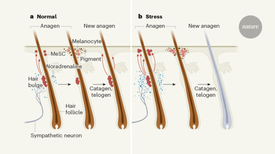
Signalling from the sympathetic nervous system of mice when subjected to stress leads to the depletion of a stem-cell population in their hair follicles. This discovery sheds light on why stress turns hair prematurely grey.
It has been said that Marie Antoinette’s hair went completely white on the night before her beheading. This story might be apocryphal, but rapid greying of the hair is now widely referred to as Marie Antoinette syndrome. It is often assumed to be caused by stress — a phenomenon perhaps best exemplified by photographs of heads of state before and after they held office. However, the relative contributions of ageing, genetic factors and stress to greying are not known — in part owing to a lack of mechanistic understanding of the process. Writing in Nature, Zhang et al.1 identify the mechanism governing premature greying in mice that have experienced stress.
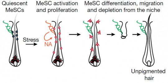
The average human scalp has 100,000 hair follicles, and a wide range of hair colours can be found across the human population. Hair colour is determined by cells called melanocytes, which produce different combinations of light-absorbing melanin pigments2. Melanocytes are derived from melanocyte stem cells (MeSCs), which are located in a part of the hair follicle called the bulge3. The normal hair cycle is divided into three stages: hair-follicle regeneration (anagen), degeneration (catagen) and rest (telogen). Melanocyte production begins early in the anagen phase (Fig. 1a). As people age, the pool of MeSCs is gradually depleted — and so pigmented hair becomes ‘salt and pepper’ coloured, and then turns to grey and finally to white after a complete loss of pigment in all hair follicles4.
Aside from ageing, there are several factors that bring about premature greying, including dietary deficiencies5, disorders such as alopecia areata or vitiligo6,7, and stress8,9. Zhang et al. set out to test the role of stress in the greying process in mice. They exposed the animals to three different stressors — pain, restraint and a model of psychological stress — during different phases of hair growth. Each stressor caused depletion of MeSCs from the bulge region, eventually leading to the development of patches of white hair.
Prevailing theories posit that stress-induced greying involves hormones (such as corticosterone) or autoimmune reactions10. Zhang and colleagues examined these potential mechanisms, first by preventing corticosterone signalling and next by stressing animals that had compromised immune systems. In both cases, greying occurred after stress, indicating that neither corticosterone nor autoimmune reactions cause MeSC depletion. However, the authors found that MeSCs express β2-adrenergic receptors, which respond to noradrenaline — a neurotransmitter molecule involved in the ‘fight or flight’ response to stress. Loss of this receptor specifically in MeSCs completely blocked stress-induced greying.
Adrenal glands are the main source of circulating noradrenaline. But, surprisingly, the researchers discovered that removing these glands did not prevent greying in response to stress in the mice.
Another source of noradrenaline is the sympathetic nervous system (SNS), which is highly active in response to stress, and which drives the fight-or-flight response. Zhang and colleagues showed that bulge regions are highly innervated by sympathetic neurons, and that ablating the SNS using a neurotoxin molecule, or blocking the release of noradrenaline from sympathetic neurons, prevented stress-induced greying. Next, the authors generated mice in which sympathetic neurons could be acutely activated, and found that overactivation of the SNS in these mice caused greying in the absence of stress. Together, these results indicate that noradrenaline released from active sympathetic neurons triggers MeSC depletion (Fig. 1b). Interestingly, Zhang et al. found that the propensity of an area to turn grey correlates with its level of sympathetic innervation.
Exactly how does sympathetic activity cause depletion of MeSCs from hair follicles? Normally, these stem cells are maintained in a dormant state until hair regrowth is required. However, when the researchers tracked MeSCs labelled with a fluorescent protein, they discovered that MeSC proliferation and differentiation increase markedly under extreme stress or exposure to a high level of noradrenaline. This results in mass migration of melanocytes away from the bulge, and leaves no remaining stem cells. To further confirm this result, the researchers suppressed MeSC proliferation pharmacologically and genetically. When proliferation was dampened, the effects of stress on MeSC proliferation, differentiation and migration were blocked.
Zhang and colleagues’ work raises several questions. For instance, is the mechanism underlying MeSC depletion in response to stress the same as that which causes greying during ageing? Future experiments modulating SNS activity over a longer period would determine whether age-related greying can be slowed or hastened. Perhaps, in the absence of sympathetic signals, MeSCs have the capacity for unlimited replenishment, pointing to a way to delay age-related greying.
Are other pools of stem cells similarly susceptible to stem-cell depletion in response to stress, if they or the cells that make up their niche express β2-adrenergic receptors? In support of this idea, haematopoietic stem and progenitor cells (HSPCs), which give rise to blood and immune lineages, reside in a bone-marrow niche that contains stromal cells, and stimulation of those cells by the SNS causes HSPCs to leave their niche11,12. Perhaps, like MeSCs, stress depletes HSPCs — which could partially explain why immune function is impaired in response to chronic stress13,14. Whether this type of relationship extends beyond MeSCs and HSPCs is an open question.
It is fascinating to consider what possible evolutionary advantage might be conferred by stress-induced greying. Because grey hair is most often linked to age, it could be associated with experience, leadership and trust15. For example, adult male silverback mountain gorillas (Gorilla beringei beringei), which get grey hair on their backs after reaching full maturity, can go on to lead a gorilla troop16. Perhaps an animal that has endured enough stress to ‘earn’ grey hair has a higher place in the social order than would ordinarily be conferred by that individual’s age.
Connecting the dots between stress, fight or flight, stem-cell depletion and premature greying opens up several avenues for future research. Beyond developing anti-greying therapies, Zhang and colleagues’ work promises to usher in a better understanding of how stress influences other stem-cell pools and their niches.
Nature 577, 623-624 (2020)
doi: 10.1038/d41586-019-03949-8
(via How the stress of fight or flight turns hair white)
also: harvard, hscrb, time, ts
#greying#aging#stress#nervous system#neurology#dermatology#science#medicine#fight or flight#noradrenaline#melanocyte#nature#marie antoinette#hair follicles#stem cell research#stem cell medicine
2 notes
·
View notes
Text
Chapitre II
IV
Natacha habitait tout près, dans un quatre et demi juché au sommet d’un escalier en colimaçon dont les rampes de fonte ornées de fioritures florales Art déco tenaient le coup en dépit de leur grinçante vétusté. Le balcon ne semblait pas plus solide et craquait sous nos pas. À l’intérieur, dans le vacillant clair-obscur en périphérie duquel s’étendait la nuit autour de la triple flamme d’un candélabre de bronze posé sur le bureau en face de l’une des fenêtres où il se répliquait tel un Cerbère, on nous enjoignit de nous asseoir au salon où de moelleux coussins de peluche s’éparpillaient sur le tapis tigré au pied de lion d’une ottomane léopard. L’amphitryonne nous servit un bordeaux qui n’avait de commun avec ce qu’elle avait bu à l’Assommoir que la couleur. Pierre s’exerçait à la patience en attendant que nous partissions. Il déchanta lorsque celle – dont les doigts aux ongles sans vernis se couvraient de sparadraps – sortit de son sac à main ocellé un sachet de plastique transparent à moitié plein de cocaïne dont elle saupoudra la table de verre où étaient posés les nôtres, scintillant de rubis liquides. Elle divisa la poudre en quatre lignes avec sa carte Visa, puis sortit de son portefeuille un billet de dix dollars qu’elle roula en cylindre et me tendit.
« Non merci.
– Sûr ? Je pensais que les écrivains expérimentaient toutes sortes de choses…
– Pour être écrivain, il faudrait que je sois vendu en librairie !
– Ça viendra. Et toi, Simon, t’en veux ?
– J’hésite… Ça fait longtemps…
– Pierre ?
– Oui ! »
Penchée sur la surface translucide où se refléta sa figure, elle se boucha de l’index une narine et de l’autre prit une longue inspiration à travers la paille de papier-monnaie mauve à l’effigie de Sir John A. Macdonald où fut aspiré, millimètre par millimètre, l’un des rails de « chimique ». Natacha céda sa place à Bouchard en reniflant et se passant la langue sur le doigt pour bien s’imprégner les muqueuses de ce qu’elle venait d’inhaler. Celui que j’avais toujours connu simple fumeur de THC, suivant son exemple, se lança à tête baissée dans la nuit de cristal. Au contraire de Simon, qui le relaya ensuite à la table en miroir, Pierre n’était point mû par sa propre volonté de jouir d’une euphorie factice, mais par le désir de plaire à Natacha en lui projetant l’image d’un amant qui l’accompagnerait dans ses bas-fonds.
La première ligne l’avait réveillée en stimulant son activité cérébrale ; la seconde l’anesthésia dans la crispation d’un sourire que n’interrompit que la rapidité du débit de ses digressions. Tremblay l’écoutait avec une attention soutenue ; Bouchard, plus à l’aise dans sa peau momentanément oubliée, la regardait comme s’il se fut agi de la seule, unique et dernière femme dans tout l’univers. Sans pousser l’audace au point de nous confesser les détails de son intimité, elle accentua néanmoins la vulgarité de ses propos, s’enorgueillissant de certains vices auxquels elle prétendait exceller : boire jusqu’au black-out, se détruire les neurones aux amphétamines, à la « mesc », à l’ecstasy, au buvard… Tout en nous vantant ses excès, elle remplissait le cendrier de mégots de Gauloises qu’elle fumait avec Pierre.
Il était environ une heure du matin lorsqu’on cogna à la porte. L’orchestre symphonique d’un ballet de Prokofiev assourdissait le bruit des toc-toc qui se faisait plus insistant. Natacha atténua le volume et alla s’enquérir de qui la réclamait. La pénombre dans laquelle était plongée l’embrasure de l’entrée où se tenait le visiteur – frêle Asiatique ou Mexicain au visage ridé – m’empêcha de distinguer de quoi était composé le bouquet de fleurs qu’il lui offrit. Elle le toisa désobligeamment.
« Qu’est-ce que tu fais ici ? »
L’être chétif bredouilla quelque chose, se confondit en excuses, déposa son présent aux pieds de celle qui le dédaignait, puis déguerpit. Elle ne ferma pas la porte ; prit les fleurs ; respira leur fragrance ; y cueillit une rose ; s’ensanglanta la paume de la main gauche avec les épines qu’elle serra d’un poing frémissant de colère ; jeta violemment la gerbe sur le sol ; la foula aux pieds ; reprit le bouquet ; en dispersa les différentes parties dans les airs comme autant de confettis lancés du haut de son balcon ; injuria l’importun qui rembarquait dans sa fourgonnette stationnée en face, et sur la tête duquel neigèrent les feuilles et les pétales tandis que les piquantes tiges, encore emballées dans leur cornet de papier, comme un avion d’origami raté, s’écrasaient pitoyablement sur la ligne jaune de la rue, en bas.
« C’est ça, décâlice ! »
En rentrant, elle claqua la porte, fonça vers la cuisine, tire-bouchonna une deuxième bouteille, s’en versa un grand verre qu’elle vida sur-le-champ, puis un second, moins généreux, qu’elle emporta sans le boire, remonta le son de la musique, ne laissa pas à Pierre l’occasion de l’aider, nous ignora pour aller à la salle de bains, enroula autour de sa main écorchée un pansement tiré de son armoire à pharmacie, repassa brièvement par la cuisine, en ramena un demi-litre de cabernet-sauvignon et vint nous le distribuer à parts égales. Constatant l’inutilité de sa démarche pour se rendre serviable, Bouchard se rassit en faisant tomber de sa poche un préservatif dont l’enveloppe dorée scintilla juste assez longtemps sur le tapis de tigre pour que celui qui l’avait égaré me décochât un regard d’incertitude chargé de reproche en le ramassant – comme si ses intentions n’étaient pas déjà limpides et que j’eusse eu besoin du hasard qui me fit seul témoin de ce bref accident pour me prouver qu’il espérait bien plus qu’une relation platonique.
Natacha reprit le sachet de cocaïne qu’elle avait laissé sur la table et répéta le rituel de tout à l’heure, accompagné de la même invitation à l’endroit de Pierre et Simon – lancée d’une voix plus morne, car imbibée de plus d’alcool –, puis, s’essuyant le nez du superflu glaireux dont elle se lécha l’annulaire, hagarde, perdue dans ses pensées, tandis que Bouchard et Tremblay se succédaient à la table verglacée de neige pour y priser un peu de déliquescence étoilée, elle pigea dans son paquet de carton bleu où était écrit le slogan : « Liberté toujours », une cigarette, la tint entre ses doigts tremblants – déjà bandés jusqu’aux jointures avant l’incident avec la rose, tant elle se rongeait les ongles –, l’alluma et s’en noircit les alvéoles pulmonaires avec volupté durant les intervalles qui espaçaient chaque gorgée du rouge qu’elle, désormais plus calme, sirotait.
Bouchard tenta de se libérer du doute qui lui glaçait le cœur.
« C’était qui, ce Chinois-là ?
– Personne. »
Ce nébuleux éclaircissement ne souffrit aucune réplique.
Étant retournés à nos discussions, nous avions oublié cet intermède quand, une heure plus tard, on cogna de nouveau à la porte.
« Quoi encore ? » s’exaspéra Natacha en se levant.
Par un retournement contraire à ce que nous anticipions, en ouvrant, elle se montra fort affable et invita même le nouveau venu à entrer. Par une imprévisible coïncidence, j’étais face au massif propriétaire du Hummer qui m’avait aspergé ! Plus étrange que la réaction de Pierre, qui parut terriblement déconcerté, fut celle de l’homme au groin tubéreux de varices dont l’accent circonflexe des sourcils fit plisser le front de béluga rubicond.
« Pierre !? Qu’est-ce que tu fais ici ?
– Je pourrais te demander la même chose, mon oncle…
– Vous vous connaissez ? s’amusa Natacha, que ce vaudeville divertissait.
– Pierre est mon neveu, répondit le collectionneur de Jeep sexagénaire.
– C’est le père d’Anne-Sophie, souffla Bouchard à Tremblay, qui opina du chef.
– Tu sniffes encore cette cochonnerie-là ? » s’indigna le globicéphale en désignant d’une grimace la table où traînaient les décombres de la débauche qu’il condamnait.
« C’est pas ça qui va t’aider avec la DPJ… Si tu veux récupérer la garde… »
Blessée au plus profond d’elle-même par cette allusion au secret de son trésor perdu, son fils qu’on lui avait enlevé, Natacha s’indigna.
« Tu es soûl ! Tes petites leçons, tu peux te les fourrer où j’pense ! T’es mal placé pour me juger, criss d’ivrogne ! Tu conduis ton char en boisson, tu empestes le cognac…
– C’est mon eau de Cologne…
– Pff… Bin oui, c’est ça…
– Tu peux ignorer mes conseils, mais je peux te payer le meilleur avocat…
– Arrête ! » s’écria-t-elle dans un murmure étranglé de sanglots qu’elle réprima.
Un silence lourd de mystères irrésolus plana sur le gâchis de cette soirée. Natacha, tristement songeuse, implosait de désespoir en ravalant ses larmes dans un mutisme héroïque. Celui qui avait provoqué en elle autant d’émotions, se tournant vers son neveu, le tira de la stupeur où la révélation des démêlés juridiques de celle dont il ignorait qu’elle fût mère l’avait précipité.
« Hé ! Pierre, ça va ? »
Celui-ci acquiesça distraitement à cette phrase dont la banalité n’avait pour but que de dissiper l’embarras qui rivait nos regards à terre. D’apprendre, par son satyre d’oncle, que la femme dont il recueillait au compte-gouttes les confidences depuis des mois s’était tue au sujet d’un problème aussi crucial que celui qui venait d’être évoqué avait réduit à néant le château de cartes de son amour où ne régnait plus que le chaos. La pesanteur qui s’était abattue sur son âme oppressait également celle de Simon, perplexe à l’idée que cet énergumène fût son éventuel beau-père. En ce qui me concernait, bien que je n’appréciasse guère la viscosité de cette atmosphère où nous étions englués, je me réjouissais des potentialités qu’offrait à la frénésie de ma plume l’existence d’un lien unissant de façon aussi inespérée qu’inouïe mon acheteur de joaillerie à tous les personnages de ce huis clos. L’éléphantesque homme d’affaires se montra cordial, serra la main de Tremblay, puis la mienne.
« Eugène Girard… Mais vous pouvez m’appeler Junior… »
Faisant un clin d’œil à son neveu :
« Je pense qu’on a déjà été présentés. »
Le rictus que Bouchard s’efforça d’afficher pour être agréable n’empêcha pas la jalousie de lui assombrir l’œil.
« Inquiète-toi pas, j’dirai rien à tes parents… Tu peux me faire confiance : motus et bouche cousue ! »
Et le cétacé des mers d’hydrocarbures mima, du majeur et du pouce, le zippage de sa gueule close, et enfonça les doigts boudinés de sa main de pachyderme dans la poche de son pantalon remplie de petit change qu’il y fit sonner et trébucher en observant le garçon de sa sœur qui avait les mêmes yeux bleus que sa fille. Simon, que cette ressemblance frappait, se rappelant qu’Anne-Sophie se présentait sous le nom de Lavoie plutôt que d’assumer d’être une Girard, s’interrogeait sur ce qui l’avait poussée à renier son géniteur jusque dans son patronyme. Pierre, qui aurait pu répondre à cette question, s’en posait beaucoup d’autres. Quant à moi, rien ne m’intriguait plus que la comédie jouée par Natacha. Acculée à ses derniers retranchements par cet imbroglio, elle se ressaisit et trancha le nœud gordien d’une oiseuse explication en ajournant l’heure des comptes.
« Messieurs, je vous aime bien, mais il se fait tard et j’ai des choses à régler avec Eugène ; je vais devoir vous mettre à la porte. »
Ce disant, elle nous la montra d’un geste indiquant nos manteaux suspendus aux crochets du vestibule au-dessus de nos chaussures cordées sur le paillasson. Le désappointement de Bouchard quant à l’issue de ce premier rendez-vous avec Natacha était d’autant plus amer que tout ce qu’il y avait appris sur le sphinx dont il s’acharnait à vouloir déchiffrer l’énigme ne l’avait rendue que plus opaque.
1 note
·
View note
Text
Biomed Grid | Nicotinic Acetylcholine Receptor-Mediated Signaling Pathways in Pluripotent Stem Cells
Mini Review
Electronic cigarettes (E-cigarettes) are battery-operated devices that transport a nicotine- containing aerosol or vapor by heating the liquid. The liquid usually contains nicotine, propylene glycol or glycerol, Acetylcholine receptors (AChRs) are membrane receptors that bind to the neurotransmitter acetylcholine. They are classified into two distinct subtypes, nicotinic AChRs (nAChRs) and muscarinic AChRs (mAChRs). nAChRs belong to the Cys-loop family of pentameric ligand-gated ion channels. They consist of seventeen subunits, various 3B1 (CHRNA1-10, but CHRNA8 is avian specific) and 3B2(CHRNB1-4) subunits with 3B4 (CHRND), 3B3 (CHRNG) and 3B5 (CHRNE) subunits [1, 2]. These subunits can be divided into neuronal-type (CHRNA2-10 and CHRNB2-4) and muscle-type (CHRNA1, CHRNB1, CHRND, CHRNG, CHRNE). Although they form various heteromeric pentamers by combination of any subunits and other subunits, some 3B1 subunits (CHRNA7 CHRNA8 and CHRNA9) function ashomomeric pentamers. Upon binding to ligands, pentameric receptors undergo conformational changes to open a central pore, causing the influx of extracellular ions and various cellular responses.
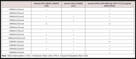
Table 1: Expression of CHRNA/Chrna and CHRNB/Chrnb genes in human and murine pluripotent stem cells.
Consistent with the findings for classification of receptor genes, nAChR-mediated signaling pathways play important roles in neuron and muscle [3]. In addition to these tissues, nAChR genes are also expressed in various non-neuronal tissues and cell types [4, 5]. Pluripotent stem cells (PSCs), so-called embryonic stem cells (ESCs) and induced pluripotent stem cells (iPSCs), represent one of such cell types. ESCs are derived from the inner cell mass of preimplantation embryo, whereas iPSCs are generated through somatic cell reprograming by the overexpression of defined transcription factors (Yamanaka factors) [6]. These are expected to be an ideal source for novel regenerative medicine [7, 8]. PSCs have unique characteristics that undergo unlimited self-renewal and retain pluripotency to differentiate into any cell types. Various signaling pathways are involved in maintaining the delicate balance between self-renewal and differentiation in PSCs [9]. They are also important for reprograming of somatic cells to establish iPSCs. Elucidation of these pathways is essential for the clinical applications of these cells. Both human and murine PSCs express various CHRNA/Chrna and CHRNB/Chrnb genes [10, 11] (Table 1). In addition, expression pattern of nAChR genes dynamically fluctuates during differentiation of PSCs into various cell lineages, including neuronal cells and myocytes. These finding indicate that nAChR-mediated signaling pathways play important roles in PSCs.
In fact, nAChR-mediated signaling pathways affect differentiation of PSCs. Triggering of nAChRs expressed in human ESCs-derived embryoid bodies by nicotine resulted in activation of MAPK and shifts of spontaneous differentiation toward hemangioblast [12]. In contrast, Gue et al. reported deleterious effects of nicotine on human ESCs-derived various lineages, including cardiomyoctes, by using single cell RNA-sequencing [13]. Consistent with the results in human ESCs, nAChR-signaling pathways inhibit differentiation of mESCs into cardiomyocytes by suppressing cardiac genes via DNA methylation [14]. Doubling time is reduced by nAChRmediated signaling via downregulation of N-myc expression during differentiation of primate ESCs into fibroblasts [15]. Taken together, it is conceivable that effects of nAChR-mediated signaling pathways on differentiation of PSCs are dependent on the cell lineages.
In addition to the differentiation processes, nAChRmediated singling pathways likely contribute to self-renewal and establishment of PSCs. Because murine and human ESCs express choline acetyltransferase and synthesize ACh [12, 16], nAChRmediated signaling pathways should be constitutively activated in PSCs. In support of this hypothesis, exogenous Ach and nicotine additions affect the proliferation and survival of PSCs via nAChRs. Nicotine increases DNA synthesis via some Chrna pathways in murine iPSCs [17, 18]. In murine ESCs, high doses of ACh and nicotine reduce apoptosis, but they inhibit proliferation [19]. In contrast, it is unknown whether nAChR-mediated signaling pathways are involved in the undifferentiated status, pluripotency and reprograming process. To achieve a comprehensive understanding of the roles of nAChR-mediated signaling pathways in PSCs, it is necessary in future studies to investigate the functions of each CHRNA/Chrna and CHRNB/Chrnb gene that are expressed in PSCs. It is also important to evaluate nAChR-mediated signaling pathways that are activated not only by exogenous ligands, but also by endogenous ACh. Such studies will provide essential insights to ensure the use of PSCs in future regenerative medicine.
Read More About this Article: https://biomedgrid.com/fulltext/volume6/nicotinic-acetylcholine-receptor-mediated-signaling-pathways-in-pluripotent-stem-cells.001061.php
For more about: Journals on Biomedical Science :Biomed Grid | Current Issue
#biomedgrid#american journal of biomedical science & research#journals on medical informatics#journal on medical science
0 notes
Photo
壓力太大一夜白頭?原來是交感神經太亢奮啦!
相傳,在法國大革命時,法國皇后瑪麗.安托瓦內特 (Marie Antoinette) 在被送上斷頭台行刑的前一晚,瞬間變得白髮蒼蒼。姑且不論這是否為民間軼事,我們在現實生活中,似乎也能發現「壓力」與「頭髮變白的速度」間,存在某種因果關係(比如說政治人物上任前後的髮色差異),如今,或許可以用科學的方法解釋這個現象了。
是什麼讓人「一夜白髮」?來點壓力試試看
哈佛大學幹細胞研究所 (HSCI) 的團隊嘗試了解「一夜白髮」背後的生理機制¹,而由於毛髮顏色的深淺與黑色素幹細胞 (melanocyte stem cells, MeSCs) 的多寡直接相關,團隊推斷,一夜白頭的原因應該與黑色素幹細胞脫離不了關係。
但首先,他們需確認:實驗鼠的毛色淡化跟牠們所受的壓力是否有關?為了確認兩者間的關聯,實驗人員將實驗鼠暴露於各種壓力源之下,像是:將籠子傾斜、開整晚的燈等等。最後發現,實驗鼠的毛色隨著生長週期明顯變淺了,證明實驗鼠也跟人類一樣,在承受一定強度及長度的壓力後,會有毛色淡化的現象。
而後,研究團隊在實驗鼠身上,施打了一種辣度超高的樹脂毒素 (Resiniferatoxin, RTX) 做為測試壓力源。數日後,他們藉由免疫螢光染色法 (immunofluorescent staining) 發現:實驗鼠毛囊中的黑色素幹細胞數量大幅減少,表示黑色素幹細胞減少會使毛色淡化。{註:免疫螢光染色法 (immunofluorescent staining) 可以藉由抗原和抗體間專一的結合反應,觀察蛋白質與胞器在細胞中的位置。}
猜猜幫兇是誰?謹慎的實驗推理之旅
另一方面,過去曾有研究推測:承受壓力後毛色淡化的原因,可能是受到免疫系統的攻擊2,3。於是,研究人員依循這個推測,將樹脂毒素注入缺乏 B 細胞及 T 細胞的實驗鼠中,卻發現實驗鼠的毛色一樣會淡化,顯示出淡化的機制與免疫系統無關。那麼,問題來了:壓力到底是透過什麼機制去影響毛色的呢?
為了找到答案,研究人員首先對實驗鼠進行了一連串的刺(ㄋㄩㄝˋ)激(ㄉㄞˋ)後,發現到:實驗鼠血液中皮質酮 (corticosterone) 及去甲基腎上腺素 (noradrenaline) 等激素的濃度明顯增加了。
那麼,這兩者跟白髮機制都有關係嗎?研究人員接著嘗試移除皮質酮受器,並注入樹脂毒素。結果毛色還是淡化了,即使研究人員提升皮質醇濃度,也並未改變實驗鼠的毛色。換句話說,這種由腎上腺皮質分泌的壓力賀爾蒙(皮質酮),並不是造成毛色淡化的主要原因。
接下來,研究人員改成移除黑色素幹細胞上的去甲基腎上腺素受器,再注入樹脂毒素,沒想到,實驗鼠的毛色淡化現象就消失了!此外,當研究人員在實驗鼠身上施打去甲基腎上腺素,施打處的毛色便開始明顯淡化,種種證據都指出:一夜白頭正是去甲基腎上腺素搞的鬼。
一夜白頭的原因找到!原來是交感神經太活躍
既然知道幫兇是去甲基腎上腺素,揪出真正幕後黑手便不是件難事,研究團隊把問題轉向腎上腺與交感神經,因為他們會在遇到壓力時,分泌去甲基腎上腺素。
嫌疑犯 1 號:腎上腺
研究人員將實驗鼠的腎上腺移除、注入樹脂毒素、觀察實驗鼠的毛色變化,結果發現實驗鼠仍有毛色淡化的現象。叭叭!真正的兇手不是它。
嫌疑犯 2 號:交感神經
當實驗鼠暴露在壓力源(樹脂毒素)中時,交感神經會被強烈活化,並分泌去甲基腎上腺素,這種機制會觸發「戰或逃反應」(fight-or-flight responses)4。而在一連串的實驗中,研究人員更發現此機制會促使黑色素幹細胞不正常地迅速增生,接著分化、遷移,最後永久地流失,導致毛色淡化。黑色素幹細胞會因為去甲基腎上腺素而不正常增生、分化、遷移而後流失。
什麼?你覺得這篇文章好像有看沒懂,讓人感覺壓力很大嗎?快去看看你的交感神經有沒有被強烈活化,讓你多出幾根白頭髮?
參考資料
Zhang, B., Ma, S., Rachmin, I. et al. Hyperactivation of sympathetic nerves drives depletion of melanocyte stem cells. Nature 577, 676–681 (2020)
Navarini, A. A., Nobbe, S. & Trueb, R. M. Marie Antoinette syndrome. Arch. Dermatol. 145, 656 (2009)
Harris, M. L. et al. A direct link between MITF, innate immunity, and hair graying. PLoS Biol. 16, e2003648 (2018)
Ulrich-Lai, Y. M. & Herman, J. P. Neural regulation of endocrine and autonomic stress responses. Nat. Rev. Neurosci. 10, 397–409 (2009)
文/nerdy 交大理科碩士畢業,半吊子的科學狂熱者,投稿是種消遣。
2020/03/20 泛科學
https://pansci.asia/archives/182003
0 notes
Text
Histone Demethylation Plays a Role in Turning Stem Cells into Blood Vessel Cells
How stem cells become specific cell types is an intricate process, yet understanding the mechanisms that trigger this process may well prove to be beneficial for use in repairing and regenerating tissue. In a recent study, scientists at the University of Illinois at Chicago investigated the molecular mechanism, histone demethylation, and how it plays an essential role in mediating the conversion of stem cells into mature endothelial cells, which then produce blood vessels. Their findings, recently published in Stem Cell Reports, focused on two different enzymes that modify histones to change gene expression related to the differentiation of embryonic stem cells to endothelial cells.
Histone modifications are an epigenetic mechanism that can alter the way genes express themselves without impacting the underlying genetic code. They occur to proteins, or histones, which are surrounded by DNA. This protein-DNA complex is referred to as chromatin, which histone modifications can open up for greater access, forming euchromatin and increasing gene transcription, or tighten to decrease access, forming heterochromatin and reducing the activity of genes. Histones may be modified by the removal or addition of methyl groups (CH3) by enzymes, similar to another popular epigenetic mechanism known as DNA methylation.
Associate Professor of Medicine and Pharmacology from UIC, Jalees Rehman, explained that more genes can be activated via histone modifications, as opposed to regulating just a single gene. Rehman and the research team focused on stem cell differentiation and aimed to pinpoint epigenetic regulators of the process. According to Rehman, stem cell differentiation is “a highly complex process, involving the transition of a cell that can form any type of tissue early on in development, into one that is locked in to producing only one cell type.”
The group of researchers led by Professor and Head of Pharmacology at the UIC College of Medicine, Asrar Malik, first used mice to investigate enzymes called histone demethylases, which act to modify histones and change gene expression by removing a methyl group. This epigenetic mechanism is referred to as histone demethylation. Specifically, the research team wanted to assess how a few enzymes could change the expression of embryonic stem cell genes during differentiation as the cells transformed into mature endothelial cells. Their study focused on KDM4A and KDM4C, two histone demethylases, which they found in high amounts during the differentiation process.
They discovered that these enzymes epigenetically regulated promoters that were specific to endothelial cells. They reported that “histone demethylases KDM4A and KDM4C independently induced demethylation at histone H3K9 to activate Flk1 and VE-cadherin expression and thus enabled the differentiation of mESCs [mouse embryonic stem cells] to endothelial cells.”
SEE ALSO: Marijuana Use May Epigenetically Impact Sperm Health
The research team then used zebrafish, an organism commonly investigated in epigenetic studies, and depleted the histone demethylases in their embryos. Without KDM4A or KDM4C, they found that the fish embryos could not create blood vessels. Removing only KDM4A had a much larger impact on the development of blood vessels than did depletion of KDM4C, hinting that it may play a role in the early stages of blood vessel creation. Overall, their results indicate that these two histone demethylases have a crucial epigenetic influence on the differentiation process of embryonic stem cells to epithelial cells.
Rehman suggests, however, that further research is needed to delve into the complexities of the blood-vessel development pathway to fully understand how it might be controlled.
“We only looked at a few of the genes activated by the epigenetic switches that guide stem cells into becoming endothelial cells,” Rehman said. “Identifying additional genes activated by these switches, as well as gene pathways that are turned off during these transitions, will help shed more light on how stem cells carefully orchestrate a complex array of molecular signals which ultimately decide their fates.”
Bailey Kirkpatrick
Source: Wu, L., Wary, K.K., Revskoy, S., Gao, X., Tsang, K., Komarova, Y.A., Rehman, J., Malik, A.B. (2015). Histone demethylases KDM4A and KDM4C regulate differentiation of embryonic stem cells to endothelial cells. Stem Cell Reports. Advance online publication.
Reference: Parmet, S. Researchers uncover epigenetic switches that turn stem cells into blood vessel cells. University of Illinois at Chicago News Center. 25 June 2015.
Related Posts
Cancer Genes May Have Been Epigenetically Silenced Over Time
The Epigenetic Benefits of Your Thanksgiving Feast
Could Epigenetics Explain the Origins of Allergic Disease?
Early Epigenetic Nutrition ‘Memory’ Could Program You for Obesity Later in Life
from WordPress https://ift.tt/3cfmiIn
via IFTTT
0 notes
Text
Sequestration of microRNA-mediated target repression by the Ago2-associated #RNA binding protein FAM120A [Report]
Argonaute (Ago) proteins interact with various binding partners and play a pivotal role in microRNA (miRNA)-mediated silencing pathways. By utilizing immunoprecipitation followed by mass spectrometry to determine cytoplasmic Ago2 protein complexes in mouse embryonic stem cells (mESCs), we identified a putative RNA-binding protein FAM120A (also known as OSSA/C9ORF10) as an Ago2 interacting protein. Individual nucleotide resolution Cross-Linking and ImmunoPrecipitation (iCLIP) analysis revealed that FAM120A binds to homopolymeric tracts in 3' UTRs of about 2,000 mRNAs, particularly poly(G) sequences. Comparison of FAM120A iCLIP and Ago2 iCLIP reveals that greater than one-third of mRNAs bound by Ago2 in mESCs are co-bound by FAM120A. Furthermore, such FAM120A-bound Ago2 target genes are not subject to Ago2-mediated target degradation. Reporter assays suggest that the 3' UTRs of several FAM120A-bound miRNA target genes are less sensitive to Ago2-mediated target repression than those of FAM120A-unbound miRNA targets and FAM120A modulates them via its G-rich target sites. These findings suggest that Ago2 may exist in multiple protein complexes with varying degrees of functionality. http://bit.ly/2XIzKRH
0 notes
Text
Organoids: from traditional 2D cell Culture to 3D culture models
In recent years, organoid culture technology is developing rapidly. Organoids has been awarded 'Method of the Year 2017' by Nature Methods, for their immense potential as tools to study human biology in health and disease.
Previously, the term ‘organoid’ has been used to encompass all 3D organotypic cultures derived from primary tissues, pluripotent stem cells (PSCs), including embryonic stem cells (ESCs) and induced pluripotent stem cells (iPSCs), established cell lines, as well as whole or segmented organs such as organ explants consisting of multiple tissue types. The ‘organoid’ was defined by Fatehullah et al. As an in vitro 3D cellular cluster derived exclusively from primary tissue, ESCs or iPSCs, capable of self-renewal and self-organization, and exhibiting similar organ function as the tissue of origin[1-3].
The development of organoids
Organoid technology is built upon the foundation of stem cell technologies, classical developmental biology and cell-mixing experiments. In the early 20th century, Wilson (1907) demonstrated that dissociated sponge cells can self-organize to regenerate a whole organism. Stem cell research began to thrive when murine ESCs (mESCs) were first isolated and established in 1981[4-6]. In 2009, Hans Clevers lab established a long-term primary culture to generate the intestinal organoid culture system. It was an outstanding technological leap for the stem cell field[5][7].
Lately, organoids have been successfully generated for an increasing variety of organs, including but not limited to gut, stomach, lung, kidney, liver, pancreas, mammary glands, prostate, thyroid, retina, inner ear, taste bud and brain[8].
Organoid model is a major technological breakthrough that acts as a valuable model for the study of tissue development, disease modeling, drug screening, personalized medicine and cell therapy[1].
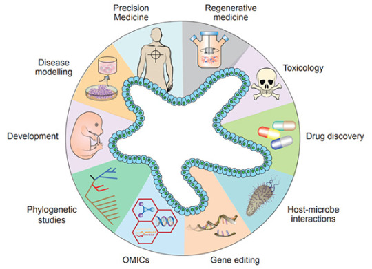
Figure 1. Diverse applications of organoid technology
[5]
.
Recently, organoids have been widely used in many areas, including developmental biology, disease modeling, precision medicine, regenerative medicine, toxicology, drug discovery studies, host-microbiome interactions, gene editing, multi-omics, and phylogenetic studies[5].

Figure 2. Generation of Reproducible Kidney Organoids
[9]
.Mouse kidney cells are suspended in Matrigel liquid precursor and fabricated organoid beads by microfluid machine and 3D printer. Organoid beads are cultured supplement with
Noggin
,
R-spondin 1
,
FGF-4
,
FGF-basic
,
SB-431542
,
CHIR-99021
. The size, shape, and composition of the kidney organoids are highly reproducible.Advantages of organoid model
Organoids represent an important bridge between 2D cultures and in vivo mouse/human models. They are more physiologically relevant than monolayer culture models and are far more amenable to manipulation of niche components, signaling pathways and genome editing than in vivo models [1][10]. Some of the advantages of the organoid models are;
1) Compared to traditional two-dimensional (2D) cell culture, organoids are similar to primary tissue in both their composition and architecture, harboring small populations of genomically stable, self-renewing stem cells that give rise to fully differentiated progeny comprising all major cell lineages at frequencies similar to those in living tissues[11].
2) Organoids can be expanded enormously, cryopreserved as biobanks, and easily manipulated using techniques similar to those established for 2D monolayer culture[11].
3) Primary-tissue-derived organoids lack mesenchyme/stroma that provides a separate system for studying a specific tissue of interest without being influenced by the local microenvironment[1].

Figure 3. Comparison of Organoid Cultures with Two-Dimensional Cell Cultures and Studies in Animals
[10]
.
Compared with the traditional patient-derived cancer cell line (PDC) and patient-derived xenograft (PDX) model, the PDO model has unparalleled advantages. In the screening of drugs for tumor therapy, tumor organoid models derived from patient tumors have higher sensitivity, heterogeneity, and stability and can restore the genuine attributes of tumors more effectively. In addition, tumor organoids can be preserved, resuscitated, passed infinitely, and mechanically cultured on a chip for drug screening[13]. Therefore, organoid technology exerts enormous potential in evaluation of efficacy and toxicity of drugs, regenerative medicine, and precision medicine. Organoids have been established successfully for multiple cancer types, including but not limited to stomach cancer, colorectal cancer, liver cancer, pancreatic cancer[14].

Figure 4. Establishment of patient-derived organoids as in vitro tumor models for colorectal cancer
[15]
.
Many pathogenic viruses that infect humans display species specificity and animal models can’t be used to study those viral infections. Hence, studying viral biology and identifying potential treatments benefits by developing in vitro cell systems (organoids) that closely mimic human physiology. In the current COVID-19 pandemic, organoids have emerged as powerful tools for SARS-CoV-2 research, bridging the gap between cell lines and in vivo animal models[16-17].
The "Magic" in organoid culture medium
Organoids can be generated from tissue-resident adult stem cells (ASCs) or from PSCs. Under appropriate conditions, supplementation of proper culture medium, growth factors and small molecules, stem cells embedded in Matrigel can undergo continuous self-renewal and differentiation, and self-organize into 3D-structures. The methods of culturing different organoids are similar[17].
1) Multiple sources:
1.1). ASCs-derived organoids: Primary tissue that is dissociated into functional sub-tissue units containing stem cells. These functional units are further digested into single cells and FACS-sorted to enrich for stem cells.
1.2). ESCs/iPSCs-derived organoids: stem cells undergo directed differentiation towards the desired germ lineage, eventually generating floating spheroids that are subsequently embedded in extracellular matrix (ECM) to initiate organoid culture[1].

Figure 5. Organoid generation and culture from primary tissue and ESCs/iPSCs
[2]
.
2) Manipulability of niche components:
Organoids are typically cultured in an ECM surrounded by culture media supplemented with specific niche factors (different from air-liquid interface (ALI) method which is introduced recently)[18]. Organoids can either differentiate spontaneously or be induced to differentiate towards desired lineages or cell types by adding suitable differentiation factors and/or withdrawing factors that promote stemness. Common niche and ECM factors include R-spondin, EGF, Noggin, Activin A, and Collagen. Specific small molecules are added such as TGF-β inhibitor A-83-01, GSK3β inhibitor CHIR99021, and ROCK inhibitor Y27632[1].
Stem cells are maintained and perpetuated in organoids, continually giving rise to differentiated progeny. In addition, organoids can be dissociated and plated onto membrane supports coated with Matrigel or Collagen to form 2D monolayer organoid models[17].
MedChemExpress offers a variety of high-quality recombinant proteins and small molecules for organoid culture.
Related products
Cytokines
Human EGF
A well-known growth factor for epithelial tissues; binding to EGF receptors, induces hyperplasic changes.
EGF can be used for the generation of Gastrointestinal tract, liver, thyroid, brain organoids.
Human FGF-2/4/9/10
FGFs play crucial roles in a wide variety of cellular functions, including cell proliferation, survival, metabolism, morphogenesis, and differentiation,
as well as in tissue repair and regeneration. In a 3D extracellular matrix, FGF-2, FGF-7, FGF-9, and FGF-10 promote lung organoid formation.
Human HGF
HGF is a known hepatocyte mitogen that can be used for the liver organoid culture.
Human Wnt3a
Wnt is a master regulator in regulation of cell development, proliferation, differentiation, adhesion, and polarity.
Wnt3a is an essential niche component for maintaining the proliferation of Lgr5-positive stem cells in various organoids such as the small intestine,
large intestine, stomach, pancreas and liver.
Human BMP-4
BMPs play crucial roles in embryogenesis and development, and also in maintenance of adult tissue homeostasis.
BMP-2 and BMP-4 are widely used in in vitro protocols of generation of hepatic cells from induced pluripotent stem cells (iPS) and from embryonic stem cells (ESC).
Human Noggin
Noggin is an inhibitor of bone morphogenetic proteins that modulates cellular differentiation, proliferation, and apoptosis.
Noggin is one of the most important components of organoid media are growth factors.
Human DKK-1
DKK-1 is a canonical WNT inhibitor that can induce retinal progenitors to self-organize.
Small-molecule Inhibitor
Y-27632 dihydrochloride
Y-27632 is a Rho Kinase (ROCK) inhibitor; Has been used to increase the proliferation and reduce apoptosis of progenitor cells grown in vitro.
A 83-01
A 83-01 is an inhibitor of TGF-β type I receptor ALK5, the Activin/Nodal receptor ALK4, and the nodal receptor ALK7.
References:
[1] Aliya Fatehullah, Si Hui Tan, Nick Barker, et al. Organoids as an in vitro model of human development and disease. Nat Cell Biol. 2016 Mar;18(3):246-54.
[2] Marina Simian, Mina J Bissell. Organoids: A historical perspective of thinking in three dimensions. J Cell Biol. 2017 Jan 2;216(1):31-40.
[3] HansClevers. Modeling Development and Disease with Organoids. Cell. 2016 Jun 16;165(7):1586-1597.
[4] Madeline A Lancaster, Juergen A. Knoblich. Organogenesis in a dish: modeling development and disease using organoid technologies. Science. 2014 Jul 18;345(6194):1247125.
[5] Claudia Corrò, Vivian S.W. Li, et al. A brief history of organoids. Am J Physiol Cell Physiol. 2020 Jul 1;319(1):C151-C165.
[6] G R Martin. Isolation of a pluripotent cell line from early mouse embryos cultured in medium conditioned by teratocarcinoma stem cells. Proc Natl Acad Sci U S A. 1981 Dec;78(12):7634-8.
[7] Sato T, Vries RG, et al. (2009). “Single Lgr5 stem cells build crypt-villus structures in vitro without a mesenchymal niche.” Nature 459(7244): 262–265.
[8] Elisa Suarez Martinez , Amancio Carnero, et al. 3D and organoid culture in research: physiology, hereditary genetic diseases and cancer. Cell Biosci. 2022; 12: 39.
[9] Chengyong He, Shaohua Ma, Zhenghong Zuo, et al. Black Phosphorus Quantum Dots Cause Nephrotoxicity in Organoids, Mice, and Human Cells. Small. 2020 Jun;16(22):e2001371.
[10] Mo Li, Juan C Izpisua Belmonte. Organoids-Preclinical Models of Human Disease. N Engl J Med. 2019 Feb 7;380(6):569-579.
[11] Mariangela Scalise, Fabiola Marino, Daniele Torella, et al. From Spheroids to Organoids: The Next Generation of Model Systems of Human Cardiac Regeneration in a Dish. Int J Mol Sci. 2021 Dec; 22(24): 13180.
[12] Xialin Nie, Zhixing Liang, Linsen Ye, Yang Yang, et al. Novel organoid model in drug screening: Past, present, and future. Liver Research 5 (2021) 72-78.
[13] Chen Liu , Chaoyang Sun , et al. Drug screening model meets cancer organoid technology. Transl Oncol. 2020 Nov; 13(11): 100840.
[14] Hanxiao Xu, Kongming Wu, et al. Organoid technology and applications in cancer research. J Hematol Oncol 11, 116 (2018).
[15] Lisi Zeng, Shuzhong Cui, Shengwei Jiang, et al. Raltitrexed as a synergistic hyperthermia chemotherapy drug screened in patient-derived colorectal cancer organoids. Cancer Biol Med. 2021 Mar 12;18(3):750-762.
[16] Maarten H.Geurts, Jeltevan der Vaart, HansClevers, et al. The Organoid Platform: Promises and Challenges as Tools in the Fight against COVID-19. Volume 16, Issue 3, 9 March 2021, Pages 412-418.
[17] Jelte van der Vaart, Mart M. Lamers, Hans Clevers, et al. Advancing lung organoids for COVID-19 research. Dis Model Mech. 2021 Jun 1; 14(6): dmm049060.
[18] Soumya K Kar, et al. Organoids: a promising new in vitro platform in livestock and veterinary research. Vet Res. 2021 Mar 10;52(1):43.
[19] Yaqi Li, Guoqiang Hua, et al. Organoid based personalized medicine: from bench to bedside. Cell Regen. 2020 Dec; 9: 21.
0 notes
Text
The stomatogastric nervous system of the medicinal leech: its anatomy, physiology and associated aminergic neurons [RESEARCH ARTICLE]
Karen A. Mesce, Magda Alania, Quentin Gaudry, and Joshua G. Puhl Blood feeding is an essential and signature activity of the medicinal leech species, Hirudo verbana. Despite keen interest in understanding the neuronal substrates of this behavior, a major component of the nervous system associated with feeding has remained overlooked. In this study, for the first time, we report on the presence and characteristics of five stomatogastic ganglia (STGs) comprising the visceral stomatogastric nervous system (STN) of the leech. Although Hanke (1948) provided a brief report that a ring of three ganglia (not five) was associated with the cephalic ganglia, this information was never integrated into subsequent neurobiological studies of feeding. Here, the anatomical features of the STGs are described as are the morphological and electrophysiological characteristics of neurons originating in them. We also determined that two of the five STGs (STG-1 and STG-3) each contained two relatively large (ca. 40 µm diameter) serotonergic neurons. The STN was also richly invested by dopaminergic and serotonergic arborizations, however, no intrinsic dopaminergic somata were observed. The trajectory of the serotonergic LL neuron, a command-like cell for feeding, was documented to project directly to the STN and not to the jaw and pharyngeal musculature as previously reported, thus reopening the important question of how the LL cell activates and coordinates biting activity with pharyngeal swallowing. Additional studies revealed that the LL cell is excited by blood serum applied to the lip and is strongly inhibited by dopamine. These findings provide a new foundation for understanding the regulation and modulation of feeding neural networks. The stomatogastric nervous system of the medicinal leech: its anatomy, physiology and associated aminergic neurons [RESEARCH ARTICLE] Source: http://jeb.biologists.org/cgi/content/short/jeb.175687v1?rss=1 © copyright 1994 Medellin Abogados Comerciales llamanos 320 542-9469 Colombia Medellin Abogados Comerciales llamanos 320 542-9469 Colombia Posted by Abogados Comerciales Medellin llamanos 320 542-9469 Colombia on Saturday, January 6, 2018 http://dlvr.it/QVzbNG
0 notes
Last Seen Blogs

donniedayz
Slow artist; I'll post soon I swear

sansyir
san

engliadwi
Afsheen

beesandviolets
THEY'RE CANONNNNNNNNNN

mycrazydaydreams
Daydreams