#synergid
Text
Brunette twinks anal sex with cum in ass
Emily Willis submissively fucked in upside down position with two handsome man with their huge cock
Guy rails hot blonde neighbor chick Fiona Cheeks in braids
Gordinha chupando meu pau gostoso
Sexy Teen Eden Sin Gets Nailed By Stepfather
CUTE TWINK HAVE GREAT GAYSEX
cumming in pants
RICOS GEMIDOS DE GORDIBUENA ME DICE YA YA
Na Rapbeh ni Boy
Deep and rough anal and DP with big dildo give milf long orgasm
#topognosia#SOHO#astrictiveness#dextroglucose#embossman#heterology#gasholder#breastsummer#lurg#overprosperously#magnificentness#Olvan#synergid#overambitioned#amphibian#Hermesianism#Arctos#Sinsinawa#ghawazi#shamableness
0 notes
Text
Based on live imagine of fluorescently tagged sperm cells, sperm cell behavior in Arabidopsis can be divided into three stages (Figure 21.17).

"Plant Physiology and Development" int'l 6e - Taiz, L., Zeiger, E., Møller, I.M., Murphy, A.
#book quotes#plant physiology and development#nonfiction#textbook#fluorescent tagging#arabidopsis#plant cells#antipodal cells#egg cells#synergids#pollen tube
0 notes
Text
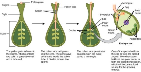
When the pollen tube senses chemical attractants secreted by the synergids, the tube grows through the micropyle, penetrates the embryo sac, and enters one of the synergid cells.
"Plant Physiology and Development" int'l 6e - Taiz, L., Zeiger, E., Møller, I.M., Murphy, A.
#book quote#plant physiology and development#nonfiction#textbook#pollen tube#chemicals#attractants#micropyle#embryo sac#synergids#plant cells#fertilization
0 notes
Text
Enzalutamide painted it microspheres because product labels regarding cancer biomarker alpha-fetoprotein detection
concolor [5] have been confirmed to be essential for micropylar PT guidance, the very last step in the PT trip. Many of us document right here in which ZmEA1 draws in maize PTs inside vitro and also arrests their particular growth in greater levels. Additionally, it adheres for the subapical region of maize Therapist suggestions within a species-preferential method. To get over hybridization obstacles on the a higher level gametophytic Therapist guidance, many of us expressed ZmEA1 within Arabidopsis synergid cells. Secreted ZmEA1 enabled Arabidopsis ovules to compliment maize PT within vitro inside a species-preferential method to the micropylar starting of the ovule. These results show your egg-apparatus-controlled reproductive-isolation hurdle associated with Rehabilitation guidance can be overcome perhaps in between irrelevant place people.Prolactin (PRL) is really a endocrine with more than 3 hundred #Link# biological activities. Even though signaling process downstream in the lengthy type of its receptor (RL) has become well characterised, tiny is well known regarding PRL actions on service in the short type (Urs). Below, we all show that rodents indicating merely Urs display a good ovarian phenotype associated with faster follicular recruitment as well as massive follicular dying resulting in early ovarian failing. Consequently, RS-expressing ovaries of teenagers are reduced involving practical roots and also created mainly simply by interstitium. We also reveal that initial regarding RS represses the particular appearance in the transcription factor Forkhead container O3 (FOXO3) knowning that of the compound galactose-1-phosphate uridyltransferase (Galt), 2 proteins considered essential for typical follicular development. Our discovering that FOXO3 handles the actual term involving Galt as well as enhances the transcriptional exercise suggests that it's the repression of FOXO3 through PRL operating by means of Urs that prevents Galt expression from the ovary to cause follicular demise. Coexpression regarding RL along with Players helps prevent PRL hang-up regarding Galt, and also the ovarian trouble is not affecting Players transgenic rats that coexpress RL, recommending which RL prevents RS-induced ovarian problems. In conclusion, we reveal that PRL indicators through RS and causes, without RL, an intense ovarian pathology by simply repressing the actual expression regarding FOXO3 which of the company's target gene Galt. Additionally we offer proof of one of the links involving the rapid ovarian failure noticed in rats articulating Players #Link# along with rats using FOXO3 gene deletion plus in human together with Galt mutation.Over the past many years, ion range of motion spectrometry (IMS) as being a more successful technique inside job areas involving military services along with stability offers gained a growing number of awareness for neurological and also health-related programs. This kind of very delicate and fast splitting up method has been crucially enhanced by the multi-capillary order (MCC), pre-separation for complex samples. As a way to unambiguously discover substances in the complex test, like inhale, through IMS, a research repository is mandatory. To get a initial group of reference information, Of sixteen selected erratic natural and organic elements have been analyzed by MCC-IMS and relatively reviewed from the normal strategy for air research, thermal desorption-gas chromatography-mass spectrometry. Experimentally identified MCC and GC retention points during the these types of 07 substances have been aligned as well as their regards has been depicted inside a #Link# numerical perform.
0 notes
Note
30, 41, 64, 81, 82, 94, 97, 100 I LOVE YOU
whats your favorite candle scent?
uhh i like cinnamon
top ten favorite songs?
ok these are just gonna be the ones that pop in my head fastest bc i could spend weeks trying to make a refined top ten list so this also doesn't have a particular order
1. Sick Love- RHCP
2. Lord willin' -Logic
3. Pedro Navaja- Rubén Blades
4. Living single- Big Sean ft chance
5. Little Wing- Jimi Hendrix
6. Fly me to the moon- Frank sinatra
7. Shaolin's Theme/Pray- 6LACK
8. Medusa in Chains- The Fratellis
9. Ultralight Beam- Kanye
10. Lemme Freak-Lil dicky
(These are all amazing songs but like god it's hard to make a top ten list)
Favorite dipping sauce?
probably honey mustard
last person you called?
My girl
favorite ice cream flavor?
Cookie dough
favorite lyrics right now?
> (Im not putting in the accent marks because im lazy)
dark milk or white chocolate?
Milk Chocolate all the way
who was the last person you cried in front of?
I haven’t cried in front of anyone since probably like 7th grade homie
2 notes
·
View notes
Text
Difference Between Synergid and Egg Cell
The key difference between synergid and egg cell is that the synergid cell is one of the two cells that accompanies the egg cell while the egg cell is the female gamete or the female germ cell of angiosperms. Synergid and egg cell are two types of cells in the female angiosperm gametophyte.
0 notes
Photo


i said i wouldnt post but @nostlgicwnderlust @synergids
4 notes
·
View notes
Text
Post-fertilization changes within the Ovule-digieduco
Post-fertilization changes within the Ovule :Post-fertilization changes within the Ovule is a series of changes take place within the ovule after fertilization resulting finally in the formation of seed. The zygote i.e. oospore (2n) gives rise to (a) embryo, cells of which are diploid (2n):(b) the triploid (3n) nucleus or so-called endosperm nucleus gives rise to endosperm, the cells of which are triploid (3n) ; (c) the ovule together with embryo and endosperm as a whole gives rise to seed which remains covered by the seed coat developed from integument(s).
DEVELOPMENT OF EMBRYO (EMBRYOGENY)-
In angiosperm ,the zygote develops into embryo simultaneously with the development of the endosperm. In most cases, the first division of the zygote is followed by the laying down of a transverse wall but in a few cases, e.g. in Piperad type such division of the zygote takes place by the formation of a more or less vertical wall. Of the two cells thus formed, the one which lies towards the centre of the embryo sac is called the terminal cell and the one nearer the micropyle is called the basal cell. Theterminal cell, next may divide longitudinally or transversely ; the basal cell usually undergoes transverse division forming the suspensor, sometimes the basal cell does not divide and becomes hypertrophied forming a large vesicular structure.
(i) Typical dicotyledonous type i.e. Crucifer type-Typical dicotyledonous type of embryo development is found in Capsella bursa-pastoris of the family Cruciferae (Brassicaceae). Zygote divides first transversely into a basal cell cb and a terminal i.e. apical cell ca. Next cb divides transversely into ci and cm cells, and ca divides longitudinally resulting in the formation of an inverted T (i.e. ‘_I_')-shaped proembryo composed of four cells. Now each of the two terminal cells divides again longitudinally at right angle to the first longitudinal division forming a quadrant (4-celled) structure. Quadrant cells then become octant (8-celled structure) by a transverse division. The lower four cells of the octant give rise rise to stem tip and cotyledons while the upper four cells to the hypocotyl and the core of the radicle. Now all the eight cells of the octant divide periclinally (i.e. parallel to the outer surface) forming outer and inner layers.
In the mean time cells of and cm of the four-celled proembryo divide transversely forming a row of several (6-10) suspensor cells-the uppermost cell of which becomes swollen, and vesicular (v) which probably serves a haustorial function ; the lowermost cell (h) functions as the hypophysis which later divides further and gives rise to epidermis and cortex of the radicle and root cap. The long suspensor pushes the embryo deep into the embryo sac. Further development of the embryo proper also takes place at two points in the cells of the lower tier forming more or less a cordate structure. By transverse cell divisions, cotyledons and hypocotyl elongate-the growing cotyledons become horse shoe-shaped structure due to horse shoelike curvature of the ovule. As the embryo matures, suspensor cells gradually wither.
(ii) Typical monocotyledonous type of embryo development is seen in Luzula forsteri of the family Juncaceae. Zygote divides into a basal cb and a terminal ca cells. Then the terminal cell ca divides longitudinally into two cells while the basal cell cb divides transversely into ci and m. cells. Next two cells of ca divides vertically at right angle to the first and give rise to quadrant q, cell m divides longitudinally into two cells while of divides transversely into n and n ’cells. By successive divisions the cells of quadrant become differentiated into I and l’ portions-l gives rise to lower part of cotyledon while l upper part of the cotyledon, hypocotyl and plumule; m and n give rise to root cap and n' to the short suspensor (o and p cells)
(b) ENDOSPERM FORMATION-Endosperm is the main source of the food for the embryo, hence it is very important. In angiosperms, endosperm tissue is triploid (3n) as it is the product of triple fusion [as a result of a fusion of two polar nuclei (n + n) and one of the male gametes (n)] involving double fertilization. In angiosperms, except the two families viz. Orchidaceae and Podostemonaceae, there are three general types of endosperm formation, e.g. (a) nuclear type, (b) cellular type and (c) helobial type.
Development of both the zygote and endosperm takes place simultaneously in angiosperms.
(1) Nuclear type-The endosperm nucleus divides and redivides forming numerous free nuclei . These free nuclei are normally unaccompanied by walls, at least in the beginning. The nuclei are pushed towards the periphery of the embryo sac, as a result a vacuole is formed in the centre. Sometimes nuclei are aggregated towards the micropylar and chalazal ends ; free nuclei remain suspended peripherally in the cytoplasm lining the wall of the embryo sac. In later stages wall formation occurs by the laying down of cell plates which begin from periphery of the sac towards the centre or from the apex of the sac towards the base. This type of endosperm formation is known as nuclear type. While the embryo and endosperm formation are taking place within the embryo sac, synergids and antipodal cells are disorganised. Nuclear type of endosperm formation is found in many plants like species of Primula, Mangifera, Malva, Juglans, Asclepias, Calotropis, etc.
(2) Cellular type-In this type, the division of the primary endosperm nucleus is immediately followed by the formation of walls within the sac, so that the endospem: becomes cellular from the beginning. The first wall is generally transverse but may Vertical or oblique sometimes. Each cell contains one or more than one nucleus. Cellular type of endosperm formation is noted in species of Adoxa, Peperomia, Centranthus, etc and in many other members of Annonaceae, Gentianaceae, Boraginaceae, Aristolochiaceae etc.
(3) Helobial type-This type is intermediate between the nuclear and the cellular types. Helobial type of endosperm formation is noted in species of Eremurus (Liliaceae) Scheuchzeria (Scheuchzeriaceae), Vallisneria (Hydrocharitaceae) etc. Here after the first division of the primary endosperm nucleus, transverse wall is formed between the two daughter nuclei so that the embryo sac is divided into two, i.e. micropylar and chalazal chambers. Further divisions are free nuclear type and may take place in both the chambers. But usually the main body of the endosperm is formed by the micropylar chamber only.
(c) FORMATION OF SEED-The ovule i.e. megasporangium develops into seed after the complete development of the embryo. The integuments develop into seed coat-the outer integument into testa while inner integument into tegmen. Stalk of the seed deveIops from the funicle ; hilum, micropyle, raphe etc. of the ovule develop into the corresponding parts of the seed.
If the endosperm and nucellus are completely absorbed by the developing embryo, the seed is called exalbuminous ; if endosperm is retained partly, then the seed is called albuminous. Sometimes nucellus is also retained as perisperm outside the endosperm- then the seed is called perispermic.
Besides testa and tegmen, a third fleshy covering or outgrowth known as aril or arillus is often developed in some seeds after fertilization e.g. Litchi chinensis, Nephelium longana, etc. of Sapindaceae. Aril is always found more loosely arranged than the true integuments, and generally extends only partially over them ; it grows from the outer cell layers of the outer integument or from the base (chalaza or the distal part of the funicle) of the ovule.
It is not infrequent to find outgrowths from various parts of the testa (outer integument) which may be confused with the aril-such an outgrowth formed from the micropylar rim of the testa is called a false aril or arillode ; this type of outgrowth is noted in nutmeg (Myristica fragrans, Myristicaceae) which is usually described as an aril-it originates. however, from both the hilum and micropyle ; it forms a scarlet covering to the seed and forms the valuable spice ‘mace’ of commerce.
Outgrowths of an irregular character are often developed from outer parts of the testa-where the outgrowths are confined to crest as long as the raphe, they are called strophioles, and where restricted near the base ( Viola tricolor, Violaceae) or apex (Ricinus communis, Euphorbiaceae) of the seed are called caruncle,-these structures are always developed subsequently to fertilization, and accordingly are not found on the ovule.
via Blogger https://ift.tt/2OGM3Fw
0 notes
Photo

New Post has been published on https://ramneetkaur.com/apomixis-flowering-plants/
Apomixis - Flowering Plants

(adsbygoogle = window.adsbygoogle || []).push();
APOMIXIS IN FLOWERING PLANTS
Reproduction in plants is of two types:
Sexual or Amphimixis
Asexual or Apomixis
Angiosperms have a Diplontic life cycle. The main part of the lifecycle is Sporophyte & the Gametophyte is reduced.
APOMIXIS
Apomixis mimics sexual reproduction but produces seeds without fertilization. Seen in some species of Family Asteraceae & Grasses.
It is of 3 types:
Apogamy: Formation of sporophyte/embryo from any cell of the gametophyte/embryo sac without fertilization. Cells of the embryo sac i.e., antipodal cells or the synergids. The embryo formed is haploid.
Apospory: Formation of gametophyte i.e., the embryo sac from any cell of the sporophyte without meiosis. Cells of the sporophyte are cells of nucellus & integuments. The embryo sac formed has diploid cells. The egg is also diploid. Diploid egg either undergo fertilization or undergoes Parthenogenesis.
Parthenogenesis: Formation of the embryo from the egg without fertilization.
Apomixis can be divided into different types according to occurrence:
Non-Recurrent: The plant produced due to apomixis is unable to reproduce further.
Seen in the case of:
Haploid Parthenogenesis: Development of embryo from the egg without fertilization. The egg formed is haploid.
Apogamy.
(adsbygoogle = window.adsbygoogle || []).push();
2. Recurrent: The plant produced due to apomixis can reproduce further.
Seen in the case of:
Diplospory: The megaspore mother cell forms embryo sac without meiosis. The egg formed is diploid.
Apospory.
3. Adventive embryony: Also referred as Sporophytic budding. The embryo is formed from the cells of nucellus or integuments. Examples are Mango, Citrus, Opuntia. Due to this Polyembryony occurs.
POLYEMBRYONY
Polyembryony is the formation of many embryos in the seed.
Adventive Polyembryony is the formation of many embryos in the embryo sac from the cells of integuments and nucellus.
Cleavage Polyembryony is the formation of many embryos due to division in the zygote. Seen in Gymnosperms.
Simple Polyembryony is the formation of many embryos due to fertilization of many eggs. Seen in Grasses.
ALSO WATCH: Apomixis in Flowering Plants
(adsbygoogle = window.adsbygoogle || []).push();
#adventive embryony#aiims#aiims 2018#aipmt#apogamy#apomixis#apomixis for neet#apospory#asexual reproduction in flowering plants#biology#biology mcqs#biology mnemonic#biology mnemonics#diplospory#MCAT Biology#MCQs#NEET 2018#neet biology#NEET MCQs#neet mnemonics#NEET question paper#NEET questions#neet ug#parthenogenesis#polyembryony#Premed#premed biology#Reproduction in flowering plants#sporophytic budding#Reproduction in Flowering Plants
0 notes
Text
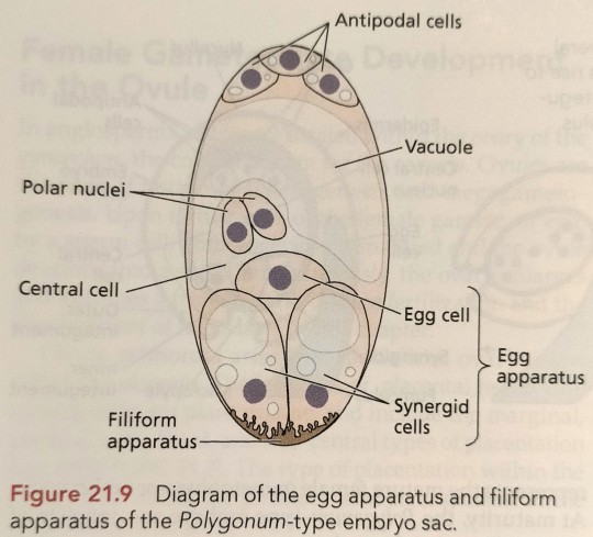
The egg cell (the female gamete that combines with a sperm cell to form the zygote) and the two synergid cells are located at the micropylar end of the embryo sac and are collectively referred to as the egg apparatus (Figure 21.9).
"Plant Physiology and Development" int'l 6e - Taiz, L., Zeiger, E., Møller, I.M., Murphy, A.
#book quotes#plant physiology and development#nonfiction#textbook#egg cell#gamete#zygote#synergid cells#micropylar#embryo sac#egg apparatus#plant cells#polygonum#filiform
0 notes
Text
An ultrastructural study of early endosperm development and synergid changes in unfertilized cotton ovules
http://dlvr.it/PGQLZH
0 notes
Text
Embryo sac:
v P.Maheshwari classified the embryo sac on the basis of no. of megaspore nuclei
Ø Monosporic embryo sac: one nucleus. Eg. Polygonum, Oenothera
Ø Diasporic embryo sac: two nuclei. eg. Allium, nuclei fromEndymion.
Ø Tetrasporic embryo sac: four nuclei.
Development of embryo sac
The functional megaspore (chalazal) divides mitotically giving rise to 2 nucleate stage. It is followed by two more division to form 4 and 8 nucleate stage.Each nucleus from pole comes to the middle and form polar nuclei. These mitotic divisions are strictly free nuclear followed by cell wall formation.
This is called Polygonum type of embryo sac studied by Starsburger in Polygonum.
a) The three nuclei gets organized at chalazal end called antipodal
b) While other three nuclei gets organized at micropyle called synergid and egg cell.
c) The polar nuclei are present in the cenral cell.
This constitutes a 7 celled and 8 nucleate embryo sac.

Organization of embryo sac:
a) Synergid: These cell possess micropyle nucleus and chalazal vacuole. Synergid lacks cell wall at the chalazal end and one synergid degenerate just with pollination. They have filiform apparatus which guides the pollen tube. They secrete chemotropic substance that helps in the growth of pollen tube inside the embryo sac.
b) Egg cell: They show cytoplasmic polarity and have cell wall thicker at the micropyle end. They have microplye vacuole and chalazal nucleus. Plasmodesmata connection is present b/w synergid and egg cell.
c) Antipodal:
d) Central cell: Largest cell.

Pollination: Types of pollination-
i. Autogamy: transfer of pollen grain b/w anther to stigma of the same flower.
Adaptation:
· Homogamy: maturation of anther and stigma at the same time.
· Bisexuality: presence of both the whorls in the same flower.
· Cleistogamy: The flower do not open at all. The anther and stigma lie very close to each other and ensure pollination in absence of pollinating agent and produce assured seed set.
· Bud pollination: When pollination takes place in bud stage before opening. Eg. Pea, Rice .
ii. Geiotnogamy: The transfer of pollen grain from anther to stigma of one flower to another.
iii. Xenogamy: Transfer of pollen grain from anther to stigma in different flower.
Agents of pollination:
· Abiotic agent:
1. Wind(Anemophily):

i. The pollen grains are light and non-sticky
ii. The stamen are well exposed to disperse pollen grains.
iii. The stigma are large and feathery to capture the pollen grains.
iv. Presence of single ovary.
v. Flowers packed in inflorescence.
It’s common in grass.
2. Water (Hydrophily):
Pollination by water is quite rare in flowering plant and is limited to 30 genera mostly monocots.
i. On the surface: (epihydrophily)
Eg.Vallisneria
The female flower has long pedicel which reaches the surface and the pollens are released by the male flower and carried passively by water current which is received by the stigma.
ii. Beneath the surface: (hypohydrophily)
Eg. Zostera
The female flower is submerged under water where the pollen –ribbon shaped is released under water .
In majority, the flowers emerge above the water level surface and is pollinated by bees or wind. Eg. Water Haycinth or Water Lilly.
Biotic agent:
Insect (entmophily):
Birds-Orinthophily
eg. Bombax, Callistemon
Bats-Chiropterophily
eg. Anthocephalus
Snakes-Ophiophily
eg.Santalum, Machelia
Snails-Malacophily
eg. Arisaema
Outbreeding devices:
a. Unisexuality:
· Monocious : male and female are in same plant. It prevents Autogamy and but not geiotnogamy.
· Deicious:
b. Self incompatibility: inhibits the growth of pollen tube genetically.
c. Dichogamy:
Pollen pistil interaction:
The stigma is able to recognize the right type of pollen by constant chemical dialogue b/w pollen grain and stigma. Its acceptance is followed by germination of pollen on stigma to produce pollen tube in style. The content of pollen grain move through the stigma. The generative cell divides mitotically to produce two male gamete.
Entry of pollen tube into ovule takes place through:
· Micropyle (Porogamy): Most common type.
· Chalaza(Chalazogamy): eg. Casurina
· Integuments (Mesogamy) eg.Cucurbita.
The pollen pistil interaction is a dynamic process.
0 notes
Text
Ovule primordia first appear along the placenta as comical projections with rounded tips (Figure 21.7). (...) The inner integument forms a ridge some distance behind the apex of the nucellus, followed by the outer integument layer (see Figure 21.7).

"Plant Physiology and Development" int'l 6e - Taiz, L., Zeiger, E., Møller, I.M., Murphy, A.
#book quotes#plant physiology and development#nonfiction#textbook#ovule#primordia#placenta#morphogenesis#plant cells#cell differentiation#integuments#chalaza#funiculus#synergids#egg cell#epidermis#nucellus#antipodal cell#embryo sac#megaspore
0 notes
Photo


i was tagged by @swearworld to do 6 selfies from 2017 this year was a mistake i tag @breastless @210pm @cryingblogger @synergids and @camilabadwolf
20 notes
·
View notes
Text
What is ovule and its structure-digieduco
The ovule may be characterized as a juvenile seed or an unripened integumented megasporangium. It may also be designated simply as “the egg containing organ within ovary.” After fertilization the ovule develops into seed. In angiosperms, ovules are protected by megasporophyll which forms a closed structure (ovary), that is why seeds remain enclosed. In gymnosperms, ovules are not protected but exposed on megasporophyll i.e. carpel resulting into the formation of naked seeds.
STRUCTURE OF AN OVULE
(1) The stalk with which the ovule remains attached to the placenta is called funicle
or funiculus
(2) The point of attachment of the funicle to the body of the ovule is called hilum.
(3) The basal part of the ovule from where the integument or integuments arise is
known as chalaza.
(4) The extension of the funicle beyond the hilum alongside the body of the ovule is known a raphe. A raphe may be ventral or dorsal in an ovule, and may extend up to the base i.e. chalaza of the ovule.
(5) The main body of the ovule consists of a central mass of tissue-the nucellus Winch Is surrounded by one or two envelopes called integuments ; the integuments enclose the nucellus except at the apex, where a small opening is left called micopyle.
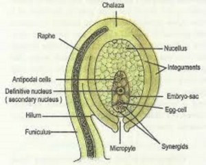
(6) Near the micropylar end, a sac-like structure known as embryo sac (female gametophyte), lies embedded into the nucellus. Within the embryo sac towards the micropyle there are three naked cells. The middle one is the largest and is known as oosphere or ovum i.e. egg cell ; two lateral ones are the synergids. Oosphere and the synergids constitute what is known as egg-apparatus. At the centre of the embryo sac lies a composite nucleus, formed by the union of two polar (one nucleus from each pole of the embryo sac) nuclei-this is known as secondary or definitive nucleus. Towards the chalazal end of the embryo sac there are three walled-cells known as antipodal cells.
via Blogger https://ift.tt/2MKo5rY
0 notes
Last Seen Blogs
citreousgoldenrod-blog
#Fuck Y9urself

gosonicprecure-blog
カオス・コントロール!

ninaluba
ninaluba FASHION DESIGN

novirinuno
Без названия

sang-el
The World of El