#Magnetic resonance imaging
Text
The biggest mystery in the XMCU isn't how Darwin managed to die in First Class, but how Wolverine survived being put in what is essentially a big, magnetic tube without his skeleton getting ripped from his body.
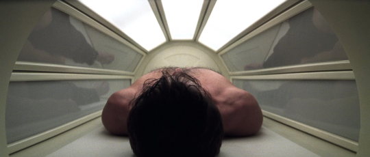
#wolverine#xmen#x men movies#hugh jackman#mri#Mris are just giant magnets#magnetic resonance imaging#Marvel explain#Is it a magic mri machine?#How did the machine not explode?#You can't have metal in an MRI#i'm loosing my mind
14 notes
·
View notes
Text
MRI tomorrow. I'm so scared and nervous, I know nothing bad is gonna happen, it's just so scary
Wish me luck guys
8 notes
·
View notes
Text

Next Generation Images
A new 7 Tesla ultra-high resolution MRI scanner takes human neuroscience imaging to the next level
Read the published research article here
Erwin Hahn 7T MRI Laboratory, Henry H. Wheeler Brain Imaging Center, Helen Wills Neuroscience Institute, University of California, Berkeley, Berkeley, CA, USA
Image originally published with a Creative Commons Attribution 4.0 International (CC BY 4.0)
Published in Nature Methods, November 2023
You can also follow BPoD on Instagram, Twitter and Facebook
#science#biomedicine#neuroscience#biology#mri scan#mri#magnetic resonance imaging#medical imaging#next generation#brains#brain scan
7 notes
·
View notes
Text
*activates MRI machine*
“Commence primary ignition!”
#dougie rambles#personal stuff#mri scan#MRI machine#cat scan#magnetic resonance imaging#medical imaging#radiology#biomagnetics#no context#my poor attempt at a joke#what#this sounded funnier in my head#shitpost#weaponised#radiation#this kills the man#star wars#death star
4 notes
·
View notes
Text
youtube
Nyello! I'm B.L.Radley, also known as @radley-writes, also known as 'that person who decided to become a radiographer on a whim literally so they could introduce themselves as Rad the Rad but then got really fucking obsessed with radiography and ology and now wants to commit themselves to several more years of specialised study and student debts to become an Advanced Practitioner in.... MRI, probably, but who the fuck knows'
Important info:
They/it/xe pronouns plz.
I am a student.
This means:
I am not an expert! I might be wrong about stuff! If you know more than me, please feel free to correct me! I love this topic and want to learn more!
I am not qualified to give out medical advice, and will only say 'go see your doctor if this concerns you!' on this blog.
It's also worth noting that, though I do work part-time in a hospital, doin' the ol' radiography, I am not going to provide funny/embarrassing patient stories. Or any patient stories at all, beyond, perhaps, very, very anonymised anecdotes to help illustrate points. I strongly disagree with mocking patients or posting anything about them without their consent - particularly if it could be traced back to them! This is just a blog where I can scream excitedly about how cool it is that I can look at all the Mushy And Bony Stuff inside you without cutting you open!
(Although sometimes you'll get cut open too <3 Just for funsies <3)
Please feel free to ask me anything, and I will do my best to reply!
Or, uh. Infodump about my hyperfixation. Not sorry.
#radiography#radiology#medblr#studyblr#x ray#mri#ct#computed tomography#magnetic resonance imaging#cath lab funsies
11 notes
·
View notes
Text

This is an image derived while doing reconstructions of an MR neurography 3D dataset (displaying vessels highlighted by contrast medium, among other features).
I love the details and its overall aesthetics, though such images don't aid in the diagnosis. Just a little eyecatcher :)
#VeluVerse#Everyday life#MRI#Magnetic Resonance Imaging#Siemens#Siemens Magnetom Skyra#3 Tesla Scanner
3 notes
·
View notes
Photo

Scientists present a new method for imaging individual electrons
Imagine going for an MRI scan of your knee. This scan measures the density of water molecules present in your knee, at a resolution of about one cubic millimeter—which is great for determining whether, for example, a meniscus in the knee is torn. But what if you need to investigate the structural data of a single molecule that's five cubic nanometers, or about 10 trillion times smaller than the best resolution current MRI scanners are capable of producing?
That's the goal for Dr. Amit Finkler of the Weizmann Institute of Science's Chemical and Biological Physics Department. In a recent study, Finkler, Ph.D. student Dan Yudilevich and their collaborators from the University of Stuttgart, Germany, have managed to take a giant step in that direction, demonstrating a novel method for imaging individual electrons. The method, now in its initial stages, might one day be applicable to imaging various kinds of molecules, which could revolutionize the development of pharmaceuticals and the characterization of quantum materials.
Current magnetic resonance imaging (MRI) techniques have been instrumental in diagnosing a vast array of illnesses for decades, but while the technology has been groundbreaking for countless lives, there are some underlying issues that remain to be resolved. For example, MRI readout efficiency is very low, requiring a sample size of hundreds of billions of water molecules—if not more—in order to function. The side effect of that inefficiency is that the output is then averaged. For most diagnostic procedures, the averaging is optimal, but when you average out so many different components, some detail is lost—possibly concealing important processes that are happening on a smaller scale.
Read more.
13 notes
·
View notes
Text
Tips from the MRI:
I've just had my 19th MRI of the year (yes, this year of 2022 lol) and I have some tips that you won't get online or from your doctor for anyone feeling nervous about it.
- Try not to move (but accept that you will move and forgive yourself). If you are getting an MRI anywhere from the thoracic spine upwards, make sure to limit swallowing. If you have to sneeze or cough, let them know. Top tip, I find that I move more with my eyes closed than my eyes open. The best thing to do is distract yourself by thinking of something else. If you think about not moving, you will move more.
- You can customize your experience however you want. If you want them to check in with you every 10 minutes over the speaker, they will do it for you ( I find that this helps get an idea of how much time is going by). If you want wedges to prop up your arms, ask. If the pads holding your head in place are too tight, ask for smaller ones. You will be lying still for at least 30 minutes up to 2 hours, so make sure you're as physically and mentally comfortable as possible.
- For first timers, make sure you know what makes you comfortable before you go. Lying flat for a long time is fine. Lying flat and completely still is a different thing. If youre worried, you can practice beforehand by lying completely still on a flat, hard surface for 30 minutes and placing pillows where it feels comfortable (under the legs and/or arms, etc.). I always ask for wedges for my arms so I can fold my hands over my belly. I also ask for a blanket to cover my legs and for the staff to speak to me approximately every ten minutes. I tell them which vein I would like my IV in as well.
- If your MRI is longer than an hour, you might overheat. You might notice that the room with the machine is super cold. That's because the magnetic energy it produces when running for a long produces a lot of heat. Sometimes this heat is absorbed by the body and can actually sometimes raise the body temp up to 2 degrees Celsius. They keep track of the room temperature, but if you start overheating, let them know.
- You can ask to be sedated if you're claustrophobic. I've never needed to which leads me to my next point:
-The fear of claustrophobia can be avoided ( for those not typically fearful of this). I've learned two things from the MRI: fear and a large part of pain is all in the mind. If you imagine yourself trapped in a loud, coffin-like tube, then you will be very uncomfortable. If you close your eyes and imagine you're in your room on your bed, then you can relax a little. Here are some other scenarios I like to imagine: I'm the goth veraion of a vampire (get it cause the room is white instead of black lol) chilling in my coffin, I'm one of the Apollo 11 crew heading back home to Earth after having seen some pretty cool moonrocks, I'm a spelunker during am earthquake, I'm the first human to listen to an alien symphony, and plenty others.
- Learn the lyrics/tunes of the following songs: 9 to 5 by Dolly Parton, the William Tell overture, and TikTok by Kesha. It will make sense with all the noises of the machine.
-If you have contrast dye be careful getting up when you are done. Some of the side effects include dizziness, nausea, and mild headache.
- Have fun! Life is short and can be scary when you're sick. An MRI is a relatively short experience in the long term of your life and we should not try and dread these experiences. Today I met a man getting an MRI to check up on his brain tumour. We were chatting about how we were on clinical trials and how if we weren't born today and experiencing the beauty of modern science he would be dead and I would be blind. An MRI scan is a gift that not everyone can experience or access. Replace dread with joy and fear with wonder and you will have a much better time both in and out of the machine.
2 notes
·
View notes
Text
youtube
What is MRI? In this short video, we discuss the principles of magnetic resonance imaging. Covered topics include: How does MRI work? How Loud Is an MRI? Closed vs Open MRI and more! Click to watch on our YouTube Channel.
0 notes
Text
What is the major concern with MRI (magnetic resonance imaging)
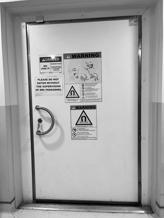
The major concern with MRI (magnetic resonance imaging) revolves around safety considerations, particularly related to the strong magnetic fields and radiofrequency radiation used during the procedure. Here are the key concerns associated with MRI:
Magnetic Field Hazards: MRI machines generate powerful magnetic fields, which can pose risks to individuals with certain types of metallic implants, devices, or objects. Ferromagnetic materials can become projectiles in the strong magnetic field, leading to serious injury or even death if not properly screened before entering the MRI room.
Implant and Device Safety: Certain metallic implants or devices, such as pacemakers, cochlear implants, or certain types of joint replacements, can be affected by the magnetic field or radiofrequency energy of an MRI machine. It's crucial for patients to disclose all relevant medical history and implanted devices to ensure safe MRI scanning.
Contrast Agent Risks: Some MRI scans may involve the use of contrast agents (such as gadolinium-based contrast agents) to enhance image quality. While generally considered safe, these contrast agents can rarely cause allergic reactions or nephrogenic systemic fibrosis (NSF), particularly in patients with kidney problems. Newer gadolinium-based contrast agents have been developed to minimize these risks.
Claustrophobia and Anxiety: The confined space of the MRI machine can trigger claustrophobia or anxiety in some individuals, leading to discomfort or even panic during the procedure. Open MRI machines or techniques to help manage anxiety (such as music or sedation) can mitigate these concerns for some patients.
Pregnancy Concerns: Although MRI does not use ionizing radiation like X-rays or CT scans, some precautions are still advised for pregnant women, particularly during the first trimester. While the risks to the fetus are considered low, MRI is generally avoided during pregnancy unless absolutely necessary, and alternative imaging methods may be preferred.
Acoustic Noise: MRI machines produce loud noises during scanning, which can be distressing for some patients. Proper ear protection is essential to prevent hearing damage or discomfort.
Overall, while MRI is considered a safe and valuable imaging tool for diagnosing various medical conditions, careful attention to safety protocols, patient screening, and appropriate use is necessary to minimize potential risks and ensure patient well-being during the procedure.
0 notes
Text
Case Presentation where MRI shows Superiority as A Modality for Breast Cancer Screening | Abstract
View On WordPress
0 notes
Text
Magnetic Resonance Imaging
An MRI (Magnetic Resonance Imaging) scan utilises magnetic forces and radio waves to take pictures of the body. It requires no radioactive sources to acquire the pictures.
Visit - Magnetic Resonance Imaging
0 notes
Photo

Baby Brains
The human body carries on developing after birth – but the rate of these changes is still in question for developmental biologists. Here scientists take magnetic resonance imaging (MRI) scans of sleeping children’s cerebellums over the first two years of life, showing months 1–5 (top row) and regular intervals afterwards (bottom) from three different angles. While the young brain is constantly growing, it’s growing at a faster rate (warmer colours) during the first six months. The researchers investigated further, analysing the scans from 235 healthy children and looking at how 27 individual regions of their cerebellums change – finding, for example, that changes in specific ‘lobules’ corresponded with milestones in fine motor skills. This data could be used to pinpoint developmental windows for further study, or even to diagnose health issues or injuries during a toddler’s early life.
Written by John Ankers
Image from work by Ya Wang and Liangjun Chen, and colleagues
Department of Radiology and Biomedical Research Imaging Center, University of North Carolina at Chapel Hill, Chapel Hill, NC, USA: UNC/UMN Baby Connectome Project Consortium
Image originally published with a Creative Commons Attribution 4.0 International (CC BY 4.0)
Published in Cell Reports, April 2023
You can also follow BPoD on Instagram, Twitter and Facebook
#science#biomedicine#brains#brain development#developmental biology#babies#brain#mri#magnetic resonance imaging#motor skills
6 notes
·
View notes
Text
Portable MRI machine?
Idk
#dougie rambles#personal stuff#bad ideas#ideas#mri machine#magnetic resonance imaging#magnetic resonance
2 notes
·
View notes
Text
How does one identify the location of the pituitary gland in the brain?
The pituitary gland, also known as the hypophysis, is a small, pea-sized gland located at the base of the brain. It is often referred to as the “master gland” because it controls the function of other endocrine glands in the body. Despite its small size, the pituitary gland plays a critical role in maintaining a wide range of bodily functions, including growth and development, metabolism, and…
View On WordPress
#adenohypophysis#Anatomy#brain#computed tomography#diagnosis#endocrine system#Health#hormones#imaging techniques#magnetic resonance imaging#nasal endoscopy#neurohypophysis#pituitary disorders#Pituitary gland#symptoms#treatment
0 notes
Last Seen Blogs

travel-tours-guide-blog
Travel Tours Guide

harnessht
Safety harness


notbotleon
Not Bot Leon

smitten-smithie-blog
chloë at smith