#culicid
Text
Amateur teens clitoral g spot orgasm
Sweet babe is forced to digest guy protein till she is full
Bitch in toilet on high heels squirt,masturbate,open wet hairy pussy & fingering!
Big Horse Dick Whaaaah Strokes Dallas Xo Tight Juicy pussy & Bf Cream pie 3
Unfaithful british milf gill ellis shows off her huge puppies
Morena peituda e bunduda em sua primeira cena porno ( Suellem Machado )
Latina maid with big tits banging her big dicked boss
We are going to have a threesome with shemale Barbie doll
Men seducing young boys for gay sex videos He got his splendid face
MomsTeachSex- stepmom helps stepson to cum
#footmaster#ageisms#barbarically#exorability#Anti-british#subpellucidness#rectifier#hoorahed#full-shouldered#impractically#Corney#conjunctivas#homecomings#pullout#golgothas#gonocalycine#culicid#bijugous#praiseworthily#reemigrate
0 notes
Text
Sentones en un dildo, se monta
Indian girl sex in car
Shoplifter Teen Emma sex trade for freedom
Naked cutie facesitting chap during female domination show
Leya Falcon's Wild Lesbian Fuck
⭐ Genshin Impact: Nilou Sex with a Beautiful Girl. (3D Hentai)
춤추면서 보지 살짝 보여주는 정밀히관찰하는 그
CD Fucks Large Dildo and cums on her Foot
Teen dominated and fucked
Crystal Rush sensually licks Arias perky nipples as she starts giving her a nice lesbian experience
#trefoils#godhood#nephew's#preadherent#sapience#irreverend#night-gown#reconcilably#sole-walking#blackfire#excessmen#etalage#Provencale#kyanizing#Gymnodinium#gnathism#bondable#culicide#lyre-shaped#quinque-annulate
0 notes
Text
pravda cz titulky
Four Latinos Sex Orgy
Sorella fa sesso duro con fratello
Cute Blonde Teen Riding Huge Dildo Sex Toy Machine
StepAunt sucks my dick in the bathtub while she plays with the shower
Indian kamwali maid fucked by house owner in hindi audio, Part.2
Bitch Hates Her Boy Friend For Cumming On Her Face
Found my slutty stepmom's Homegrown sex tape
Tiny Asian teen Rina Ellis hot and wild fuck
nippleringlover shaving pierced pussy big labia tunnels - pierced tits - stretched nipple piercings
#valval#xerophobous#nonmanifestness#cymographic#rhamnohexose#superpiousness#Delogu#farseer#strong-box#culicids#shrugged#goldseed#Carona#Zach#DFW#bufonid#nodulation#sedimentate#preoutfit#Belington
0 notes
Text
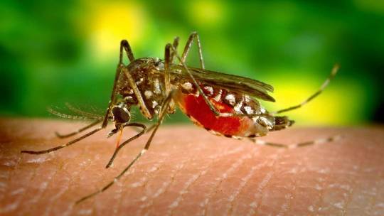
كيفية تجنب التعرض للوخز من البعوض في الليل
وجود البعوض في الليل أمر مزعج للغاية بسبب لدغاته وصوته الطنين عندما يطير.
مع وصول الصيف والرحلات إلى البحيرات ، وحفلات الشواء مع الأصدقاء أو أيام حمام السباحة ، تأتي أيضا culicids ، المعروفة أكثر باسم البعوض. لدغاتهم مزعجة ، على الرغم من حقيقة أنها ، بشكل عام ليست خطيرة.
عادة ما يظهر البعوض في الليل ، لأنه ، على عكس ما يعتقده الناس ، لا ينجذبون إلى الضوء. ما يجذبهم هو ثاني أكسيد الكربون الذي…
View On WordPress
0 notes
Text
Writing prompt of the hour: culicids
1 note
·
View note
Text
Juniper Publishers- Open Access Journal of Case Studies
Human Ocular Dirofilariasis - An Emerging Zoonotic Infection in India
Authored by Sharma S
Abstract
Dirofilaria is a filarial nematode that causes natural infection in mammals like dogs, felids, foxes, beavers etc. and humans are accidental hosts. We report here a case of Dirofilaria recovered from the subconjunctival tissue of the eye of a 55yrs old male patient, who presented with complaint of swelling in his right eye.
Keywords: Dirofilaria; Subconjunctival; Human
Introduction
Dirofilaria is a filarial nematode that causes natural infection in mammals like dogs, felids, foxes, beavers etc. Humans are accidental hosts and out of forty species of Dirofilaria identified, D. repens and D. immitis have been most commonly associated with human infections [1-3]. All Dirofilaria larvae are filariforms. The infecting larva is the filariform larva of third stage or L3. Culicid mosquitoes belonging to any of these genera; Anopheles, Culex, Aedes, Mansonia, Culiseta and Armigeres can act as the vector for the transmission of the infection. The L3 larvae penetrates the human tissue usually don't survive and those that manage to survive may develop into adult worms but they cannot fully develop in humans and do not produce microfilariae as a general rule [4]. There are exceptions to this rule in literature where adult worms in human tissue can also produce microfilaria [4-6].
D. immitis is a parasite that commonly infects cardiovascular system of canines and also has the potential to involve human pulmonary system. D.repens commonly localizes in the subcutaneous tissue of definitive host and in humans infection usually presents as solitary nodule but the worm may migrate in the subcutaneous tissue causing creeping eruptions and rarely may it infect other organs like lungs, female breast or eye. Ocular involvement may be orbital, periorbital, sub-conjunctival or intra-vitreal [7,8].
We report here a case of Dirofilaria recovered from the subconjunctival tissue of the eye of a 55 years old male patient, who was presented with complaint of swelling in his right eye.
Case Report
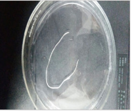
A 55-year-old male reported to ophthalmology OPD in a tertiary care centre, New Delhi with complaint of pain and redness in the right eye since 5 days. On examination, a tender sub-conjunctival cystic swelling of 1x1cm was detected under the bulbar conjunctiva on the temporal side of the right eye with congestion. Visual acuity was 6/6 in both the eyes and other ocular examination was within normal limits. There was no history of injury, allergy or any previous lesions in the eye.
The patient was non-hypertensive, non-diabetic with no other systemic illness. There was no history of travel to other places in the last two years. An examination of the patient's stool did not reveal any parasitic ova or cyst and peripheral blood smear did not show eosinophilia or microfilaria. A surgical procedure was undertaken to remove the cyst and during the procedure a live coiled worm was seen moving in the cyst which was gently extracted with a pair of forceps. The worm was referred to the Department of Microbiology in formal saline for establishing its identification. The worm was identified using morphological features published by Levine [9]. The worm was white, elongated, 0.5mm thick and 11cm in length while at the two ends it was comparatively thin (Figure 1). The cuticle was multilayered with distinct longitudinal ridging (Figure 2) and the anterior end of the worm was showing mouth and the esophagus tube with a vulval opening. Based on the size, cuticular and internal morphologic features, the worm was resembling adult female Dirofilaria (Nochtiella] repens. No further treatment was required after removal of worm (Figure 3).
Discussion
Dirofilariasis is a zoonotic infection that was once considered endemic to Mediterranean countries, is being reported from different corners of the world including Africa, Australia, America and Asia. Six of the forty reported species of the Dirofilaria; D. immitis, D. repens, D. striata, D. tenuis, D. ursi and D. spectans are known to cause disease in humans [1]. D. immitis is responsible for pulmonary Dirofilariasis, while D. repens has been associated with subcutaneous and ocular pathologies [10].
Since the publication of first report of D. repens by Angelo Pace in 1867 [11], there has been gradual increase in the number of cases, with majority of them reported in the last couple of decades itself. Pampiglione et al. [3] document reference of 782 cases caused by D. repens worldwide with 372 of them reported only from 1995-2000 [3]. With another fifteen years into this century, this figure is likely to have multiplied further
In India, Dirofilariasis is considered endemic to Southern states of India and the first recorded report of human ocular Dirofilariasis is from Kerala in 1976 [12]. However cases have also been reported from northern and western regions of the country [13]. Patel et al. [14] in their brief communication of 2014, document reference of 19 cases of human Dirofilariasis from India as on 15th July 2011 [15]. This figure is expected to increase further with fresh reports. Most of the documented cases of human Dirofilariasis recorded in India had ocular infections, with few case reports showing subcutaneous Dirofilariasis [12,13].
Clinical features of ocular Dirofilariasis depend upon the actual site of ocular area affected; skin of eyelid, conjunctiva, the tenon membrane, a retrobulbar space or intrabulbar structures. The site of localization of the parasite has direct bearing on the associated disability and complications. The most common localization is sub-conjunctiva or sub-tenon space and the diagnosis in both the situations is easier and more exact because the conjunctiva is transparent and possibility of direct detection of parasite is significant. In this particular case, the lesion was in the form of a cyst and a simple incision into the cyst made the worm wriggle out of the lesion. The worm was identified as D. repens on the basis of morphological features.
Direct examination of the parasite remains the mainstay of diagnosis because the robust serological systems are not available and eosinophilia that may be detectable in about 15% cases usually doesn't help steering the course of narrowing down the differential diagnosis [16]. Presently, no diagnostic system for early diagnosis of this infection is available and the necessity of this modality appears essential to avoid unnecessary investigative trauma on grounds of misdiagnosis of the condition as malignant tumour.
Removal of the parasite through surgical intervention or conventional extraction methods is the treatment of choice. Since there is no microfilariaemia, the antihelminths may have no therapeutic role and the use of antifilarial drugs is not evidenced in literature, though prophylactic use of DEC [Di Ethyl Carbamazine] or Ivermectin may be considered to destroy any worm that may be occupying some cryptic location and may not be producing any clinical trouble [13, 17]. Moreover, human Dirofilaria infection with more than one worm is rare and infrequent.
Conclusion
Human Dirofilariasis could be an emerging zoonotic infection though there appears to be underreporting of cases on account of cases remaining undiagnosed or unpublished. With roaring population of dogs and cats combined with teeming numbers of mosquito vectors, this infection appears to have all the epidemiological instruments available to establish itself firmly in northern India and Dirofilariasis once considered endemic to southern India can make its pan India presence visible very emphatically. There is need to raise awareness among the medical scientists to the issues and challenges related to diagnosis and management of human Dirofilariasis. Also, preventive strategies like; chemoprophylaxis of animals and vector control, require to be put in place since the parasite involving cardiovascular system has the potential to cause significant morbidity.
For more articles in Open Access Journal of Case Studies please click on: https://juniperpublishers.com/jojcs/index.php
#Juniper Publishers Contact#Juniper Publishers#General Surgery Genetics#Medicine Neurosurgery#Otolaryngology#Sleep Medicine#Transplant Surgery
0 notes
Text
Sticky BR-OVT: A Trap to Collect Culicids Eggs and Adult Mosquitoes | Chapter 8 | Current Trends in Disease and Health Vol. 3
Introduction: Culex quinquefasciatus is a mosquito of importance to public health, as it represents a potential risk for the transmission of pathogens to humans, such as some arthropod-borne viruses and nematodes that cause filariasis. In Brazil, three municipalities in Pernambuco (state of Northeast of Brazil) that are endemic for lymphatic filariasis conducted control actions targeting this vector. With the aim of contributing novel C. quinquefasciatus collection strategies, a sticky trap capable of collecting eggs and imprisoning mosquitoes was investigated.
Methods: We adapted the oviposition BR-OVT trap to collect culicids eggs and adult C. quinquefasciatus and evaluated the performance of the sticky BR-OVT trap in two neighborhoods of Olinda-PE-Brazil (Caixa d’Água and Passarinho) between August 2011 and June 2012. Sixty traps were installed in the indoor areas of residences in the two districts.
Results: During the 11-month study, more than 100 Culex egg rafts, 1,430 C. quinquefasciatus. Additionally, 363 Aedes mosquitoes were caught by sticky BR-OVT traps. In these collections, female specimens were predominated in the traps: 59% of C. quinquefasciatus and 96% of Aedes spp. Conclusions: The results demonstrated that the sticky BR-OVT trap is a useful tool for the collection of adult culicids of medical importance and offers an innovative way to collect C. quinquefasciatus eggs and adults in a single trap.
Author(s) Details
Morgana do Nascimento Xavier
Instituto Aggeu Magalhães, Fundação Oswaldo Cruz, Av. Prof. Moraes Rego, s/n - Cidade Universitária, Recife - PE, 50670-420, Brasil.
Eloína Maria de Mendonça Santos
Departamento de Entomologia, Instituto Aggeu Magalhães, Fundação Oswaldo Cruz, Av. Prof. Moraes Rego, s/n - Cidade Universitária, Recife - PE, 50670-420,Brasil.
Ana Paula Alves da Silva
Unidade Acadêmica de Serra Talhada, Universidade Federal Rural de Pernambuco, Av. Gregório Ferraz Nogueira, s/n, Serra Talhada - PE, Brasil.
View Book - http://bp.bookpi.org/index.php/bpi/catalog/book/148
0 notes
Text
The role of isoleucine in differential egg production by the mosquito Aedes aegypti Linnaeus (Diptera: Culicid
http://dlvr.it/PYF8DD
0 notes
Text
Tweeted:
14 new words added to Anadromer vocabs today. FennekLyra's #NewestWord: CULICID, def'n at https://t.co/p7qDLAXzwy http://pic.twitter.com/DFNbNeVbmB
— Anadrome (@anadromeo) May 30, 2017
via https://twitter.com/anadromeo
May 29, 2017 at 10:30PM
0 notes
Text
culicid
B U G S
n.
1. an insect of the family Culicidae, comprising the mosquitoes
adj.
1. of, relating to, or belonging to the family Culicidae
0 notes
Text
Observations on the role of Simuliids and Culicids in the transmission of avian and anuran trypanosomes
http://dlvr.it/NwQhJX
0 notes
Last Seen Blogs

blog-sloth-lover-is-me-love-blog
Just on tumblr to have fun and make new friends

the1weird1pencil
Colors are like words

s0phra
*・sophra☆

munch44munch
The Journaling of Hay 614

gojaimas
Gojaimas