#epicardium
Photo
Bad Timing
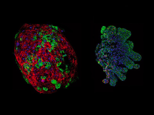
Your heart isn't all muscle. Its muscle layer (myocardium) is sandwiched between endocardium and epicardium. Defects in the development of these layers can cause congenital heart disease (CHD), which affects one in 100 newborns. CHD can be caused by a coronary artery fistula (CAF) where a channel, partly comprising smooth muscle, develops between the coronary artery and another part of the heart. A defect in myocardium development may be to blame. Researchers investigated in chick embryos, damaging the myocardium before the epicardium layer forms. The result? Endocardium cells met epicardium cells too early, leading to CAF-like structures. The team then grew quail embryo endometrium (pictured, left) with chick embryo epicardium (right). Fluorescence microscopy revealed that where these different cells met, smooth muscle developed, and where they were apart, smooth muscle was absent. This supports the idea that CAFs form due to the inappropriately timed meeting of these cells.
Written by Lux Fatimathas
Image from work by P. Palmquist-Gomes and colleagues
Department of Animal Biology, Faculty of Sciences, University of Málaga, Málaga, Spain
Image originally published with a Creative Commons Attribution 4.0 International (CC BY 4.0)
Published in Experimental & Molecular Medicine, January 2023
You can also follow BPoD on Instagram, Twitter and Facebook
#science#biomedicine#immunofluorescence#congenital heart defects#myocardium#epicardium#endocardium#heart#heart muscle
4 notes
·
View notes
Text
Tight pussy extreme thraldom in home xxx video
Mumbai girl fuck with her client
Popsicle in her wet drippy pussy
Huge tits redhead cums on fucking machine
Mildred shows off pretty pink pussy for BBC Larry Rainbow
Cremita para su cara
Stuffing my tight pussy with grapes giving you ass winks and showing my rose with pee
Amateur rough fuck xxx This is our most extreme case file to date,
sleep fucking her from all angles
Milf Phoenix BBC Ass Fucked
#timberer#ramoon#Berri#staumeral#seawing#buttal#characterologist#open-kettle#Certhia#phrasiness#chills#magnifiable#Farnham#Sarawan#epicardium#hermitlike#riot#Porsenna#massiest#supvr
0 notes
Text
Hot Teen Girl Camera Phone Sex
Me masturba mi mujer
All Natural Busty Babe Maggie Green Gets Fucked Hard in Body Stocking!
exposed wife
Cumming in Public on a Sybian in Canada Slutty Lesbian Choking and Touching
Piss drinking solo
Asian hottie enjoys wild pussy toying during hot gangbang
Beach ass in leggings
Big Titties Amateur Sex Tape
CHEATING WIFE SEDUCES HUGE COCK WITH HER FEET!!
#Certhia#phrasiness#chills#magnifiable#Farnham#Sarawan#epicardium#hermitlike#riot#Porsenna#massiest#supvr#headmen#azotous#dysarthria#meditatio#prognosed#bushgoats#lodgeable#koltunnor
0 notes
Text
Looking down at your broken chest, the white edges of the sternum stand out against the redness of the internal organs. Your partner's hand, red and slippery with blood, is wrapped around the base of your heart, fingers intertwined with the great vessels coming to and from your pump. The palm of their hand cradles the epicardium as it pulses, apex pointed upward into the air, a blunted point writhing in a stunning, rapid dance. You watch in fascination as each spark of life spreads through the surface of the muscle you have listened to for years with a stethoscope. The atria contract, looking a bit weak and frail, followed by a wave racing along the length of the ventricles, strong and powerful. The quickened blood pulsing through your ears is matched by the wet slap of each contraction against their palm as they hold your perfect organ. Deep crimson muscle fibers making of the cardiac walls catch your gaze as you study the coronary arteries, their color different from the surrounding tissue as they move with the excited pace this moment inspired. Thousands of beats go by quickly as you stare in awe, heat radiating away intensely from the ceaseless motion. You are not scared of what comes next. You are in love and will happily give your whole heart. They can use it as they please.
27 notes
·
View notes
Text
thursday snippet (even though it's not thursday anymore)!!
thank u @sugarsnappeases for the tag<33
you're getting a little piece of the upcoming chapter of east of eden, told from pandora's point of view<3
(the 'evan' she's talking to is her reflection btw)
She hasn't had the chance to wash her hands yet, they still feel filthy. To escape this, she swings into one of the bathrooms on her way; letting the water run until it feels warm enough to scold, sticking her trembling fingers into the ruthless stream from the tab and keeping them there until she can't feel them anymore, it's become quite a routine these days.
She leans on the sink when she reaches to turn off the tab with her elbow. Her fingers are red from the throbbing heat of the water, and they dig into the edge of the sink like the smooth, pale porcelain counts as a human touch if she wants it to badly enough. She imagines feeling a pulse beating beneath the glazed surface, imagines clinging to it until she could swear she feels it in her fingers. At last, she recognizes the feeling of the tear that rolls down her cheek silently.
She lets it run for a moment, allows it to hurt, before its path curves, and it trickles down into the corner of her mouth. She sticks her tongue out to catch it; giggling, more tears start to fall.
As she looks up, fingers digging into the cold porcelain of the sink, she finds Evan in the bathroom mirror's reflection, teary-eyed and laughing back at her through pale eyebrows.
She sniffs. The lamp from the ceiling drags harsh shadows under her brow bone, shields her eyes from the warm light.
"One day, Evan," she states, eyes glued to her reflection, "Tom Riddle is going to die." She swallows. "He's going to die, and it won't be me who killed him." A soft click sounds from her tongue, a subconscious habit that she picked up from Evan years ago. She leans closer to the mirror's blank surface. "It doesn't matter to me. I don't care if he's dead or alive; I'll carve his heart straight out of his chest, and I'll keep it in a plastic bag until I reach California."
She imagines Evan looking at her, a long look that drags out for minutes before he blinks. The heart, he will know about; he will know, instinctively, that they have three beating hearts, now. There will still be guts spilled up to Pandora's elbows; she'll be squeezing the epicardium a hundred and twenty times a minute just to keep it beating through her travel, and when she hands it to Evan, he will put it in a glass jar of liquid nitrogen and place it on her nightstand with a proud smile.
"Hello, sister," is what he'll say. "Welcome back to California."
No pressure tags to @sophyayayayaya (dunno if you want the thingie you showed me to see the light of day or not but if you'd like) and also @grimsneverendingfuneral because i'm actually obsessed with your rosekiller snippets 🤭🫀
#'it's unedited' i chant to myself in the mirror like anyone's listening😭#LISTEN fallen angel pandora#(the alexandre cabanel painting i mean)#pandora said there will be guts spilled up to her elbows and i felt that#someone sedate this safety hazard of a woman#pandora rosier#east of eden tag
10 notes
·
View notes
Text
Epicardium
Full disclosure I read this kidlaw fic where Law steals kids heart and lauds it over him the whole time. OF COURSE now I can’t find it or I would link to it. But the whole time I was reading it I was like “put it in your mouth put it in your mouth put it in your mouth.”
It’s still really good- but he doesn’t put it in his mouth. So, I wanted to play around with my own take. The heart doesn’t belong to anyone in specific, but if I developed it out further it would.
I welcome comments and edits b/c I'm still pretty self conscious about writing and have a hard time developing anything past little scenes and into a full story
Added ambiance
--
Law nuzzles softly into the side of the heart sitting in his hands. It’s warm and leaves a stain against the hollow of his cheek. Law is breathing deep, eyes closed and mouth falling open, trying to steady the rhythm of his own heart still safely in his chest.
Law’s heart is beating slowly and steadily and he can hear the persistent thumping in his ears, loudly echoing through him as if he were hollow.
The heart, cradled between his hands and his face, convulses at a sickening pace. He wishes its owner were there, watching him nestle snugly against the only thing truly keeping him alive. He can picture the abject horror, the disgust, the fear all pulling into tight lines across the other’s face.
It makes his own chest heavy with want, makes his stomach tighten as warmth pulls in his groin. He breathes in once, long and shaking, steadying himself. He rolls the organ slowly against his skin from his cheek to his lips.
The outer muscle is pulled impossible tight, slick and warm. Law can feel blood pulsing into the coronary veins and out of the descending arteries thickly against his lips. He lets his tongue slip past his lips and press against the musculature. He relaxes his tongue so it’s wide and flat, soaking up as much possible contact. It’s warm and wet and alive.
Law groans, steeling himself against the impulse to sink his teeth down into the musculature and filling his mouth with the thick copper taste inside.
Every kiss he presses into the still beating organ, whether a chaste peck or the languid roll of his tongue slow and thick against the epicardium, he can feel it clench, feel the pulse grow quicker and stronger. God, it’s so alive.
Still pressed firmly to his lips, mouth open and breathing ragged, he lets a finger drift upward, dipping tentatively into the superior vena cava. The tissue is smooth, carved cleanly and precisely out of its owner. He teases the rim, muscle constricting tightly with each wave of life pumping in and out. A shiver runs down Law’s spine. It’s so wet; impossibly wet. He holds himself knotted together with restraint, only allowing his finger to gently press and prod into the opening. He wants to press harder, wants to let his fingers curl into the fragile organ and squeeze, the satisfying gush of viscera oozing between his fingers and down the back of his hands. He wants to pull it apart with his teeth and press his lips against the heart’s inner walls and let his tongue drag against the rough, tight ventricle webbing.
He lets his hand still, breath hitching in his throat, concentrating hard on just letting himself taste, just letting himself tease, not letting himself buckle and end all of this fun too soon.
2 notes
·
View notes
Text
Heart
Your heart is the main organ of your cardiovascular system, a network of blood vessels that pumps blood throughout your body. It also works with other body systems to control your heart rate and blood pressure. Your family history, personal health history and lifestyle all affect how well your heart works.
Function
What is the heart’s function?
Your heart’s main function is to move blood throughout your body. Your heart also:
Controls the rhythm and speed of your heart rate.
Maintains your blood pressure.
How does your heart work with other organs?
Your heart works with other body systems to control your heart rate and other body functions. The primary systems are:
Nervous system: Your nervous system helps control your heart rate. It sends signals that tell your heart to beat slower during rest and faster during stress.
Endocrine system: Your endocrine system sends out hormones. These hormones tell your blood vessels to constrict or relax, which affects your blood pressure. Hormones from your thyroid gland can also tell your heart to beat faster or slower.
Anatomy
Where is your heart located?
Your heart is located in the front of your chest. It sits slightly behind and to the left of your sternum (breastbone). Your ribcage protects your heart.
What side is your heart on?
Your heart is slightly on the left side of your body. It sits between your right and left lungs. The left lung is slightly smaller to make room for the heart in your left chest.
How big is your heart?
Everyone’s heart is a slightly different size. Generally, adult hearts are about the same size as two clenched fists, and children’s hearts are about the same size as one clenched fist.
How much does your heart weigh?
On average, an adult’s heart weighs about 10 ounces. Your heart may weigh a little more or a little less, depending on your body size and sex.
What are the parts of the heart’s anatomy?
The parts of your heart are like the parts of a house. Your heart has:
Walls.
Chambers (rooms).
Valves (doors).
Blood vessels (plumbing).
Electrical conduction system (electricity).
Heart walls
Your heart walls are the muscles that contract (squeeze) and relax to send blood throughout your body. A layer of muscular tissue called the septum divides your heart walls into the left and right sides.
Your heart walls have three layers:
Endocardium: Inner layer.
Myocardium: Muscular middle layer.
Epicardium: Protective outer layer.
The epicardium is one layer of your pericardium. The pericardium is a protective sac that covers your entire heart. It produces fluid to lubricate your heart and keep it from rubbing against other organs.
Heart chambers
Your heart is divided into four chambers. You have two chambers on the top (atrium, plural atria) and two on the bottom (ventricles), one on each side of the heart.
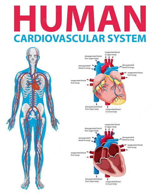
Right atrium: Two large veins deliver oxygen-poor blood to your right atrium. The superior vena cava carries blood from your upper body. The inferior vena cava brings blood from the lower body. Then the right atrium pumps the blood to your right ventricle.
Right ventricle: The lower right chamber pumps the oxygen-poor blood to your lungs through the pulmonary artery. The lungs reload blood with oxygen.
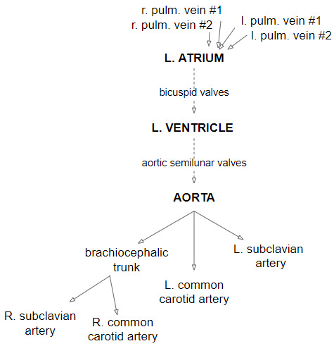
Left atrium: After the lungs fill blood with oxygen, the pulmonary veins carry the blood to the left atrium. This upper chamber pumps the blood to your left ventricle.
Left ventricle: The left ventricle is slightly larger than the right. It pumps oxygen-rich blood to the rest of your body.
Heart valves
Your heart valves are like doors between your heart chambers. They open and close to allow blood to flow through.
The atrioventricular (AV) valves open between your upper and lower heart chambers. They include:
Tricuspid valve: Door between your right atrium and right ventricle.
Mitral valve: Door between your left atrium and left ventricle.
Semilunar (SL) valves open when blood flows out of your ventricles. They include:
Aortic valve: Opens when blood flows out of your left ventricle to your aorta (artery that carries oxygen-rich blood to your body).
Pulmonary valve: Opens when blood flows from your right ventricle to your pulmonary arteries (the only arteries that carry oxygen-poor blood to your lungs).
Blood vessels
Your heart pumps blood through three types of blood vessels:
Arteries carry oxygen-rich blood from your heart to your body’s tissues. The exception is your pulmonary arteries, which go to your lungs.
Veins carry oxygen-poor blood back to your heart.
Capillaries are small blood vessels where your body exchanges oxygen-rich and oxygen-poor blood.
Your heart receives nutrients through a network of coronary arteries. These arteries run along your heart’s surface. They serve the heart itself.
Left coronary artery: Divides into two branches (the circumflex artery and the left anterior descending artery).
Circumflex artery: Supplies blood to the left atrium and the side and back of the left ventricle.
Left anterior descending artery (LAD): Supplies blood to the front and bottom of the left ventricle and the front of the septum.
Right coronary artery (RCA): Supplies blood to the right atrium, right ventricle, bottom portion of the left ventricle and back of the septum.
Electrical conduction system
Your heart’s conduction system is like the electrical wiring of a house. It controls the rhythm and pace of your heartbeat. It includes:
Sinoatrial (SA) node: Sends the signals that make your heart beat.
Atrioventricular (AV) node: Carries electrical signals from your heart’s upper chambers to its lower ones.
Your heart also has a network of electrical bundles and fibers. This network includes:
Left bundle branch: Sends electric impulses to your left ventricle.
Right bundle branch: Sends electric impulses to your right ventricle.
Bundle of His: Sends impulses from your AV node to the Purkinje fibers.
Purkinje fibers: Make your heart ventricles contract and pump out blood.
Conditions and Disorders
What conditions and disorders affect the human heart?
Heart conditions are among the most common types of disorders affecting people. In the United States, heart disease is the leading cause of death for people of all genders and most ethnic and racial groups.
Common conditions that affect your heart include:
Atrial fibrillation (Afib): Irregular electrical impulses in your atrium.
Arrhythmia: A heartbeat that is too fast, too slow or beats with an irregular rhythm.
Cardiomyopathy: Unusual thickening, enlargement or stiffening of your heart muscle.
Congestive heart failure: When your heart is too stiff or too weak to properly pump blood throughout your body.
Coronary artery disease: Plaque buildup that leads to narrow coronary arteries.
Heart attack (myocardial infarction): A sudden coronary artery blockage that cuts off oxygen to part of your heart muscle.
Pericarditis: Inflammation in your heart’s lining (pericardium).
1 note
·
View note
Text
Best Cardiac Hospital in India
The thick focal piece of the heart is comprised of cardiac muscle. It makes up one of three types of muscle in the body, the others being skeletal and smooth. For a better treatment of cardiac, you can think about the Best cardiac hospital in India. My Care India stresses the appropriate consideration and best treatment for its patients as a help association. A slim external covering called an epicardium and an inward endocardium circles the myocardium. Coronary supply routes give blood to the heart muscle, while cardiovascular veins channel it.
The fundamental cells that spread the word about the heart muscle are cardiomyocytes. Cardiomyocytes' chief intention is to contract, producing the tension expected for circling blood all through the circulatory framework. My Care India is an Indian clinical help organization. We help to find the best cardiac hospital in India. As a medical services help association, we want to help you find the best specialists, treatments, and establishments for your specific medical problem. My Care India is an Indian medical help service-providing organization. As a medical services help association, we want to help you find the best specialists, treatments, and foundations for your specific medical problem.
read more- https://mycareindia.com/best-cardiac-hospital-in-india.html
0 notes
Text
SEPT. 29 '23
... gifs for decoration tbd x)
it's funny how this works; myself and the two people i was sitting between were left with whiplash after this morning's 2hr H&AB class and it felt like i was never going to be able to begin to understand wtf that lady was talking about, but then afterwards with a free afternoon i took 3? hours to just take it slow, type out my simple handwritten notes from this morning, stare intensely at diagrams & read thru the lecture slides and U know what.... i'm not afraid anymore<3
NATURE OF NURSING: no classes today but started the beginnings of some digital notes on intro nursing theory, here's what my unfinished sheet on ADLs looks like so far lol!!

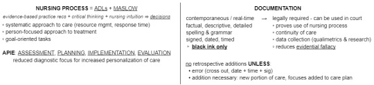
HEALTH & APPL. BIOSCIENCES: CARDIOVASCULAR SYSTEM TODAY AUGHHHH i'm TELLING YOU they're releasing NEW BIOLOGY bro WTF IS THIS SHIT !!!!!
blood pressure: force of blood pushing against inside walls of vessels
heart position: slightly left of midline deep to the sternum in the mediastinum (compartment of thorax)
pericardium: tough sac restricts ♡ mvmt -> only moves slightly in thorax
fibrous p.c. = outer covering of tough dense connective tissue
serous p.c. = parietal layer (lines inner surface of fibrous p.c.) + visceral layer (epicardium, covers outer surface of ♡)
pericardial cavity = space between parietal & visceral layers
♡ walls = 3 distinct layers
epi = visceral layer of serous p.c. + connective tissue
myo = cardiac muscle
endo = lining of ♡ chamber (touches blood)
external ♡ anatomy: bottom point of ♡ = inferior apex; top point of ♡ = superior base -> 2x small superior atria + 2x larger inferior ventricles // separated by coronary sulcus (deep groove, lets veins & arts sit nicely on surface)
aneurysm: abnormal swelling of heart or artery/vein walls // varicose veins -> weakened valves
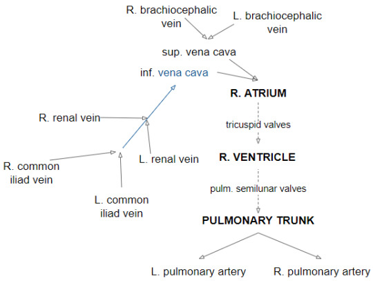
and now i present to you my desperate attempt to understand all these new tubes I JUST REALIZED I FORGOT THE CORONARY SINUS DRAINING INTO THE R. ATRIUM AUGHHHHH i also drew a diagram on paper of the heart yaaay
0 notes
Text
What is cardiology?
Cardiology is the study of the heart and treatment of the disorders relating to the heart. It is involved with the cardiovascular system (heart, blood, blood vessels). A cardiologist is someone who studies and treats the heart, but does not perform surgery or do major hands-on work with hearts. They often diagnose possible conditions, perform tests, and do less invasive procedures (See below). A cardiac surgeon does surgeries relating to the heart and they are more invasive with opening the chest cavity and working on the heart directly.
Some signs of heart issues that may make you need to see a cardiologist include: shortness of breath, dizziness, chest pains, abnormal heart rhythm or pace, high blood pressure.
Most times, you are going to see a cardiologist first and fully, not interacting with a cardiac surgeon unless you really need surgery from blockages, heart failure, or have heart valve issues. You might see a surgeon once, then return to the care of a cardiologist. A subsection of cardiology are congenital heart doctors who work with heart defects from work, but the usual cardiologist deals with issues as people age and external factors. A cardiac surgeon will monitor you after surgery, then return you back to your cardiologist and you must have a diagnosis from a cardiologist to get surgery.
Parts of the heart
What is the heart?
The heart is a muscular organ that pumps blood through the circulatory system to carry oxygen and nutrients around the body to keep it functioning. It works with your blood vessels as a path of giving blood.
Parts
There are 4 chambers to the heart (sections) that are controlled by electrical impulses (nutrients, neuro signals, and chemical reactions triggering responses in order to keep the muscle pumping). There are also walls, valves, and blood vessels.
Heart walls- they squeeze and relax in order to send blood through your system and there are 3 layers. There is also a wall in the inside of your heart to separate the right and left sides.
Endocardium- inner layer
Myocardium- middle muscular
Epicardium- protective outer
There is also a protective sac that is called the pericardium that covers your heart to keep it safe. It also self lubricates to prevent your heart from rubbing against your other organs and wearing its walls down.
Chambers- they are the internal parts of your heart that are divided into 4 sections to pass blood through successfully.
Right atrium- this is the start where oxygen deprived blood enters your heart through two larger veins. This is the upper right chamber.
Right ventricle- this section pumps the deprived blood to your lungs through your pulmonary artery where your blood is refilled with oxygen. It is the lower right chamber.
Left atrium- this gets the blood from the pulmonary artery, then pumps the fresh blood to your left ventricle. This is the upper left chamber.
Left ventricle- it pumps your blood back into the blood vessels and veins to the rest of your body. It goes to the valves first.
Heart valves- They open and close to let blood in and out of your heart. They are usually on top of your heart or out of sight. You have internal heart valves (Atrioventricular) and external heart valves (Semilunar).
Atrioventricular
Tricuspid- between right atrium and left ventricle
Mitral- left atrium and left ventricle
Semilunar
Aortic- open from the left ventricle and carries blood to the artery that has oxygen rich blood
Pulmonary- carries oxygen deprived blood and opens from the right ventricle
Blood vessels- there are 3 types and they all carry blood through your body
Arteries- carry oxygen rich blood from your heart to your other body parts
Left coronary- divides into 2 branches of artery near your heart
Circumflex artery- supplies blood to the left atrium along with side and back of left ventricle
Left anterior descending artery (LAD)- gives blood to the front and bottom of left ventricle and front of septum.
Right coronary artery- supplies blood to right atrium and ventricle and bottom portion of the left
Veins- carry oxygen poor blood to your heart
Caparillies- tiny and exchange oxygen rich blood and oxygen poor blood
Heart attack vs Cardiac Arrest
Basic definitions
Heart attack: a blocked flow of blood causes the heart to suffocate (clogging, slow)
Cardiac arrest: when the heart malfunctions and stops suddenly (rhythms, electrical impulses, etc.)
Heart attacks in depth
Occurs when an artery is blocked, not allowing oxygen rich blood through and effectively suffocating your heart (and it’s tissues)
Slow and dull
The heart usually does NOT stop beating during a heart attack, but it panics and the rhythm can go wack. It only stops when cardiac arrest happens and the electrical impulses get too weak
You can have many small heart attacks or infarctions, making small parts of your heart tissue to die before a bigger one happens that stops your heart fully
The longer without treatment, the worse the damage
Symptoms (DIFFERENT IN AFAB AND AMAB PEOPLE):
Chest pain (dull, pressure, tightness, squeezing)
Discomfort in shoulders, arms, tummy, chest
Fatigue
Heartburn
Cold sweat
Dizziness
Short of breath
Cardiac arrest in depth
Sudden and no warnings
Caused by electrical malfunctions in your muscles that disrupts the rhythm of your heart pumping
It makes the person go unconscious and makes the heart still entirely, just collecting blood, but not pumping it to the lungs and slowly suffocating the whole body
This happens a lot AFTER heart attack recovery and heart attacks increase this risk
Other diseases and terms
Heart disease- any disease or condition relating specifically to the heart
Cardiovascular disease- conditions affecting the heart and blood vessels
Atrial fibrillation (afib)- irregular impulses to your atrium (improper rhythm)
Arrhythmia- any heartbeat that is irregular, no consistency, too fast or too slow
Cardiomyopathy- unusual stiffness or thickening in the heart muscle
Congestive heart failure- when the heart muscle is too weak or stiff to pump blood
Coronary heart disease- plaque build up in the narrow arteries (clogged arteries)
Often leads to infarctions or heart attacks
Pericarditis- inflammation in your heart’s lining
Surgeries relating to the heart
Less invasive (can be done by a cardiologist)
Angioplasty- this opens clogged arteries by using a balloon like tool to widen the space
This is often combined with heart stenting that uses a tiny mesh tool to KEEP the space open with medications as well. This just widens the space, but does not remove the plaque like a surgeon would do
This is used during heart attacks to allow blood flow and to decrease the amount of damage down
Inserting a pacemaker- this is when a device is inserted into your chest and connects to your heart through wires to control your heartbeat or rhythm.
Single chamber- carries electrical impulses to the right ventricle
Dual chamber- carries electrical impulses to right atrium and right ventricle to control the timing of contractions
Biventricular- carries and controls electrical impulses to the left and right ventricles of the heart
Heart catheterizations- when a small, flexible tube is guided through blood vessels to diagnose or treat conditions. It is basically used as a look inside tool.
X Rays and other scanners- to look inside the chest without opening it. This is for the heart, lungs, and bones.
Ambulatory monitors- they are monitors that the patient can take home and is used to track arrhythmias outside of medical settings.
Stress tests- these are used in medical settings to see how your heart reacts when it works the hardest. You are taken vitals before, during, and after the “stress” period (running on a treadmill for example)
These are often for heart disease or coronary issues
EKG- taking vitals over your heart to track its beats and can show if you’ve had a heart attack and keeps things monitored.
More invasive (done by cardiac surgeons)
Coronary artery blockage removal- this is when your arteries are so clogged up with plaque that it prevents blood from flowing through effectively. This often leads to heart attacks and this surgery is often done when a heart attack or high risk is detected.
Heart valve issues: the surgeon can go in and work on your heart values/your chambers in order to make sure the blood flows in the right direction and your heart still works. This is either repairing or replacing.
Stenosis- when the valve flaps become stiff and possibly fuse together. This reduces blood flow as the passage is narrow
Prolapse- makes the blood flow backwards
Aortic aneurysm- the weakening of the aorta to the point it bursts and stops the blood flow entirely as the blood spreads to the rest of the body in not needed areas.
Abdominal- when it ruptures below the chest and it caused by hardened arteries
Thoracic- in the chest and caused by high blood pressure or injury
Some heart failure related issues, but that is discussed with a cardiologist first.
#heart attack#cardiac arrest#cardiac disease#cardiac surgery#hyperfixation#im hyperfixating again#we hyperfixating#hyperfixating? kinda check. i don't like using specific neurodivergency terms w/o a diagnosis but i sure do obsess with things completely#im hyperfixating so hard rn#help girl im hyperfixating#hyperfixiating#heart#heartcore#heart disease#mad science#cardiology#cardiophilia?#heartache#diseases of the heart#heart monitor#cardiologist#cardiac surgeon#recovery#research#my research#sciencecore#coronary#artery#anatomy#tw anatomical heart
1 note
·
View note
Text
Watch "ANATOMY OF THE HEART; EPICARDIUM; CARDIAC MUSCLES; HEART CHAMBER; ATRIA; heart diagram easy draw;" on YouTube
youtube
0 notes
Text
Anatomy & Physiology
Quick Reviews
➖Cardiovascular system
❑ A client’s electrocardiogram showing ST elevation in leads V2 , V3 , and V4 suggests an anterior-wall myocardial infarction.
❑ The left anterior descending artery is the primary source of blood for the anterior wall of the heart.
❑ The circumfl ex artery supplies the lateral wall of the heart.
❑ The internal mammary artery supplies the breast.
❑ The coronary arteries may receive a minute portion of blood during systole.
❑ Most of the blood flow to the coronary arteries is supplied during diastole.
❑ Breathing patterns are irrelevant to blood flow.
❑ Coronary artery disease accounts for 30% of all deaths in the United states.
❑ Atherosclerosis, or plaque formation, is the leading cause of coronary artery disease.
❑ A myocardial infarction is commonly a result of coronary artery disease.
❑ In atherosclerosis, hardened blood vessels can’t dilate properly; therefore,they constrict blood flow and block oxygen transport. As a result, oxygen can’t reach the heart muscle, resulting in angina.
❑ Diabetes mellitus is a risk factor for coronary artery disease that can be controlled with diet, exercise, and medication.
❑ Cholesterol levels above 240 mg/dL are considered excessive and are a risk factor for developing coronary artery disease.
❑ Total cholesterol levels below 240 mg/dL are considered below the nationally accepted levels and carry a lesser risk of coronary artery disease.
❑ A lipid panel tests the amount of total cholesterol, low-density lipoprotein cholesterol, high-density lipoprotein cholesterol, and triglycerides.
❑ Sublingual nitroglycerin is administered to treat acute angina.
❑ Coronary artery bypass surgery and percutaneous transluminal coronary angioplasty are invasive, surgical treatments for coronary artery disease.
❑ An electrocardiogram showing ST elevation in leads II, III, and aVF suggests occlusion of the right coronary artery.
❑ The right coronary artery supplies the right ventricle, or the inferior portion of the heart.
❑ Occlusion of the right coronary artery could produce an infarction in that area.
❑ The most common symptom of a myocardial infarction is chest pain, resulting from deprivation of oxygen to the heart.
❑ The correct landmark for obtaining an apical pulse is the left fifth intercostal space in the midclavicular line.
❑ The apex of the heart is the point of maximal impulse where heart sounds are heard loudest.
❑ Rescuers of adult victims should begin compressions rather than opening the airway and delivering breaths.
❑ The sequence for cardiopulmonary resuscitation is CAB (compressions, airway, breathing) rather than ABC (airway, breathing, compressions).
❑ Chest compression depth on an adult should be at least 2 inches (5 cm).
❑ All rescuers, trained or not, should deliver high-quality chest compressions by pushing hard to a depth of at least 2 inches (5 cm), at a rate of at least 100 compressions per minute, allowing full chest recoil after each compression, and minimizing interruptions in chest compressions.
❑ Trained rescuers should also provide cardiopulmonary resuscitation with a compression to ventilation ratio of 30:2.
❑ The outermost layer of the heart is called the epicardium.
❑ The epicardium is made up of squamous epithelial cells overlying connective tissue.
❑ The myocardium is the middle layer of the heart and forms most of the heart wall.
❑ The myocardium has striated muscle fi bers that cause the heart to contract.
❑ The heart’s inner layer is called the endocardium.
❑ The endocardium consists of endothelial tissue with blood vessels and bundles of smooth muscle.
❑ The serous pericardium has two layers: the parietal and the visceral layer.
❑ The pericardium surrounds the heart and the roots of the great vessels.
❑ The pericardium has two layers: the fi brous and serous pericardium.
❑ Pulmonic sounds can be auscultated at the left second intercostal space in the midclavicular line.
❑ Abnormalities of the pulmonic valve are auscultated at the left second intercostal space along the left sternal border.
❑ Aortic valve abnormalities are heard at the second intercostal space to the right of the sternum.
0 notes
Text
First-ever "epicardium" created
A team at the Technical University of Munich (TUM) has induced stem cells to mimic the development of the human heart. The result is a "mini heart" known as an organoid. It will allow the study of the earliest developmental stages of our hearts and boost disease research.
The human heart begins to form about three weeks after conception. This places the early stages of heart development at a time when women are often still unaware they are pregnant. This is one reason why we still know so little about many details of how the heart forms. Results from animal studies are not fully transferable to humans. Organoids developed at TUM may help researchers.
A sphere of 35,000 cells
A team working with Alessandra Moretti, professor of regenerative medicine for cardiovascular disease, has developed a method to create a "mini heart" using pluripotent stem cells. About 35,000 cells are spun into a sphere in a centrifuge. Over the course of several weeks, different signaling molecules were added to the cell culture according to a fixed protocol. "In this way, we mimic the signaling pathways that control the developmental program of the heart in vivo," explains Alessandra Moretti. The group has now published its work in the journal Nature Biotechnology.
The first-ever "epicardium"
The resulting organoids are about half a millimeter in diameter. Although they don't pump blood, they can be electrically stimulated and can contract like human ventricles. Professor Moretti and her team were the first researchers in the world to successfully create organoids containing heart muscle cells (cardiomyocytes) and cells in the outer layer of the heart wall (epicardium). In the young history of cardiac organoids—the first of which was described in 2021—researchers have previously created organoids using only cardiomyocytes and cells from the inner lining of the heart wall, the endocardium.
"To understand how the heart is formed, epicardial cells are decisive," says Dr. Anna Meier, first author of the study. "Other cell types in the heart, such as those connecting tissues and blood vessels, are formed from these cells. The epicardium also plays a very important role in forming the heart chambers." Named "Epicardium".
0 notes
Text
Spatially resolved multiomics of human cardiac niches
A cell's function is defined by its intrinsic characteristics and its niche: the tissue microenvironment in which it dwells. Here, we combine single-cell and spatial transcriptomic data to discover cellular niches within eight regions of the human heart. We map cells to micro-anatomic locations and integrate knowledge-based and unsupervised structural annotations. For the first time, we profile the cells of the human cardiac conduction system, revealing their distinctive repertoire of ion channels, G-protein coupled receptors, and cell interactions using a custom CellPhoneDB.org module. We show that the sinoatrial node is compartmentalised, with a core of pacemaker cells, fibroblasts and glial cells supporting paracrine glutamatergic signalling. We introduce a druggable target prediction tool, drug2cell, which leverages single-cell profiles and drug-target interactions, providing unexpected mechanistic insights into the chronotropic effects of drugs, including GLP-1 analogues. In the epicardium, we show enrichment of both IgG+ and IgA+ plasma cells forming immune niches which may contribute to infection defence. We define a ventricular myocardial-stress niche enriched for activated fibroblasts and stressed cardiomyocytes, cell states that are expanded in cardiomyopathies. Overall, we provide new clarity to cardiac electro-anatomy and immunology, and our suite of computational approaches can be deployed to other tissues and organs. http://dlvr.it/ShqVvx
0 notes
Text
HOW THE HEART WORKS
HOW THE HEART WORKS
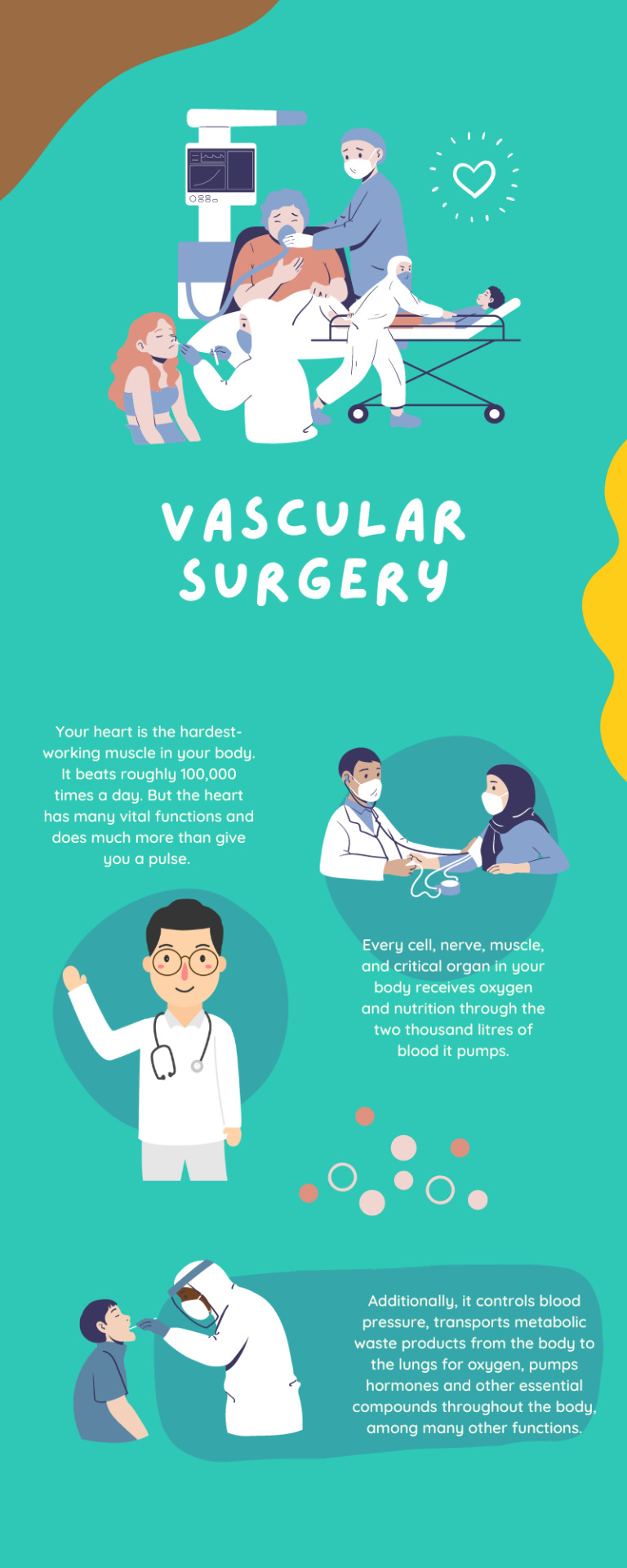
In the middle of your chest, between your lungs and behind the sternum, is the heart, an essential organ. The average adult heart weighs between 7 and 15 ounces and measures about the size of a giant fist.
The heart muscle may be tiny, but it works the hardest of any muscle in your body. It provides every cell, nerve, strength, and critical organ with blood that carries oxygen and nutrition.
The hub of your circulatory system, a network of blood vessels, including arteries, veins, and capillaries, transports blood to and from all body parts. It is made up of several layers of tissue.
Walls of the Heart
Three layers of muscular tissue make up the walls of your heart. Namely, the epicardium, myocardium, and endocardium. These muscles contract so that your body's blood can flow.
#Vascular Surgery#best vascular surgery hospital in india#vascular surgery services#vascular surgery hospital in gujrat
0 notes
Text
research: the heart
The heart is a vital organ in the centre of the circulation system, that pumps blood throughout the body in order to provide oxygen and nutrients to all parts of the body. As well as this, the blood also carries away any unwanted carbon dioxide and waste products. The heart consists of four main sections (called chambers) made of muscle and powered by electrical impulses. The brain and nervous system direct the heart's function.

the heart’s anatomy:
Heart walls: The walls of the heart are muscles that contract and relax to send blood throughout the body. The heart walls are made up of three layers - endocardium (inner layer), myocardium (muscular middle layer) and epicardium (protective outer layer).
Heart chambers: The heart is divided into four chambers, two on the left side and two on the right side of the heart, named the atria and the ventricles.
Heart valves: The heart has four valves that open and close to keep the blood flowing and moving in the right direction. These valves are called the aortic valve, the mitral valve. the pulmonary valve and the tricuspid valve.
Blood vessels: Blood is pumped through the body through three types of blood vessels:
Arteries: carries oxygen rich blood from the heart to all parts of the body.
Veins: the veins in the body carry blood that is lacking in oxygen back to the heart.
Capillaries: connect the smallest arteries to the smallest veins, and help exchange water, oxygen, carbon dioxide and other nutrients and waste substances between the blood and the tissues around them
Blood vessels are also able to widen or narrow depending on how much blood each part of the body requires.
Electrical conduct system: In order for the heart to keep pumping regularly, it requires electrical signals to be sent to the heart muscle, which tell it when to relax and contract.
bibliography:
Cleveland Clinic. (n.d.). Heart: Anatomy and Function. [online] Available at: https://my.clevelandclinic.org/health/body/21704-heart#:~:text=What%20is%20the%20heart%3F.
Reference listNHS Inform (2020). How your heart works. [online] Nhsinform.scot. Available at: https://www.nhsinform.scot/illnesses-and-conditions/heart-and-blood-vessels/about-the-heart/understanding-how-your-heart-functions.
0 notes
Last Seen Blogs

unpre-tentious
Somebody

evehealy
Eve Healy

hehedoesntdrawhere
hehe's other account

haught-pants
Probably Stoned Right Now

jennette-mccurdy
Jennette McCurdy