
scinote
SciNote Tumblog
connecting the world through science
220 posts
Don't wanna be here? Send us removal request.
Last Seen Blogs

insecure-aesthetic
a monet of my mind

valeriemassonbourde
Découpages et autres

wolf-makes-deadman-tell-no-tales
THE WOLF SANCTUM

myrtlebeachforfamilies
Myrtle Beach for Families

shittydumbstuff
sapphic
Note
Can a solar eclipse cause any medical issues in a human, or perhaps even amplify symptoms if they are already affected by a certain condition (i.e. reproductive organ problems)?
Not that I’m aware of. The only damage a solar eclipse can cause is vision problems, but that’s true of anytime really that you stare at the sun without protection.
6 notes
·
View notes
Note
Hello, sorry if this is a stupid question, but do you know how to make a homemade electrolyte that can produce a really high voltage? (Preferably, one that can produce more than 2.8 V?)
Its fairly easy to make a home made electrochemical cell using electrolytes, as all you really need are ions in solution (Na+, K+, etc) connected together.
But most of those don’t produce really high voltages. From what I’ve seen 0.5-1.5V is what you get from simple ions.
To get near 3V requires more advanced chemicals. I’m not aware of a home made electrolyte that can achieve that voltage range, but if you know, feel free to let me know.
3 notes
·
View notes
Text
How MRI works, Part III
We’re finally ready for the last leg of our journey! This one will require quite a bit of math, but it’ll be a stroll in the park after the last two posts.
Even with everything we’ve learned, how can MRI pinpoint a tissue to a specific location in the body? Today, we’ll be discussing how we can tweak the concepts we’ve talked about in order to provide spatial specificity. Everything we talk about today revolves around an idea we mentioned in Part II, so let’s review this really quickly.
For the RF pulse to be effective in rotating the proton’s magnetic moment to a desired angle, it has to come as close as possible to matching the frequency of the proton’s precession, which is called the Larmor frequency in MRI. If the RF pulse is out of sync with the Larmor frequency, it will be ineffective, kind of like pushing a swing at the wrong time. Now, a proton’s Larmor frequency depends on the strength of B0, the big magnetic field, at the specific location of the proton in question. This is a very important concept for what we’re going to be talking about in Part III, so keep this in mind!
Let’s first just worry about the z-axis of your body, so imagine slicing up your body into little z-slices from your top of head to your toes. It would be great if we could cause just the protons in one of these little slices to precess, while we keep all of the other protons aligned with B0, because we would then be able to measure the T1 or T2 relaxation of the protons in just this slice. Well, we can do so by setting up B0 as a slice-selection gradient, or GSS, that varies with location along the z-axis so that it is strongest at the top of your head and gradually gets weaker toward your feet, as shown in the picture below. Then, every slice along the z-axis will have a difference Larmor frequency, and we can use a RF pulse tuned to the specific Larmor frequency of the slice we want. In other words, our RF pulse can “select” the protons in a single slice along the z-axis of your body, like f1 in the picture, so that only those protons begin to precess. We call this process slice-selective excitation.

Remember, we’ve just selected a single slice-- only the protons in that slice, and no other protons, are precessing. Since our readout relies completely on precession (because we can only measure changes in magnetic fields, as discussed in Part I), we’ve essentially isolated the signal from this slice, and we can now manipulate the protons in this slice to give us more information without affecting other protons-- for example, we can now further isolate a single line of protons within the slice.
For this, we use a similar concept as before, but this time, we create a gradient in B0 in the y-direction, so that the magnetic field is strongest at your right ear and weakens toward your left ear, as shown in the picture below. Additionally, this time, we only turn on the gradient during readout, so that only the protons already precessing from slice-selection are affected. Since Larmor frequency depends upon B0, each line of protons from the right to the left of your body precess at a different speed. This localization technique is called frequency encoding, because this gradient encodes every line of protons within the slice with a different frequency depending on its location along the y-axis.

To measure the output from this, we use a math technique called a Fourier transform. You can read the intricacies of the math here, but essentially what you need to know is that combining waves with different characteristics and different frequencies creates a single wave with a unique structure. This also works in the reverse: we can to break down the composite wave into the waves that compose it. We pick up a single signal from all of the precessing protons, and using the Fourier transform, we can break down the signal into individuals signals from each line of protons.
The last piece of the puzzle is that we can turn on a third phase-encoding gradient in B0 in the x-direction. In MRI, the phase-encoding gradient is turned on briefly and quickly switched off before we turn on the frequency-encoding gradient. As shown in the picture above, the brief phase-encoding gradient makes the protons at the front of your body precess more quickly than the protons at the back of your body. When it’s turned off, all of the protons return to their normal speed, but their rotations are slightly delayed from each other, creating a phase shift dependent on their location along the x-axis. In the picture below, the bump in the gray line represents the gradient turning on, and the dashed line represents the rotation of a proton near the back of your head, where the phase-encoding gradient is minimal. The light blue line represents the rotation of a proton near the front of your head-- you can see that even though the two protons are precessing at the same frequency, there is a small time delay between their rotations.

So, just to summarize: All of MRI really revolves around this central idea of using magnetic fields to manipulate the magnetic fields inside your body in a way that can be measured and translated into an image. In an MRI, there’s a constant, large B0 from your head to your toes that lines up the protons in your body. We turn on a slice-selection gradient to “select” a single z-slice of your body by forcing the protons within that slice to precess. Then, we turn off this gradient and begin readout via the large current-measuring coils at the top of your head. During readout, we briefly turn on and switch off a phase-encoding gradient and then turn on a frequency-encoding gradient, effectively encoding every proton’s signal with its x- and y-position, which can be determined by its phase and frequency, respectively. (The loud sounds you hear inside an MRI are a result of all those gradients being turned on and off, which attract metals inside the machine and cause them to grate and strain against the other metals that hold them in place!) Lastly, we can parse out each proton’s signal by using a Fourier transform, and we can translate the signal to a single pixel in an MRI image, coloring it with a grayscale value that correlates to the T1 or T2 value of the tissue at that location in the body.
31 notes
·
View notes
Text
How MRI works, Part II
In our last post, we discussed the basic physics underlying magnetic resonance imaging, or MRI. But we haven’t built the complete picture yet-- even with all of the physics that we talked about last time, we’re still only left with the ability to use a magnetic field to create a smaller magnetization vector inside the body that can then be measured by the electric current it induces in a loop of wire. How do we use this to create images of different types of tissue?

If you recall from our last post, we talked about the MRI producing an excess of spin up protons in your body, the magnetic moments of which sum to the measureable net magnetization vector, or NMV. To make this easier to visualize, let’s think of each excess spinning proton as a top, just like the mind-blowing eternally spinning one from Inception. As a result of the big magnetic field B0 pointing toward your head in the MRI, each of these little tops is lined up along B0 with its stem pointing toward your head and its tip toward your feet. Let’s also think of the magnetic moment produced by the spinning proton, which we can visualize as an arrow pointing along its axis of rotation, as essentially analogous to the top’s angular momentum-- take a look at the arrow labeled L in the diagram below. In the absence of other forces, each proton acts just like a top set spinning with its stem perfectly vertical. It would keep spinning forever, precisely parallel to B0.

However, let’s say your top tips over just slightly, as it inevitably will in the real world-- now the force of gravity isn’t perfectly aligned with the top’s axis of rotation. In fact, gravity now has a component perpendicular to the top’s angular momentum, as in the diagram above. This perpendicular component applies a torque that causes the top’s axis of rotation to rotate around its tip, changing the direction of the top’s spin without changing its speed. This phenomenon, called precession, is actually what you see in that wobbling circular motion of a top-- see how its stem traces a circle, if viewed from bird’s-eye view?

We actually make use of precession for MRI. Remember how we said that the coil of wire at the head of the MRI can only measure changes in the z-component of the NVM? Well, we can change the z-components of our protons’ magnetic moments by making the protons precess. You see, when the magnetic moment is inclined so that it is no longer perfectly parallel to B0, its z-component shrinks. This is a simple matter of the Pythagorean theorem: try working it out to convince yourself that each leg of a triangle must be shorter than the hypotenuse. In fact, if we can get the proton to precess so that it is perpendicular to B0, its z-component will be 0.
We can cause precession by applying a little pulse of a magnetic force in the x- or y-direction, perpendicular to B0, to “pull” the proton’s magnetic moment (which lies along its axis of rotation) down and to the side. Let’s imagine we applied a little force to the right of the proton so that its magnetic moment tilts just a little to the right. Now, as its magnetic moment tilts forward and around to the left, we have to pull to the left in order to pull the magnetic moment further downward. So as the proton begins to precess, we have to apply an oscillating magnetic field, called a radiofrequency (RF) pulse, to pull its magnetic moment to the right as it rotates past the right, then left as it rotates past the left, then right again, left again, etc. in a swirl-- all the while pulling it downward until it lies perpendicular to B0. This can be a little hard to visualize, so if you’re lost, try watching the first half of this video (until 0:03)!
youtube
For this RF pulse to be effective in rotating the proton’s magnetic moment to a desired angle, it has to come as close as possible to matching the frequency of the proton’s precession, which is called the Larmor frequency in MRI. If the RF pulse is out of sync with the Larmor frequency, it will be ineffective, kind of like pushing a swing at the wrong time. Now, a proton’s Larmor frequency depends on the strength of B0, the big magnetic field, at the specific location of the proton in question. This is a very important concept for what we’re going to be talking about in Part III, so keep this in mind!
After we use an RF pulse at the Larmor frequency of the proton long enough to force it to precess around the z-axis at a 90 degree angle, we just have to work out how to turn this precession into something measurable that we can use to differentiate between tissues. One way to do so is to use the density of protons, usually from water, in that tissue. Another way, which we’ll be discussing in further detail, is to use relaxation times, like T1 and T2.
At the beginning of the MRI, when we just have B0 without any other magnetic forces, our magnetization vector (which is, again, just a fancy name for the sum of the protons’ little magnetic moments) lies completely along the z-axis, so it has a really large z-component. Then, we use the RF pulse to force a 90 degree precession, so all of the protons precess in the XY-plane and the z-component of the magnetization vector decreases to a much smaller value. After we stop the RF pulse, however, these protons will gradually precess back to align with B0, essentially doing the opposite of the video above. As the protons do so, the NVM’s z-component exponentially increases back to its initial value. T1, or spin-lattice, relaxation measures the time constant of this recovery, marking the time at which approximately 63% of the original z-component. Because other atoms nearby can affect how easy it is for protons to precess back upward to align with B0, T1 varies between different types of tissues. Watch the second half of the video above (after 0:03) to see T1 in action.
In addition, when we apply the RF pulse to rotate the protons’ magnetic moments into the XY-plane, all of our protons precess in sync, with all of their magnetic moments aligned so that the magnetization vector oscillates between having a really large x-component and a really large y-component. As time passes, though, the protons dephase, getting out of sync with each other so that the x- and y-components of their magnetic moments cancel out. T2, or spin-spin, relaxation measures the time that it takes for the x- and y-components of the magnetization vector to decrease by 63%. This value also depends on the protons nearby, so T2, like T1, varies between tissues. You can see a bird’s eye view of T2 dephasing in the video below, in which the translucent arrows represent individual magnetic moments, which sum to the big bright yellow arrow, or the magnetization vector.
youtube
Wow, that was a doozy. We zoomed in on individual protons, combining physics and math to figure out how to change their rotation to produce something that our wire coils can pick up and measure. In our next post, we just have to worry about the easy part-- how to localize different tissues to specific locations in your body. Stay tuned!
For further reading:
https://en.wikipedia.org/wiki/Angular_momentum
http://hyperphysics.phy-astr.gsu.edu/hbase/rotv2.html
https://en.wikipedia.org/wiki/Physics_of_magnetic_resonance_imaging
165 notes
·
View notes
Text
How MRI works, Part I
Magnetic resonance imaging (MRI) seems like a crazy idea, when you think about it. How is it even possible to use magnets to create a 3D map of your body?
First, we have to understand something about the little tiny protons in the nuclei of the atoms that make up your body. If we’re to be very reductionist, we can think of protons as essentially “spinning” positive charges, and in physics, spinning charges produce magnetic fields that can then interact with other magnetic fields.
In fact, the interaction isn’t all that different from what you would intuit from playing with bar magnets as a kid. Let’s do a thought experiment-- let’s say you had two bar magnets. How would you place it them so that, in the absence of external forces, neither one of them rotates due to the other’s magnetic field? Try it out, if you have a few magnets on hand, and see what you find!

Well, the obvious answer is that you can line them parallel to each other so that the poles on the first magnet align with the opposite poles on the other. The less obvious answer is that they can also be antiparallel to each other, so that both the North and South poles of the two magnets are lined up, as in the left panel of the diagram below. In this configuration, though, any little disturbance would cause the magnets to rotate and ultimately end up aligned in the first configuration, which is the most stable and energetically favored configuration.
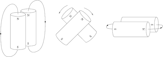
In the absence of a big magnetic field, the spins of the protons in your body go in all kinds of directions so that the little magnetic fields they produce effectively cancel each other out. But when there IS a big external magnetic field, the protons’ magnetic fields take the two configurations we mentioned above in the thought experiments. We call the energetically favored parallel configuration spin up, and the other antiparallel configuration spin down.
Because spin up is energetically favored and more stable, there are always more spin up protons than spin down protons. These excess spin up protons produce a small but measurable magnetic moment called the net magnetization vector, or NMV, that lies parallel to the big magnetic field.
Imagine you’re lying on MRI table. Let’s map your body onto a 3D coordinate system, with the x-axis going from back to front, y-axis from left to right, and z-axis from bottom to top. MRI uses a big magnetic field along the z-axis of your body, going from your feet to the top of your head. We call this field B0, and it lines up the protons in your body to produce a NMV that also goes from your feet to your head.

[Credit: MRI scan via Shutterstock]
Now, we just have to figure out how to measure this NMV. Actually, that’s what the big golden coil at the head of the patient in the illustration above is for. Remember when we said that spinning charges create magnetic fields? Well, it turns out that changing magnetic fields can also induce the movement of charges.
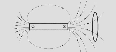
More accurately, changes in magnetic flux, or the number of magnetic field lines passing through a surface like the circle of space enclosed by the loop of wire in the diagram above, induce this movement of charge. Let’s imagine the NMV in your body as effectively a little bar magnet to the left of the wire loop. You see, this circle enclosed by the loop prefers to keep the number of magnetic field lines passing through it constant. If the the bar magnet were to hypothetically become stronger, more field lines would pass through the wire loop. Thus, the electric charges inside the wire would begin to flow in a way to produce a magnetic field inside the circle that counteracts the bar magnet’s effect on the flux. This phenomenon is called Lenz’s Law [1].
To put this all together, MRI uses a big magnetic field B0 to line up all the protons in your body to produce an NVM from your toes to your head. Whenever the z-component of your NMV inside the MRI system changes, it induces a current of a specific direction and strength within the coil at your head that can be easily measured.
So basically, even though you’re not normally a walking magnet, the MRI machine makes you into a tiny weak magnet, and then measures the strength and direction of your body’s magnetic fields to map you. Isn’t that awesome?
In Part II, we’ll talk about how to manipulate these basic concepts to map specific areas of your body. Stay tuned!
For further reading:
http://www.livescience.com/39074-what-is-an-mri.html
http://science.howstuffworks.com/mri4.htm
[1] https://en.wikipedia.org/wiki/Lenz%27s_law
171 notes
·
View notes
Text
The Biology of Brains
Our first topic for you, as requested by our readers, is the biology of brains!
Tell us everything you know about brains-- how they’re structured, and how they work. A few questions to get you thinking:
What’s the history of the biology of brains?
How are brains organized?
How do brains function?
How do brains differ across species?
Where do you think science is headed with regard to the biology of brains?
Note that all of these questions can be answered at many different levels and in thousands of different ways, not to mention that we encourage thinking outside of these guiding questions! We’re just asking you to give us a tiny bit of anything you know.
Again, the rules, for those new to our game:
Reblog and add anything that comes to mind regarding the topic– facts you know, things you learned in school, questions you have, links to an article, papers you’ve read– we mean anything!
If you don’t think you have anything to contribute, just reblog and pass the thread along to your followers.
That’s it! SciNote will be reading every single one of your responses, and given enough responses, we will combine your contributions, citing your work, in one big comprehensive article to be posted on SciNote.org.
18 notes
·
View notes
Text
SciNote Revamped

Dear Readers,
Happy 2016! It’s a new year, and SciNote’s excited to try something completely new.
In light of the enormous, amazing, and ever growing Tumblr Science Community, we’ve got an idea: we’re going to take our philosophy that anyone, and everyone, is a scientist to the next level. Let’s put all of our love for science together and tap the collective resource that our millions of brilliant, passionate minds create.
So here’s the idea (and the rules of the game):
Every week, we’re going to post a topic or a question for all of you to discuss. The topics will be pretty general, perhaps something like plants and evolution or biomimetics, and the post might come with a few guiding questions or ideas to get you started.
Then, it’s your turn. Your job is to reblog and add anything that comes to mind regarding the topic-- facts you know, things you learned in school, questions you have, links to an article, papers you’ve read-- we mean anything! It can be as long or as short as you like: you can write paragraphs if you like, but a couple of sentences would also be great. If you don’t think you have anything to contribute, just reblog and pass the thread along to your followers.
You don’t have to be an expert in the subject-- that’s the whole point of this! Our goal is just to get ideas and trains of scientific thought bouncing off one another and see where we wind up in the end. It’s like one big Tumblr brainstorm.
SciNote will be reading every single one of your responses, and given enough responses, we will combine all of your contributions, citing all of your work, in one big comprehensive article to be posted on SciNote.org.
This is as much an experiment for us as it is for you. We’re curious to see how this goes, and what you come up with. What do you say, Tumblr? Are you in?
95 notes
·
View notes
Text
Berries and Drupes and Pomes, Oh my! A Botanical Guide to True (and Untrue) Berries
Does a berry by any other name taste as sweet? Probably, since the majority of fruits that get lumped together under the term “berry” aren't actually berries, botanically speaking. So what, according to science, makes a true berry different from an untrue one? Let's start by stepping back for a second to consider how any fruit – berry or not – forms.
Fruit Formation
All fruits are the product of flower fertilization. The female reproductive organ of a flower is called an ovary; it holds one or more egg cell-producing ovules. When pollen (which contains plant sperm cells) lands on a flower's stigma (a part of the pistil), it triggers the development of one or more seeds inside the ovary, and a fruit begins to form. The ovary walls to grow into a soft, fleshy pericarp around the seeds, which forms the outer layer of a fruit. In fact, you will find that you know many botanically-labelled fruits as vegetables; when scientists create classifications, they are only concerned with structure, and not with whether you'd be more likely to put it on your cereal or your hamburger.
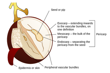
Illustration: https://en.wikipedia.org/wiki/Berry_(botany)
Berry True
A berry is a specific category of fruit. They are simple fruits, which form from a single ovary in a single flower. Because their pericarp is soft all the way through, they are further classified as fleshy simple fruits. Fleshy simple fruits are the true berries of the botanical world, and they include, among other things: grapes, tomatoes, persimmons, peppers, and eggplant.
To help organize the riotous fruit salad of botanical science, there are further subsets of berries. Pepos and hesperidium are modified berries, meaning that they have all the characteristics of berries, but also share additional characteristics amongst themselves. Pepo describes the berry structures of fruits such as squash, cucumber, watermelon, and pumpkins; these fruits have edible pericarps and rinds formed from the outer layer of their skin which may or may not be edible. Hesperidium refers to berries with a leathery skin and sectioned flesh surrounded by pith – think citrus fruits, like grapefruit, oranges, lemons, and kumquats.
When is a Berry not a Berry?
If it looks like a berry and tastes like a berry, it may nevertheless not be a berry – at least, not botanically speaking. It could, instead, be one of a number of other types of fruits – including drupes, pomes, aggregate fruits, accessory fruits, andepigynous or false berries.
A drupe is a fruit with a juicy pericarp and a hard endocarp (which is the inner layer surrounding the seeds). Drupes include stone fruits such as cherries, peaches, olives, and mangoes. Coconuts are also considered drupes, since they consist of a hard endocarp surrounding the seed embryo, which is embedded in the coconut flesh. The outer layers (the mesocarp and exocarp) are generally removed prior to sale.
A pome is a fruit that has its ovary at center, embedded in a core. Apples and pears are common pomes. You can, technically, eat the inner core that contains the seeds, but most people prefer not to.
An aggregate fruit emerges from the fusion of multiple ovaries in a single flower; unlike true berries, they do not conform to a one-ovary-one-flower rule. Blackberries and raspberries, for example, are formed from tiny individual drupes called drupelets, which each consist of a juicy pericarp surrounding a hard seed.
Related to aggregate fruits are accessory fruits. Accessory fruits, such as strawberries and pineapples, are created when tissue surrounding the plant ovary is incorporated into the formation of the fruit. You may hear of an accessory fruit referred to as a pseudocarp – a botanical term which means false fruit – which isn't accurate since accessory fruits definitely are fruits. They just aren't berries.
An epigynous or false berry is formed from a flower with an inferior ovary. This doesn't mean that the ovary is bad or unhealthy; rather, it describes its position below the rest of the flower (in contrast, superior ovaries sit above the surrounding flower tissues and semi-inferior ovaries sit at the same level as them). Epigynous berries incorporate some of the surrounding flower tissue into the bodies of their fruits. The little star patterns on blueberries, cranberries, and the ends of bananas give them away as epigynous fruit; the mark is actually a part of the flower that the fruit has held on to.
Seek out Botanical Berries
Now that you know what to look for in a true berry – and a number of not-so-true berries – you'll be able to identify them anywhere. Take a look around the next time you go grocery shopping. How are all of the berries classified in the produce section? Grocery store definitions don't always line up with botanical truths. Think about why that might be.
Once you're home, take a look at some of the fruits and vegetables you bought. Are any of them true berries, or other types of fruits labelled as berries? Cut open a pepo and a pome if you have them to see how their inside structures differ. And make sure to enjoy your tasty purchases, whether you put them in a pie or on the grill!
Grilled Eggplant
3 pounds eggplant, cut into 1/2-inch slices
3 tablespoons coarse salt, plus more for serving
1/3 cup extra-virgin olive oil
Ground pepper
Sprinkle eggplant slices with salt on both sides. Place in a colander set over a bowl; let stand 1 hour to drain. Discard liquid andrinse eggplant slices under cold running water. Place on several layers of paper towels, and press out the water.
Preheat a grill or grill pan over medium-high heat. Generously brush both sides of eggplant slices with oil and sprinkle with pepper. Place on grill and cook until browned (5 to 6 minutes). Flip eggplant slices and cook until browned on bottom. Serve hot, drizzled with olive oil and sprinkled with salt and pepper. Recipe courtesy of Martha Stewart Living.
Berry Salad (Although you should probably call this “Accessory and Aggregate Fruit Salad”)
1/4 cup fresh mint, chopped
2 tablespoons sugar
1/2 (16-ounce) container strawberries, hulled and chopped
1 (6-ounce) container blackberries
1 (6-ounce) container blueberries
1 (6-ounce) container raspberries
Crush mint and sugar in a mortar and pestle until well-blended (or place sugar and mint in a blender or food processor and pulse until well-blended). Place mint-sugar in a large bowl and add strawberries, blackberries, blueberries and raspberries. Gently toss until evenly combined. Recipe courtesy of Whole Foods.
Sources:
1. http://study.com/academy/lesson/fruit-definition-types-benefits-examples.html
2. https://www.hort.purdue.edu/newcrop/morton/banana.html
3. http://waynesword.palomar.edu/fruitid1.htm
4. http://www.loc.gov/rr/scitech/mysteries/coconut.html
5. http://www.botanical-online.com/flores1angles.htm
6. McGee, Harold (November 16, 2004). On Food and Cooking: The Science and Lore of the Kitchen. Simon and Schuster. pp. 247–248. ISBN 0-684-80001-2.
7. S.J. Meades, D. Schnare, and K. Lawrence and C. Faulkner. (2004 onwards). Northern Ontario Plant Database Website. Version 1, January 2004. Algoma University College and Great Lakes Forestry Centre, Sault Ste. Marie, Ontario, Canada.
By Jenna L, Writer
Edited by Carolyn Dallimore
63 notes
·
View notes
Text
Not All Ice is the Same...
On this water world we live on, only a small fraction is frozen—about 2 percent. But even that small amount of ice can be devastating if it melts. If all of the ice sheets melted in Greenland and Antarctica, world sea levels would rise over 200 feet, causing widespread coastal flooding.
A lot of media attention is focused on the Arctic ice, especially because so much of it melts during the summer. As the ice melts, it exposes the darker open water. This darker water soaks up much more solar heat than the white and reflective sea ice. Absorbed heat turns up the thermostat on the planet, making the problem increasingly worse.
Because of the important role it plays in preventing further warming of the oceans, Arctic ice is, for good reason, a matter of great concern to scientists. But did you know that even if all the ice in the Arctic Ocean melted, sea levels wouldn’t rise at all? This is because the ice on top of the sea is already part of the ocean, displacing its own weight in liquid water. As it melts, the total amount of water in the ocean doesn’t change, so the levels stay constant.
You might be a bit suspicious about this, but you can do a simple experiment in your kitchen to prove it. Take a glass, and fill it most of the way with water. Drop an ice cube in it, and then slowly top off the glass with tap water, taking care not to let it overflow. Watch as the ice melts. You’ll notice that the water level won’t change, and your glass won’t overflow. The ice floating in the ocean behaves the same as your ice cube, remaining part of the sea, but not adding to the total volume.
The more direct danger to the millions of people living on coastlines around the world is land-based ice. When that ice melts, much of the water released runs off into the ocean, raising the total amount of seawater. Greenland alone has enough frozen water sitting on it to increase global sea levels by 20 feet. If Antarctica, with an ice sheet covering more area than the US and Mexico, melts, sea levels will jump 200 feet! (To see what even a small rise of up to 6 feet would do, check out this interactive tool provided by NOAA that lets you dial in your own amount of coastal flooding.)
In the glass-and-water simulation, melting land ice would be like holding an ice cube in a sieve over a full glass, and waiting for the ice to melt. If you let it drip through the sieve into the water below, the glass will slowly overflow, flooding your counter.
An interesting note: sea ice generally doesn’t raise sea levels, but scientists are still concerned that widespread melting of certain kinds of sea ice will indirectly cause sea level problems. In the large bays of the Antarctic continent, large expanses of ice, like the Ross Ice Shelf float on the Southern Ocean, silently guard coastlines half a world away. How do they do this? By holding back the land glaciers, ice shelves slow their advance to the sea. When the ice shelves collapse or melt, the land glaciers accelerate their downhill flow, dumping loads of liquid water and icebergs and raising sea levels. This graphic from the National Snow & Ice Data Center illustrates the problem quite clearly:

[Effects of ice shelves on glacial flow; https://en.wikipedia.org/wiki/Ross_Ice_Shelf#/media/File:Glacier-ice_shelf_interactions.svg(NSIDC via Wikipedia)]
References and Further Reading:
Frequently Asked Questions on Arctic Sea Ice, NSIDC
Global Warming, NASA Earth Observatory
Greenland Ice Sheet Today, NSIDC
Quick Facts on Arctic Sea Ice, NSIDC
Quick Facts on Ice Sheets, NSIDC
Ross Ice Shelf, Wikipedia
By Dan L., Writer
Edited by James H., Editor
36 notes
·
View notes
Text
Kirigami Creates New Graphene Structures

[Image: "Graphen" by AlexanderAlUS - Own work. Licensed under CC BY-SA 3.0 via Wikimedia Commons - https://commons.wikimedia.org/wiki/File:Graphen.jpg#/media/File:Graphen.jpg]
Cornell researchers get crafty in their paper published in August's Nature.
A group at Cornell University has been playing with graphene and combining it with a technique called kirigami. Like origami, kirigami is Japanese paper folding, but with a twist-- you can cut the paper in addition to folding it.
Graphene is an allotrope (form) of carbon, in which the carbon atoms are arranged into stacked single-atom thick layers of hexagons. This means you can get microscopic sheets of graphene-- check out the picture above to see what a graphene sheet would look like at the atomic level! Graphene may prove very useful for scientists and engineers because it has electronic properties that may allow it to replace silicon in microchips. In order to build the graphene structures necessary for tiny nanodevices, however, scientists first needed to figure out how to manipulate graphene effectively. Noting that the mechanical behavior of the graphene was similar to that of regular old paper, the Cornell researchers decided to test the application of kirigami-inspired techniques to create their graphene structures.
The first step for the researchers was, naturally, to figure out how to move the graphene sheets. Although monolayer (single-atom) graphene sheets could be made through a process called chemical vapor deposition, which can be thought of as "condensing" a vapor-like form of carbon onto the surface of a piece of copper, moving these microsheets around was a lot more difficult-- graphene, at this thickness, very easily crumples and sticks permanently to itself. The researchers solved this problem by floating these graphene sheets in water with surfactants, which act like soap to make the sheets slippery and easier to handle.
The next step was to head to the Art Department to create full scale models of kirigami structures they wanted to build. After making the models, they began the process of building with graphene. To begin, graphene was bonded with aluminum, along with gold pads on either side of the graphene to limit direct contact from the researchers. Then, light was used to harden and strengthen only the graphene they wished to keep. Finally, unwanted graphene was removed with oxygen plasma and the aluminum removed with a weak acid. All in all, the process isn't too much unlike its inspiration; just as kirigami requires you to trace along the patterns you want to keep with scissors and discard the pieces you cut out, working with graphene in this way is a subtractive form of manufacturing.
This allowed the team to create graphene springs and pyramid shapes that work as flexible transistors, which could become a key component in wearable electronics. It could also enhance personal technology, with the creation of folding tablets and TV screens. Medically, it could be used for comfortable heart sensors and medical devices for 24/7 use by at-risk patients. Additionally, the authors write that this graphene construction has potential to create sensors that respond to the tiniest amounts of force, improving detection. The Cornell researchers intend to pursue all avenues to find applications for this amazing new method.
Paper Source: http://www.nature.com/nature/journal/vaop/ncurrent/full/nature14588.html
Press Release: http://news.cornell.edu/stories/2015/07/paper-graphene-twists-folds-nanoscale-machines
Written by Adam M., Discoverer
Edited by Peggy K.
229 notes
·
View notes
Text
Anthropology Meets Palaeontology with Prehistoric Primates
The general population often confuses archaeology and paleontology. Paleontology is the study of life on the scale of millions of years, usually by way of fossil remains. Archaeology seeks to understand human culture by way of man-made artifacts and is itself a subset of anthropology, a field of study that seeks to understand humanity. Biological anthropology seeks to understand humans from a biological and evolutionary standpoint, and as such intersects in part with paleontology to understand human descent. Prehistoric primates hold important keys to the perennial question of human evolution. Indeed, a recent study from the University of Kansas brings new information to light about prehistoric baboons, which in turn may help us understand the timeline of human evolution.
Papio angusticeps is a fossil baboon dating from the Plio-Pleistocene, roughly two million years ago. Such a fossil is important to understand when baboons diverged from other primate groups. Previous studies suggest that such a baboon occupied a woodland or savannah ecosystem and was preyed upon by other organisms (McKee and Keyser, 1994).
It is also important to understand what Africa was like during the Plio-Pleistocene. Scientists accept that this was an era of vast diversification for mammals in general, with Homo erectus arising immediately prior to P. angusticeps. The diversification of these mammals was likely due to a series of punctuated climate shifts, with the relevant shift here occurring at roughly 2.8 million years ago. This climatic shift led to arid, cooler and drier conditions that were conducive to several mammalian groups (deMenocal, 2004).
As with most paleontological work, the researchers had access to incomplete fossils, and were only able to analyze the cranium, or skull, of the organism. It is interesting to note that this incomplete skull was the only non-hominid, or non-human fossil material recovered at the excavation site, which places this early baboon as co-existing with human ancestors (Gilbert et al., 2015).

[Photographs of P. angusticeps cranium; from Gilbert et al. 2015]
P. angusticeps was part of a very different, dynamic African ecosystem. However, its placement in the timeline helps us better understand the sorts of ecological roles primates (and more specifically, non-hominid primates) played in Africa relatively recently, which in turn helps us understand the path to anatomically modern humans.
References
Gilbert, C.C., Steininger, C.M., Kibii, J.M., Berger, L.R. (2015). Papio cranium from the hominin-bearing site of Malapa: Implications for the evolution of modern baboon cranial morphology and South African Plio-Pleistocene biochronology. PLoS ONE 10(8): e0133361. doi:10.1371/journal.pone.0133361
deMenocal, P.B. (2004). African climate change and faunal evolution during the Pliocene-Pleistocene. Earth and Planetary Science Letters 220(1-2): 3-24. doi:10.1016/S0012-821X(04)00003-2
McKee, J.K. and Keyser, A.W. (1994). Craniodental remains of Papio angusticeps from the Haasgat cave site, South Africa. International Journal of Primatology 15(6): 823-841. doi:10.1007/BF02736070
Written by Joe C., Discoverer
Edited by James H., Editor
28 notes
·
View notes
Text
The Chemistry of Aquaponics
Aquaponics is a hybrid food technology system: it combines conventional aquaculture (tank rearing of aquatic animals) with hydroponics (the growing of crops in nutrient-rich water, without soil). This system is a valuable alternative both to traditional agriculture and to fishing and fish farming; its advantages include water conservation, sustainability, and the eliminated need for soil. Aquaponics allows for successful farming in areas where arable soil is not available, such as urban centers. It also has potential value for sustaining life in outer space.
The basic mechanism behind aquaponics is the recirculation of water. Fish are kept in a tank of water where their waste products (which contain nitrogen), are allowed to accumulate. Before the nitrogen concentration reaches toxic levels, the waste-filled water is cycled down to the plants, which are able to utilize the nitrogen as a nutrient. This purifies the water, which is then cycled back to the fish, allowing the process to begin again.
Aquaponics systems rely on specific nitrogen chemistry to function; in fact, the aquaponic nitrogen cycle is essential to yielding a successful harvest. Fish produce ammonia, a compound of nitrogen and hydrogen with the formula NH3, during excretion, which is turned into ammonium, NH4+, when it reacts with water in an ionizing reaction. Bacteria then convert the ammonium to nitrates, NO3-, which are the main nitrogen source for most plants. As the plants use the nitrates, nitrogen is removed from the water, purifying it for the fish. Provided that the chemistry of the system remains balanced, aquaponics makes for healthy plants and fish.
References:
http://horttech.ashspublications.org/content/21/1/6.full
http://www.usc.edu/org/quikscience/Projects/2013/HS/Kamehameha-RP.pdf
http://afsic.nal.usda.gov/aquaculture-and-soilless-farming/aquaponics
By Akshata Yalvigi, Writer
Edited by Carolyn Dallimore
#water#aquaponics#agriculture#biology#sustainability#noting science#Writer: Akshata Y#Editor: Carolyn D
261 notes
·
View notes
Text

Question:
What is the relationship between the genetic code and protein folding?
Asked by anonymous
Answer:
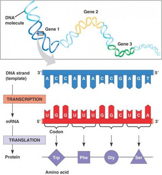
Basically, the genetic code (that is, DNA) dictates the sequence of amino acids that makes up a protein, with amino acids being the building blocks of the final protein. These amino acids then determine the folded shape of the protein by the way they make bonds and interact with neighboring amino acids, something that is dictated by the sequence in which they're chained. In this way, the genetic code determines protein folding. However, it's actually a little more complicated than that because there are many other factors that affect protein folding.
During the preliminary stage of protein synthesis, the gene transcript itself (the messenger RNA, mRNA) is edited and sections of RNA can be removed and added. Even before transcription of mRNA, there are factors that can regulate the expression of particular parts of the genetic code, meaning that there are proteins that can turn on or turn off gene expression. The DNA sequence is transcribed into mRNA by RNA polymerase and then translated by a ribosome into a chain of amino acids. Each codon, a unit of three bases in the genetic code, ends up corresponding to one amino acid. Of course, both transcription and translation are also imperfect processes where mistakes can often be made, and these will of course affect the end result, although such changes can have little or no impact on the overall shape of a protein.
[Image source: http://hubpages.com/hub/protein-production-a-step-by-step-illustrated-guide]
The ultimate shape of a protein comes from the fact that it "wants" to be in its lowest energy state, which means that the shape it takes needs to require as little energy input as possible. Immediately after a protein is translated, or synthesized, in a ribosome, it spontaneously twists and condenses to form the most energetically stable shape. Each amino acid along the chain helps to determine how it folds based on the properties of the amino acid side-chains. These side-chains are polar or non-polar, acidic or alkaline, etc., and all have different chemical properties that will influence folding. Protein structures develop in different stages of increasing complexity. A freshly synthesized, unfolded and unmodified protein has what is referred to as a primary structure. Secondary structures, such as alpha helices and beta pleated sheets, develop from amino acids side-chains interacting and forming hydrogen bonds with each other. Tertiary structure describes when all the beta sheets and alpha helices condense into the overall three-dimensional structure of the protein. Some proteins go on to bind with other proteins and gain an additional quaternary structure.

[Image source: https://commons.wikimedia.org/wiki/File:Protein_structure.png]
Most of these properties come from the pattern of hydrophilic (polar) and hydrophobic (nonpolar) amino acids. Generally speaking, the most stable structure is going to be the one that folds hydrophobic acids away from the solution the protein is in, hiding them away on the inside of the structure and closing them off with hydrogen bonds while wrapping its hydrophilic acids around its exterior where they can interact with the solution. Amino acids that can form hydrogen bonds with others also influence much of the protein folding. In that sense, the amino acid sequence is what causes the protein to fold the way it does, and that sequence is what the genetic code dictates.
Other factors can also influence the formation of protein structures. For instance, the environment that the folding process takes place in has a huge effect on the final structure. Most protein synthesis takes place in cytoplasm of the cell, which is almost entirely water, but the pH, the temperature, and the presence of other enzymes interacting with the protein as it develops can change its structure. This is also one of the reasons why proteins may form "incorrectly," or differently than they usually do. Since a protein's chemical function is based almost completely on its shape, abnormal biological protein folding can result in a protein that is useless or harmful to the body that created it.
Additionally, some proteins require the assistance of other proteins to fold them because their ability to randomly reach the most stable state has too high of an activation energy. This means that the energy required for them to fold into their most stable form is too great to accomplish by random folding alone and these proteins require some assistance from enzymes called chaperone proteins. Without chaperones, some proteins form improperly. But for most proteins, the folding process occurs randomly and spontaneously, and the main determinant of folded structure is the primary amino acid sequence dictated by the genetic code.
Sources and more information:
The Science Behind Foldit, a game used to apply human intuitive skills to protein folding simulation
Nature Publishing Group’s Horizon Symposia’s page on protein folding by Joachim Pietzsch
A video on the process of transcription and translation
A longer video from Crash Course about protein synthesis
Answered by Lauren W., Expert Leader
Edited by Carrie K.
#protein#biology#DNA#genetic code#science#Seeking SciNote#Expert: Lauren W#Expert: Chloe L#Editor: Carrie K
57 notes
·
View notes
Text
The Prehistoric Bat Family
Bats are an iconic part of the cultural consciousness. The World’s Greatest Detective has taken a bat for his own sigil, and he leads a group of like-minded crime fighters. But this discussion detracts from how purely interesting bats are in their own right. As mammals gifted with unique forms of locomotion, several bat species play interesting ecological roles.
A study from earlier this year published in PLoS One outlined a newly discovered bat fossil dating to roughly 19 to 16 million years ago from New Zealand. The fossil was classified as Mystacina miocenalis, and has a modern relative that occupies comparable geographic regions: the New Zealand short-tailed bat (Mystacina tuberculata). While the geographic overlap is undeniable, it should be noted that because M. miocenalis lived so long ago, New Zealand’s local environment was vastly different. We can use known information about both of these bats in order to understand them more completely.

[Anatomical drawings of Mystacina tuberculata; https://commons.wikimedia.org/wiki/File:MystacinaTuberculataFord.jpg]
M. miocenalis lived during the Miocene, as its name might suggest. The Miocene was a period of vast global change from roughly twenty to ten million years ago, perhaps best summarized as the transition from a world without ice caps to one with them. New Zealand is of particular interest to geology and paleontology due to its isolated nature. In fact, some geologists employ a separate geologic timescale to highlight New Zealand’s unique geologic history. The names utilized in the New Zealand Geologic Timescale are drawn from prominent locations and formations across New Zealand. For example, this fossil dates to the Pareora Series, roughly 19 to 16 million years ago, which roughly correlates to the Early Miocene. This timescale is continually calibrated, and significant work is done to ensure it is a meaningful marker of time, comparable to the timescale used in other countries (Kennett 1967; Scott 1972).

[Teeth comparisons; from Hand et al. 2015]
The primary fossil used in discussion for M. miocenalis are its teeth, which provide important information regarding its diet. The size of the teeth also holds important information regarding the size of the organism: it was roughly thrice the size of its modern relatives. It is best to understand these findings regarding M. miocenalis in relation to the modern New Zealand Short-Tailed Bat.
The New Zealand short-tailed bat is a ground-dwelling, burrowing bat (Daniel 1979). Likely in part due to its size, M. miocenalis was also a terrestrial bat, and foraged along the forest floor similarly to its modern counterpart (Hand et al 2015). A large part of the New Zealand Short-Tailed Bat’s diet is fruit and other plant matter, supplemented by arthropods like insects and spiders (Daniel 1979). Based upon its teeth, we can conclude that M. miocenalis likely had a comparable diet of nectar and pollen, also supplemented by insects and the like (Hand et al 2015). Pairing these two features, we realize that the bats of New Zealand have a very unique role in their forest ecosystems: these bats act as pollinators and seed dispersers, in addition to managing the populations of smaller consumers.
In short, the M. miocenalis fossil tells us about the changes in local climate in New Zealand during the Miocene, and may be best understood in reference to its modern relative. Perhaps most importantly, the connection between these bat species across millennia cements these bats as an important part of the New Zealand ecosystem as far back as 16 million years ago.
References
Hand, S.J., Lee, D.E., Worthy, T.H., Archer, M., Worthy, J.P., et al. (2015). Miocene fossils reveal ancient roots for New Zealand’s endemic Mystacina (Chiroptera) and its rainforest habitat. PLoS ONE, 10(6): e0128871. doi: 10.1371/journal.pone.0128871
Daniel, M.J. (1979). The New Zealand short-tailed bat, Mystacina tuberculata; a review of present knowledge. New Zealand Journal of Zoology, 6(2): 357-370. doi: 10.1080/03014223.1979.10428375
Kennett, J.P. (1967). Recognition and correlation of the Kapitean Stage (Upper Miocene, New Zealand). New Zealand Journal of Geology and Geophysics, 10(4): 1051-1063. doi: 10.1080/00288306.1967.10423208
Scott, G.H. (1972). Revision of Hutchinsonian, Awamoan and Altonian Stages (Lower Miocene, New Zealand) – 2. New Zealand Journal of Geology and Geophysics, 15(1): 49-70. doi: 10.1080/00288306.1972.10423946
Written by Joe C., Discoverer
Edited by James H., Editor
27 notes
·
View notes
Text
Is using an electronic device before sleep bad for your health?
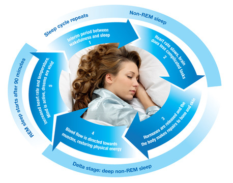
[The brain goes through a process of different levels of sleep throughout the night, and it has different physiological effects, some of which are still not completely understood. Image source: http://www.katkremp.com/wp-content/uploads/2015/03/Sleep-Cycle.jpg]
If you are reading this online close to or after your bedtime, I suggest you go to sleep and come back to this in the morning. A study published in Proceedings of the National Academy of Sciences (PNAS) shows that it may be beneficial to reduce reading from back-lit devices, like computers and smartphones, before sleep.
A quick word about the importance of sleep. Most of us are aware that sleep is important for a healthy brain function and physical health. Sleep promotes learning (i.e. you might be better off sleeping the night before a big exam) and repairing of heart and blood vessels, and controls of growth and development.
When it comes to sleep, both quantity and quality matter. Quantity-wise, if you are a teenager between ages 12-18, you need at least 8.5 hours of sleep. For adults over age 18, you need at least 7.5 hours of sleep. Even just losing 1 or 2 hours of sleep per night for several days can severely impair how you perform during the day.
The study done by PNAS found that reducing reading of backlit screens before bedtime increases the quality of sleep of its participant. This is particularly pertinent as a recent survey showed that nearly 90% of Americans use some type of electronic device at 2-3 nights per week within 1 hour before bedtime. Light, including that from electronic devices, is one of the major cues that influences the human circadian clock and may contribute to sleep deficiency.
The study showed that from subjects who read from an iPad before bedtime showed:
1. Nearly 50% reduction in plasma melatonin level. Melatonin is an important hormone in regulating the circadian rhythm. Circadian rhythm is the body's internal clock that regulates when we sleep, when we wake, and indirectly controls when we are hungry.
2. Longer time to fall asleep. While this time was only 10 minutes, this can easily add up to large numbers after a long time. Also, subjects’ circadian clocks were delayed by more than 90 minutes the following day, which may compound the problem by causing individuals to fall asleep at a later time.
3. Significantly less rapid eye movement (REM) sleep. REM sleep is the phase of sleep that stimulates the brain regions responsible for learning. Scientists have found that individuals who have REM sleep after learning a new task have increased retention of what they have learned, versus those individuals who do not experience REM sleep.
4. Taking a longer time to feel “alert” the following morning. The researchers speculate that the negative effect is possibly due to short-wavelength blue light emitted from electronic devices, but further study needs to be done to confirm and elucidate the potentially long-term effects.
References
http://www.helpguide.org/articles/sleep/how-much-sleep-do-you-need.htm
http://www.pnas.org/content/112/4/1232.full.pdf
http://www.nhlbi.nih.gov/health/health-topics/topics/sdd/why
For additional information, check out Tuck Sleep!
--
By Lee H., Discoverer
Edited by Peggy K. Editor.
32 notes
·
View notes
Text
Seeking SciNote, Astronomy: Landing on the Moon

Question:
Do you think we should land humans on the moon again, if so, why? Also, if so, where on the moon should we land them?
Answer:
Let me start with a disclaimer: I'm setting aside the issue of whether the mission to the Moon should be funded by the government or private industry. That's a really interesting question in its own right, but I'm not going to address it here. Let me also say that I'm not an expert on this sort of thing, though I find it really interesting and try to learn more when I can.
Disclaimer aside- yes, I think humans should absolutely land on the Moon again. The rationale for going fall into three broad categories: the philosophical/evolutionary, the historical, and the scientific.
First, the philosophical/evolutionary reason for returning to the Moon. We as a species have a unique desire to dive headfirst into the unknown with optimism and courage. The Moon may not be the unknown, sure, but it's certainly not fully known, either. We have this spirit implanted in us that seeks a frontier in order to push it back. We see a boundary and wonder how we can get past it.[2] We should go to the Moon because we're exploratory by nature, and landing on the Moon is exactly the sort of thing that we do.
Next, the historical reason we should go back. When America landed men on the moon in 1969, it wasn't for science or fulfill an evolutionary urge to push boundaries. Our spaceship was a giant missile with men strapped to it- we wanted to show the Soviets that our giant missiles were better than their giant missiles. We should land people on the Moon now, not as a show of nationalism but of cooperation. The journey should be an international effort with everyone sharing in the enterprise, the science, and the benefits.
The International Space Station is a great first step along this path but we should set our sights higher. The Apollo program and its successors gave us a worldwide perspective; they gave us image after image of Earth as a single unit, not as a discrete set of divided landmasses. I think that perspective is invaluable and something that an international collaboration that sends humans back to the moon would engender.
The scientific reason for sending humans back to the Moon is pretty straightforward: humans and rovers do science differently. Humans go off the beaten bath; we meander our way to the goal; we test gravity by dropping a hammer and a feather at the same time; we stop to see how far we can throw a rock, hit a golf ball, and jump. Rovers have led to fantastic advances in science and we've learned extraordinary amounts from them, but the human brain notices what cameras might not and the human body interacts with objects in ways rovers certainly don't. I think that there is more to learn on the moon and I think that human explorers – human scientists – are going to be a key part of the discovery.
Now to the second part of your question, which is where we should land on the surface. Given that I just Googled "map of the lunar surface" (I told you I'm no expert on the Moon [3]), I'm instead going to tell you what kind of place I think we should look for. These are my personal opinions, so there may be perfect places professionals have picked out that I just don't know about. But, first, I think we should land close to where rovers have been but not follow directly in their footsteps – or their tire tracks, as it were. Second, it would be great to land near a relatively recent crater, though "relatively recent" means something very different for astronomers than it does for you and me. (The newest crater to have appeared on the moon seems to be created by a meteor that hit the surface ~2 years ago. New craters could lead to some really fascinating new science, so that's where I think we should aim.
Thanks for asking!
-------
[1] Two quick links about the issue of privately-vs-government-funded space exploration. First, a debate that I went to last year at the Museum of Natural History in New York City on that very subject. Second, Neil deGrasse Tyson's book, "Space Chronicles". He's very strongly pro-NASA, but the book is interesting and well-written.
[2] There's something to be said for this being a "Western" mindset, I'm sure, but I don't think it's exclusively a Western, imperialist way of thinking. Humans spread across the Earth over the course of thousands of years, hardly stopping because of ice ages and surely not stopping because there were oceans in the way. Space is no more a barrier to us than oceans were to early civilizations who chose a direction and went until they found an island or a continent. Carl Sagan wrote much better about this than I do (he has gorgeous phrases like "even vicarious exploration has social utility" that I can only dream of writing), so I'd really encourage you to go to these links [1, 2, 3] and listen. I'd also really encourage you to read everything he ever wrote and watch all of the original Cosmos series, but that's just me.
[3] Though, if you're curious, I can tell you why there are so many more craters on one side of the Moon than the other.
Answered by Expert Brandon C.
Edited by Peggy K.
30 notes
·
View notes
Text
Feel the Burn: How UV Rays Can Damage Our Skin
If spending time out in the sun has ever resulted in red, painful, blistering or peeling skin, then you’ve probably had sunburn.
Sunburn is caused by a particular set of light wavelengths emitted from sun. Much of the light produced by the sun is outside the range of human sight, including infrared and ultraviolet (UV) light. You can sense infrared light—you feel it as heat on your skin when you step into sunshine. Stay in the sun unprotected for too long, however, and ultraviolet light will leave its mark in the form of a sunburn.
Ultraviolet Radiation
Ultraviolet light is extremely damaging to the DNA of living things. Luckily for us, the Earth’s atmosphere and ozone layer do a great job of blocking a lot of the ultraviolet radiation the sun throws our way. UVA and UVB rays are notable because some of them get through. About 3% of sunlight is made up of UVA/UVB radiation, and of that 3%, about 95% of that is UVA. UVB is what tends to cause you grief every time you get a sunburn, burning the upper layers of your skin, known as the epidermis, to cause the redness we associate with catching the sun. UVA doesn’t cause burns, but unfortunately it does penetrate the skin’s layers much more deeply, getting all the way to the tissues underneath. It is responsible for “photoaging,” the process that accelerates the breakdown of collagen and connective tissue in skin.
Solar Erythema or, Darn it, I got Sunburned!
The effects of a sunburn, or solar erythema, may not be noticeable right away; the full extent of the burn takes anywhere from 6 to 48 hours to appear. However, once the symptoms start, they are difficult to ignore. Sunburn triggers an immune response reaction as the skin tries to heal itself. This is characterized by redness or excessive heat that radiates from the skin, caused by the dilation of blood vessels near the skin’s surface as the body increases blood flow to the burned area, in order to help with the healing process. The pain that accompanies sunburn is caused by cytokines, protein messengers that signal to the body that damage has occurred. White blood cells soon arrive on the scene to attack and remove the harmed skin cells. This part of the healing process is what causes the itching, peeling, and blistering that occurs over the hours or days following getting burned.
Skin Damage and Exposure
UVA and UVB radiation both cause skin damage, but in different ways:
UVA triggers a tanning response in skin, which is a sign that the skin had been exposed to too much UV radiation. Specialized skin cells called melanocytes, or pigment cells, work to guard the skin from UV exposure. If these cells get overwhelmed, they ramp up production of a substance called melanin to produce a tan in order to protect the skin from additional future damage. UVA lights are used in tanning beds for this reason. This is also why there is no such thing as a safe tan, since it is a sign that the skin has already been injured. It is also important to note that UVA rays are constant throughout the year, so people who only apply sunscreen in the summer will still be exposed to UV light over a long period of time.
UVB exposure can vary greatly depending on the time of year. It is easiest to be burned midday in the summer, when the sunlight is most direct and concentrated. However, UVB rays can damage skin year-round, especially at high altitudes (the thinner atmosphere allows more UV radiation through) or in areas with highly reflective surfaces, such as snow or ice. Because UVB damage occurs on the topmost layer of skin, it is a major factor in the development of skin cancer. Skin cells are short lived, with new cells replacing old in a 28 day cycle. This rapid cell growth makes it much more likely that if a mutation occurs, it will get passed on instead of destroyed.
Skin repeatedly damaged by sunburn or tanning lamps is vulnerable to cell mutations which can lead to skin cancers, including basal cell carcinoma, squamous cell carcinoma, and melanoma.
UV Protection
Your skin does provide some natural protection in the form of the melanin produced by melanocytes. The amount of melanin naturally produced by your skin determines skin color: the more melanin in your skin, the darker it is. People with lighter skin will burn much more quickly than people with darker skin, but a higher melanocyte count simply means that person has a greater tolerance to UVB radiation, and it does not make them immune to the damage of UVA radiation.
Seeking shelter between the hours of 10am and 4pm is best if you want to avoid UV rays at their strongest. Keep in mind that windows only block UVB, not UVA rays. Hats and clothing made out of tightly woven materials in darker fabrics can help block or absorb UV rays; some clothing is even manufactured with a UPF (ultraviolet protection factor) rating. UV light can damage your eyes as well, so wear sunglasses that offer UV protection.
Make wearing sunscreen a habit and reapply often! Sunscreens act as a chemical absorber or a physical filter to UV radiation. Only sunscreens labeled with broad spectrum or UVA/UVB protection contain ingredients that protect against both the surface damage of UVB and the deeper tissue damage caused by UVA radiation.
Calculate a sunscreen’s Sun Protection Factor (SPF): The sun protection factor number is a multiplier you combine with how long it takes for you to burn naturally. For example, if you burn after five minutes without protection, putting on SPF 30 will theoretically give you 150 minutes of burn-free fun in the sun. There are a lot of caveats to consider: sunscreen won’t last as long if you swim or sweat a lot, you need to apply it evenly and you need to use at least one and a half ounces (a shot glass full) every time you apply.
Video: Animation - Short and Long Term Effects of UV exposure
http://www.fda.gov/Radiation-EmittingProducts/RadiationEmittingProductsandProcedures/Tanning/ucm135889.htm
References:
U.S. Food and Drug Administration. The Risks of Tanning. http://www.fda.gov/Radiation-EmittingProducts/RadiationEmittingProductsandProcedures/Tanning/ucm116432.htm#1
JAMA Patient Page | July 02, 2015. Suntan and Sunburn. http://jamanetwork.com/article.aspx?doi=10.1001/jama.2015.8045
Skin Cancer Foundation. Understanding UVA and UVB. http://www.skincancer.org/prevention/uva-and-uvb/understanding-uva-and-uvb
Wired Magazine. Big Question: How Does Sunscreen Shield Your Skin With Science? http://www.wired.com/2015/07/big-question-sunscreen-shield-skin-science/
By Jenna L., Writer.
Edited by Anna G.
165 notes
·
View notes