#Brainimaging
Text
Analysis of: "From Brain to AI and Back" (academic lecture by Ambuj Singh)
youtube
The term "document" in the following text refers to the video's subtitles.
Here is a summary of the key discussions:
The document describes advances in using brain signal recordings (fMRI) and machine learning to reconstruct images viewed by subjects.
Challenges include sparseness of data due to difficulties and costs of collecting extensive neural recordings from many subjects.
Researchers are working to develop robust models that can generalize reconstruction capabilities to new subjects with less extensive training data.
Applications in medical diagnosis and lie detection are possibilities, but risks of misuse and overpromising on capabilities must be carefully considered.
The genre of the document is an academic lecture presenting cutting-edge neuroscience and AI research progress to an informed audience.
Technical content is clearly explained at an advanced level with representative examples and discussion of challenges.
Ethical implications around informed consent, privacy, and dual-use concerns are acknowledged without overstating current capabilities.
While more information is needed, the presentation style and framing of topics skews towards empirical science over opinion or fiction.
A wide range of stakeholders stand to be impacted, so responsible development and governance of emerging neural technologies should involve multidisciplinary input.
Advancing both basic scientific understanding and more human-like machine learning is a long-term motivation driving continued innovation in this important field.
Here is a summary of the key points from the document:
The speaker discusses advances in using brain signal recordings (fMRI) to reconstruct images that a person is viewing by training AI/machine learning models.
An example is shown where the top row is the actual image viewed and the bottom row is the image reconstructed from the person's brain signals.
Larger datasets with brain recordings from multiple subjects are allowing better models to be developed that may generalize to new subjects.
Challenges include the sparseness of brain signal data due to the difficulty and costs of collecting it from many subjects.
A model is presented that maps brain signals to a joint embedding space of images and text, allowing reconstruction of novel images from new brain signals.
Examples are shown where the reconstructed images match fairly well or not as well depending on image details and semantics.
Issues around ethics, risks of misuse, and questions of explaining and improving the models are discussed.
Ongoing work aims to address challenges around transferring models between subjects and measuring reconstruction performance.
Based on the content and style of the document, it appears to be an academic lecture or presentation.
Key evidence points include:
The document consists primarily of a speaker talking and presenting slides/examples to an audience, as indicated by phrases like "Let me just start with this" and an applause at the end.
Technical topics from neuroscience and machine learning/AI are discussed in detail, such as fMRI brain recordings, reconstructing images from brain signals, modeling approaches, dataset descriptions, and challenges/questions in the field.
Academic concepts like human subjects studies, building models, transferring learning, and measuring performance are referred to.
The presentation of information is intended to educate the audience on the state of the field and cuttting edge research, not just entertain.
There are no narratives, characters or creative storytelling elements - it adheres strictly to explaining complex technical content.
Therefore, based on the instructive/educational style and focus on explaining advanced technical research, I would classify the genre of this document as an academic lecture/presentation intended to inform an audience about progress in the domain of neuroscience and artificial intelligence.
Here are some of the key stakeholders that could be affected by the content and topics discussed in this document:
Neuroscience researchers - Directly interested and impacted by advances reported from their field of study. Progress informs future research directions.
AI/machine learning researchers - Models and challenges discussed could inspire new research at the intersection of AI and neuroscience. Transfer of model techniques.
Medical/clinical communities - Potential applications to diagnosis, personalized medicine if models can accurately reconstruct brain states. Raises ethical issues.
Government research agencies - Fund future related research based on understanding of opportunities and challenges conveyed. Evaluates societal impacts.
General public - Increased understanding of science helps engagement but topics like brain reading raise public concerns around privacy, consent if translated beyond research.
Research participants - Summary does not disclose identities but motivates others to contribute data for progress. Well-being depends on responsible use of data.
Technology companies - Commercial opportunities from real-world applications but also reputational risks if misused. Data ownership questions.
Academic institutions - Collaborations, education of next generations of researchers on cutting-edge topics is enabled through open dissemination of knowledge.
Overall the document provides value to a wide range of stakeholders by communicating the state and potential implications of this multidisciplinary area of neuroscience and AI research. Responsible stewardship is important given several ethical considerations.
Here is a summary of the key points about advances in using brain signal recordings from the document:
Researchers are using fMRI to record brain signals as people view images, in order to reconstruct the viewed images from the brain activity patterns alone.
Machine learning models, especially neural networks, are allowing researchers to train mappings from brain signals to representations of semantic content like images and text.
Examples show the capability of reconstructing fairly accurate images that a person viewed, based solely on their brain activity recordings during viewing.
Larger datasets with multiple subjects are important to develop models that may generalize beyond individual subjects. However, fMRI data is very difficult and expensive to collect at scale.
Advances now allow reconstruction of more complex stimuli like full stories a person reads, not just static images.
Ongoing work aims to better understand brain learning processes and apply those principles to more human-like machine learning architectures.
Analyzing brain recordings also has potential applications for medical diagnosis, lie detection, personalized medicine as models improve.
Ethics must be carefully considered to ensure technologies for reading neural signals are developed and applied responsibly and avoid misuse or privacy violations.
So in summary, the document outlines significant progress using machine learning on brain recordings to reconstruct sensory experiences, with applications and open questions that can further both neuroscience and AI.
Here is a summary of the key points about the sparseness of brain signal data:
Collecting fMRI brain signal recordings from large numbers of subjects is extremely challenging and costly due to the resources and time required.
Each recording session with a single subject takes considerable time, as the number of images/stimuli that can be presented is limited within a session.
Collecting data from a single subject sufficient for training complex models may require recording sessions over multiple days or weeks.
As a result, publicly available datasets typically contain recordings from only a small number of subjects, often under 10.
The sparseness of data makes it difficult to develop robust models that will generalize well to new subjects that lack extensive recordings.
Researchers aim to better leverage smaller datasets by training models that can reconstruct novel stimuli from just a subset of training data per subject.
If successful, this could enable more efficient data collection, such as obtaining usable models from a subject after a single shorter session rather than many long sessions.
However, overcoming the sparseness of brain signal data remains a major challenge holding back progress in the field. Larger collaborative datasets are needed.
So in summary, sparse data severely limits progress but also motivates techniques to optimize learning from limited samples per subject.
Here is a summary of key points regarding risks of misuse from the document:
Advances in reconstructing cognitive states like images viewed from brain recordings raise potential dual-use concerns if technologies are not regulated.
Applications in medical diagnosis and lie detection could help many, but lead to issues if used without consent or for non-medical surveillance.
Overly ambitious claims about capabilities need to be avoided to prevent misleading expectations that could fuel inappropriate usage assumptions.
Privacy and security of sensitive neural data must be prioritized to prevent potential misuse for profiling, identification or manipulation without oversight.
Commercial pressures may incentivize premature deployment before understanding implications, so continued research is important.
Public education about limitations and complexities involved can help curb fears that fuel misuse if capabilities are overestimated.
Multidisciplinary cooperation ensures societal and ethical issues are considered proactively rather than reactively addressed later.
Governance frameworks may be needed to steer applications toward public benefit rather individual or state abuse of personal mental information.
The speaker acknowledges misuse risks to inform discussion and underscore the importance of ongoing examination of new neural technologies through multidisciplinary lens including ethics.
Based on the content and style of presentation, my evaluation of the document status is:
Science vs Fiction: Clearly grounded in science as it discusses ongoing empirical research studies using neuroimaging and machine learning techniques. No fictional or hypothetical elements.
Empirical vs Anecdotal: Empirical in nature, rooted in dataset collection from human subjects and quantitative modeling/evaluation, not personal experience. While data quantities are limited, research follows scientific method.
Fact vs Opinion: Primarily presents technical details and research findings as established facts from the literature. Does not advance strong personal opinions beyond realistic discussion of challenges. Maintains an objective tone.
Objective vs Subjective: Remains objective in explaining research accomplishments and questions objectively without emotive language or bias. Any subjective experience like dataset limitations are clearly labeled as such. Presentation aims for factual information transfer.
In summary, while farther research is still ongoing, the document presents the current state of a scientific research domain factually based on empirical evidence and quantitative analysis published in the field. It does not intermingle non-factual elements or stray from an objective reporting of the topic at hand. The status therefore skews heavily toward science, empiricism, fact and objectivity.
I did not identify any logical fallacies in the content or arguments presented in this document.
The speaker objectively summarizes established research results and proposals, without making assertions that require logical scrutiny.
Some key reasons why logical fallacies are not present include:
No causal claims are made without proper evidence or empirical support from published studies. Progress is tied to concrete research efforts.
No arguments are constructed through flawed reasoning or non sequiturs. Concepts are explained clearly and build upon each other logically.
Comparisons to other approaches are fair without strawman misrepresentations of opposing ideas. Related work is acknowledged.
Limitations and open challenges are candidly outlined rather than ignored or obscured through fallacious defense.
No emotional, exaggerated or politically motivated language is used that could constitute emotional appeals. Tone remains factual.
No false dilemmas or false attributions of complexity are presented. Discussions appropriately scope multi-factor research questions.
No hasty generalizations are made from limited evidence. Data constraints are clearly communicated.
Overall, through objectively summarizing established neuroscience and AI research domains and techniques, without engaging in argument or debate, I did not find any logical fallacies employed in this academic information-sharing document. The focus appears to be factual reporting of progress and issues, avoiding fallacious reasoning.
Here is an evaluation of the ethics addressed and considered in the document:
Informed consent is implied but not explicitly discussed for research participants. Given the sensitivity of brain data, proper protocols were likely followed.
Privacy and anonymity of participants is a concern, but cannot be fully assessed without more details on the dataset and review process.
Potential dual-use issues around brain reading/reconstruction technologies are identifed by discussing applications but also worries about misuse or lack of oversight. This shows awareness of ethical implications.
Limitations and challenges and openly discussed, avoiding overpromising on capabilities. This establishes credibility and sets appropriate expectations.
Societal impacts and usage beyond research (e.g. diagnostics) are flagged as requiring careful consideration of risks like surveillance, discrimination if not regulated properly.
No claims are made without empirical evidence, showing results are driven by facts rather than desires which can bias judgment. Objectivity helps ethical analysis.
Multidisciplinary collaboration is emphasized , suggesting diverse viewpoints were incorporated into the research process.
Overall, while full review details are not provided, the document demonstrates an awareness of important ethical considerations around privacy, consent and responsible development for these sensitive types of neural data and technologies. A balanced assessment of opportunities and risks is conveyed.
Here are the usual evaluation criteria for an academic lecture/presentation genre and my evaluation of this document based on each criteria:
Clarity of explanation: The concepts and technical details are explained clearly without jargon. Examples enhance understanding. Overall the content is presented in a clear, logical manner.
Depth of technical knowledge: The speaker demonstrates thorough expertise and up-to-date knowledge of the neuroscience and AI topics discussed, including datasets, modeling approaches, challenges and future directions.
Organization of information: The presentation flows in a logical sequence, with intro/overview, detailed examples, related work, challenges/future work. Concepts build upon each other well.
Engagement of audience: While an oral delivery is missing, the document seeks to engage the audience through rhetorical questions, previews/reviews of upcoming points. Visuals would enhance engagement if available.
Persuasiveness of argument: A compelling case is made for the value and progress of this important multidisciplinary research area. Challenges are realistically discussed alongside accomplishments.
Timeliness and relevance: This is a cutting-edge topic at the forefront of neuroscience and AI. Advances have clear implications for the fields and wider society.
Overall, based on the evaluation criteria for an academic lecture, this document demonstrates strong technical expertise, clear explanations, logical organization and timely relevance to communicate progress in the domain effectively to an informed audience. Some engagement could be further enhanced with accompanying visual/oral presentation.
mjsMlb20fS2YW1b9lqnN
#Neuroscience#Brainimaging#Neurotechnology#FMRI#Neuroethics#BrainComputerInterfaces#AIethics#MachineLearning#NeuralNetworks#DeepLearning#DataPrivacy#InformationSecurity#DigitalHealth#MentalHealth#Diagnostics#PersonalizedMedicine#DualUseTech#ResearchEthics#ScienceCommunication#Interdisciplinary#Policymaking#Regulation#ResponsibleInnovation#Healthcare#Education#InformedConsent#Youtube
2 notes
·
View notes
Text
Advancing Autism Awareness: Exploring the Role of Brain Imaging 🔬
Brain imaging techniques, such as functional magnetic resonance imaging (fMRI) and electroencephalography (EEG), have revolutionized our understanding of autism. These tools allow researchers to examine the structural and functional differences in the brains of individuals on the spectrum.
Studies utilizing brain imaging have revealed distinct patterns of connectivity, neural activity, and brain organization in individuals with autism. These findings provide valuable insights into the neural basis of autism and offer potential biomarkers for early detection and intervention.
Furthermore, brain imaging research is shedding light on how different regions of the brain contribute to specific symptoms and behaviors associated with autism. This knowledge helps inform the development of targeted interventions and therapies.
As researchers continue to explore the intricate workings of the autistic brain through imaging studies, we gain a deeper understanding of the neurological underpinnings of autism and pave the way for innovative approaches to diagnosis and treatment.
0 notes
Text

🧠✨MIND-READING AI REVEALS STORIES IN YOUR HEAD!📚🔮 #MindReading #AI
Remember those epic sci-fi movies where characters could read minds? Like in Chronicles of Riddick, Star Wars, The Matrix, and Dune? 🎥🚀 Well, scientists at The University of Texas at Austin are turning fiction into reality with a semantic decoder that translates brain activity into text! 😱
🔬 The system uses fMRI scanners and AI (similar to ChatGPT & Google's Bard) to transform brain activity from listening to or imagining stories into text - just like mind-reading powers in our favorite sci-fi universes! 🧠💥 No surgical implants needed! 🏥✨
🧪 This groundbreaking research brings us closer to the telepathic abilities we've seen in Chronicles of Riddick, Star Wars, The Matrix, and Dune! 🎬💫 Imagine communicating like a Jedi or a Bene Gesserit! 🤯
🚫 Fear not, sci-fi fans! The system only works with cooperative participants who've willingly trained the decoder. Untrained or resistant participants won't produce intelligible results, so no evil empires will misuse this technology! 🚷🔐
🔜 The system still relies on fMRI machines, but this research could pave the way for more portable brain-imaging systems, bringing us closer to the sci-fi world we've always dreamed of! 🌐💼
#UTAustin#ScienceMeetsSciFi#Noninvasive#FutureTech#Neuroscience#LanguageDecoder#PrivacyMatters#ConsentIsRequired#FutureOfCommunication#BrainImaging
0 notes
Text
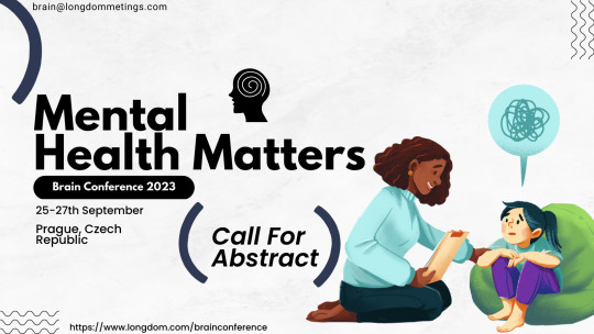
Mental Health
The impact of social media on mental health: With the widespread use of social media, it's important to examine the potential effects of these platforms on mental health, including the development of anxiety, depression, and other disorders.
Trauma and the brain: Understanding the neural mechanisms underlying trauma can help inform more effective treatments for PTSD and other trauma-related conditions.
Mindfulness-based interventions for mental health: Mindfulness has gained popularity in recent years as a way to reduce stress and improve overall well-being. Research on the efficacy of mindfulness-based interventions could be an interesting topic for a brain conference.
The gut-brain connection and mental health: Recent research has shown that the gut microbiome may play a role in mental health. Exploring the link between the gut and the brain could be a fascinating topic for a conference on the brain.
Advances in neuroimaging and mental health: As technology continues to improve, new tools for understanding the brain and its role in mental health are emerging. Discussing these advances and their potential applications could be a valuable topic for a brain conference.
Mental health and artificial intelligence: As AI becomes more integrated into mental health care, exploring its potential benefits and limitations could be an interesting topic for a conference on the brain.
Genetics and mental health: Advances in genetic research have shed light on the potential genetic underpinnings of mental health disorders. Discussing the latest research in this area could be a valuable topic for a brain conference.
#alzheimers#braindisorder#braininjuryconference#brain 2023#mental health#mentalillness#mental health awareness#neuroscience#brainimaging
0 notes
Text


aote moodboards 2023 - wryn masters
god, I'm so sophisticated / I might fall in love with you / a hundred ticks upon the clock / and I'll be through, through, through
“Are you all right?” the boy said after a long, uncomfortable silence spent sitting next to her without further comment. Blinking tears away she took him in.
“You’re kind of young to be a student,” she said, after clearing her throat. She wished she had not spent quite so much time crying. A bell tolled; a class ended; the courtyard filled with students again. She shouldn’t have left Leander, she thought. That wasn’t fair to him or Theresa.
“So are you,” he said. It was stupid and deceptive and unfair, the implication that he could have ever mistaken her for one, but she didn’t say anything. When he did not gain a reaction, the boy cleared his throat and said, quite formally, “Would I be remiss in assuming you’re searching for an expert in the Ildaanic Wastes?”
#*shamelessly invents last names for everyone*#yassified rim is so fucking funny but IT'S HARD TO FIND IMAGES THAT MATCH MY BRAINIMAGE ON PEXELS#anyway that's a wrap on all eight. might do next gen gang tomorrow.#aesthetics#this has been an exercise in 2016isms.
0 notes
Text
Brain disease
#brain, #brains, #humanbrain, #thebrain, #braintumor, #braincancer, #braindevelopment, #brain101, #oldbrain, #newbrain, #bloodbrainbarrier, #brainbank, #pornbrain, #youngbrain, #brainmodel, #brainscans, #musicbrain, #brainlobes, #brainhealth, #braintumour, #teenagebrain, #brainscience, #brainanatomy, #brainimaging, #brainsurgery, #thehumanbrain, #brainfunction, #brainresearch, #musicandbrain, #brainaffected, #musicianbrain, #brainstructure, #depressedbrain.
0 notes
Text

Did You Know?
Brain and different types of imaging techniques.
###medical#medicine#radiology#brain#brainimaging
1 note
·
View note
Link

#brainmapping#brainimaging#methodsofbrainmapping#chronicstress#talkingtherapy#cognitivetherapy#nervoussystemhealth#mentalillness#erasethestigma#mentalhealth#technology
1 note
·
View note
Photo
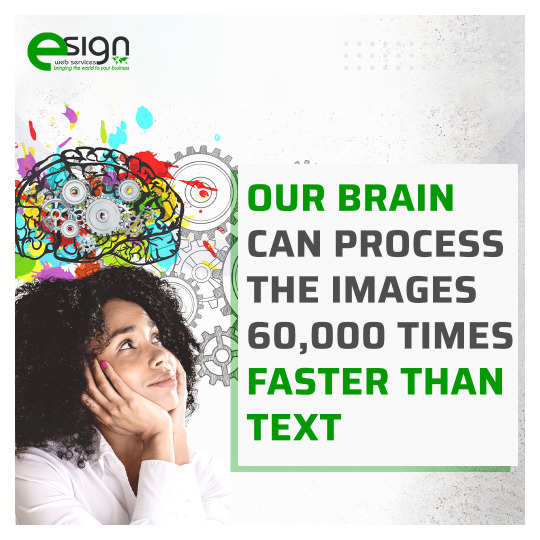
The research concluded by 3M Corporation concluded our brain is able to process images 60,000 faster. So, when you increase your text-to-image ratio, you are sure to have better digital marketing results. At eSign Web Services, we optimize the website to increase the text-to-image ratio.
To know more about our SEO service, please visit: https://bit.ly/3sJdjIq
Call us : +91-9718099999
#seocompany#digitalmarketingcompany#brain#intelligence#textratio#braincode#websitedesign#brainimaging
1 note
·
View note
Photo

When you’re looking forward to a #hospital appointment cause it means you leave #work early, you know it’s been one of those weeks 👏🏻😴#selfie #snapchat #snapchatfilters #undercut #brain #brainscan #hammersmith #septum #followforfollow #sepia #tattooedgirls #altgirls #piercedgirls #girlswholift #bodymods #brainimaging
#work#hospital#snapchatfilters#septum#followforfollow#piercedgirls#girlswholift#altgirls#brainscan#bodymods#snapchat#hammersmith#brainimaging#selfie#tattooedgirls#brain#undercut#sepia
1 note
·
View note
Text
Brain Patterns Differ Between Partisans and Political Non-Partisans

MedicalResearch.com Interview with:
Darren Schreiber JD PhD
Senior Lecturer
Exeter
MedicalResearch.com: What is the background for this study?
Response: My co-authors and I saw an opportunity to match existing functional brain imaging data with publicly available voter registration data so that we could look for patterns that distinguish brain activity in nonpartisans from partisans. While a number of studies have found differences in both brain structure and function between partisans on the left and right and there is a massive amount of scholarship in political science on partisans and polarization, no brain imaging work had focused on nonpartisans. Around 40% of Americans do not affiliate with a political party and one important campaign strategy has been to persuade these voters to support party candidates. However many political scientists are skeptical about voters claims to be nonpartisans and will instead treat them as if they were merely covert partisans.
MedicalResearch.com: What are the main findings? Is it possible to change from partisan to nonpartisan?
Response: If the covert partisans view of nonpartisans was true, we would have expected to find no differences between the partisans and nonpartisans. However, we find that there are distinct patterns of brain activity in regions that are typically involved in social cognition.
Read the full article
0 notes
Photo

Our brain, and its vascular architecture 🔭 . A birdseye view into the very vessels we strive to preserve through diet 🍏, exercise ⛹, and avoidance of harmful toxins like smoking 😷 . 📸: Michael Bernier, PhD - MGH Martinos Center for Biomedical Imaging . . Follow @atomstalk . . . . #brainimaging #brainscan #brainpower #vascular #biology🔬 #biologists #neurosciences #neurologist #neurology #brainscience #amazingimage #interestingpicture https://www.instagram.com/p/CCaupslj8Zy/?igshid=oaju7afl282m
#brainimaging#brainscan#brainpower#vascular#biology🔬#biologists#neurosciences#neurologist#neurology#brainscience#amazingimage#interestingpicture
0 notes
Photo

March 17: Honolulu Science Cafe at Terry's Place (formerly HASR Bistro). Featured speaker Dr. Jonas Vibell (Dept of Psychology, UH Manoa) will present "Brain Imaging: Mapping an Imploded Universe." #brainimaging #brainscience #sciencecafe #honolulusciencecafe #honolulu #science (at Terry's Place) https://www.instagram.com/p/B9BXoW2DjR0/?igshid=1lbl8tvusl5xb
0 notes
Photo

#AI Predicts #Autism From Infant #Brain Scans Twenty-two years ago, #researchers first reported that adolescents with autism spectrum disorder had increased brain volume. During the intervening years, #studies of younger and younger children showed that this brain “overgrowth” occurs in #childhood. Now, a team at the University of North Carolina, Chapel Hill, has detected #braingrowth changes linked to autism in children as young as 6 months old. And it piqued our interest because a #deep-learning #algorithm was able to use that data to predict whether a child at high-risk of autism would be diagnosed with the disorder at 24 months. The #algorithm correctly predicted the eventual #diagnosis in high-risk children with 81 percent accuracy and 88 percent sensitivity. That’s pretty damn good compared with behavioral questionnaires, which yield information that leads to early autism diagnoses (at around 12 months old) that are just 50 percent accurate. “This is outperforming those kinds of measures, and doing it at a younger age,” says senior author Heather Hazlett, a #psychologist and brain development researcher at UNC. As part of the Infant #BrainImaging Study, a U.S. National Institues of #Health–funded study of early brain development in autism, the research team enrolled 106 #infants with an older sibling who had been given an #autismdiagnosis, and 42 infants with no #familyhistory of autism. They scanned each child’s brain—no easy feat with an infant—at 6-, 12-, and 24 months. The researchers saw no change in any of the babies’ overall brain growth between 6- and 12-month mark. But there was a significant increase in the brain surface area of the high-risk children who were later diagnosed with autism. That increase in surface area was linked to brain volume growth that occurred between ages 12 and 24 months. In other words, in autism, the developing brain first appears to expand in surface area by 12 months, then in overall volume by 24 months. https://spectrum.ieee.org/static/ai-vs-doctors (at New York, New York)
#infants#braingrowth#ai#researchers#health#brain#childhood#deep#algorithm#studies#psychologist#familyhistory#autismdiagnosis#autism#diagnosis#brainimaging
1 note
·
View note
Link
Did you know our Glossaries also include real clinical images? Check out this one of Cavernous Sinus Aneurysm
#ditki#neurology#neuroanatomy#neuropathology#pathology#cme#brainimages#human brain#cavernous sinus#internal carotid artery#learnmedicine#learnneuroanatomy#learnpathology#medicalschool#medicalstudent#medicalscience
1 note
·
View note
Text
Modern Day #MentalHealth : #BrainImaging Identifies Different Types of #Depression @TheBrainDriver #tDCS #Neurology #BrainStimulation http://flip.it/cVfMga
1 note
·
View note
Last Seen Blogs


champagneloverboy
Champaña🍾

reinen5astro
Reinen's Corner

dqrships
a Lover

iconshaper
𝐢𝐜𝐨𝐧𝐬 & 𝐡𝐞𝐚𝐝𝐞𝐫𝐬