#Gut-brain axis
Text
The Gut-Brain Connection: Benefits of Creatine and Probiotics on Cognitive Function
The Gut-Brain Connection: Benefits of Creatine and Probiotics on Cognitive Function #nutribliss #probiotics+
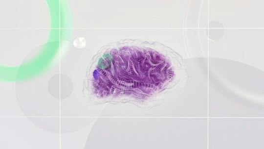
Creatine for Brain HealthAs we age, it’s common to start experiencing changes in our cognitive function. Many people notice a decline in memory, focus, and concentration as they get older. But what if there was a way to support and enhance brain health? Recent research suggests that creatine, a popular supplement in the fitness world, may have a positive impact on cognitive function.Creatine and…
View On WordPress
#benefits of dha#benefits of magnesium#benefits of mushrooms#brain and gut connection#brain function#brain gut connection#cognitive health#cognitive health improvement#connection#creatine#creatine supplementation#digestion and brain#effects of ammonia#functional medicine#functional medicine grand rounds#functional medicine gut health#get brain connection#gut and brain connection#gut brain connection#gut brain connection and mental health#gut brain connection frequency#gut brain connection ted talk#gut-brain axis#health benefits of mushrooms#inspiration#lion&039;s mane benefits#mind and gut connection#myprotein creatine monohydrate#second brain#side effects of prednisone
0 notes
Text

Probiotics And Prebiotics: What You Should Know?
Discover the importance of probiotics and prebiotics for gut health in this detailed guide. Learn about their roles, sources, benefits, and how they work together to promote a healthy digestive system. Explore the latest research findings and practical tips for incorporating probiotics and prebiotics into your diet for improved overall well-being.
#Probiotics#Prebiotics#Gut health#Digestive system#Microbiome#Probiotic foods#Prebiotic foods#Gut flora#Gut bacteria#Gut microbiota#Healthy digestion#Nutritional supplements#Fermented foods#Gut-brain axis#Immune system#Dietary fiber#Health benefits#Digestive disorders#Balanced diet#gutfoundation
1 note
·
View note
Photo

Foods that Make You Feel Good, Plus a Pomegranate Rose Smoothie
What we choose to eat and what we choose not to eat directly affect the way we feel both physically and mentally. Food choices are connected to the health of our mood and overall outlook on things due to how our gut microbes either benefit or suffer from the dietary choices we make, a connection known as the gut-brain axis. For example, the probiotics in fermented foods, such as kefir, are increasingly showing that they are vital for optimal mood support by supporting a healthy microbiome. Other foods provide important nutrients to feed the gut and brain which contribute to […]
Read more: https://lifewaykefir.com/foods-that-make-you-feel-good/
#diet and mood#foods for your mood#gut-brain axis#healthy recipe#healthy smoothie#heart healthy#kefir and mood#kefir recipe#mood support#smoothie#valentine's day recipe#Digestion#Health#Healthy Living#Living#Recipe
1 note
·
View note
Text
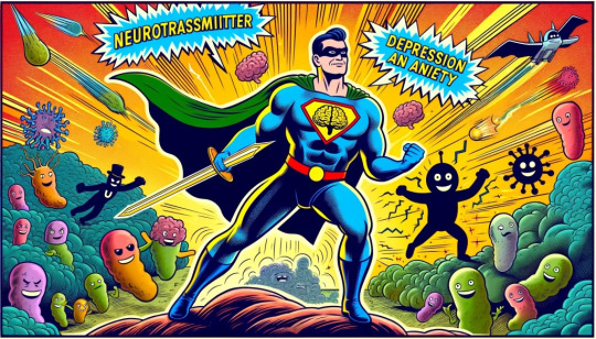
Exploring the Gut-Brain Connection: New Insights into Mental Health
Introduction:
Depression and anxiety are not only common but also complex mental health challenges, affecting millions globally. Cutting-edge research is now shining a light on an unexpected influencer of mental health: our gut microbiota, often dubbed the “second brain.” In this comprehensive guide, we’ll explore the intricate ties between gut health and mental health conditions such as…
View On WordPress
#anxiety#depression#diet#Gut health#gut-brain axis#immune response#inflammation#lifestyle modifications.#Mental Health#neurotransmitters#nutrition#prebiotics#probiotics#stress response
1 note
·
View note
Text
Sleep Disorders - Gastroesophageal Reflux Disease(GERD)
Gastroesophageal reflux disease (GERD), a chronic digestive condition, can adversely affect sleep quality due to increased abdominal pressure and relaxed upper intestine muscle tone. Addressing GERD can improve individuals' quality of life.
And you may wonder, “How does that happen?” Well, it’s important to note that GERD is not primarily a sleep disorder in itself. However, it often occurs during sleep due to a combination of increased pressure in the abdominal cavity and relaxed muscle tone in the upper intestine. As a result, this sequence leads to sleep disturbances and compromises the quality of sleep. So let’s start by looking…

View On WordPress
#acid reflux#apnea#GERD#gut-brain axis#H2 blockers#insomnia#PPIs#proton pump inhibitors#reflux disease#restful sleep#sleep#sleep apnea#sleep disorder#sleep disorders#sleep disturbance#sleep health#sleep hygiene#sleeping#slept#snoring#stress#stress management
0 notes
Text
The Profound Connection Between Gut Health, Body Health, and Mental Health
Gut health has increasingly become a focal point of medical research in recent years. As health professionals, we often see the manifestation of physical symptoms due to various health conditions. However, it’s essential to understand that the root of many of these conditions can be traced back to the gut. Not only does gut health influence body health, but it also has a profound impact on mental…

View On WordPress
#absorption#body health#chronic inflammation#digestion#Gut Health#gut microbiota#gut-brain axis#immune cells#Mental health#neurotransmitters#nutrient metabolism#pathogens#probiotics#short-chain fatty acids
0 notes
Text
The Gut-Brain Axis Recent
View On WordPress
1 note
·
View note
Link
The classical symptoms of PD are characterized by progressive motor dysfunction, including rest tremor, bradykinesia, rigidity, and postural instability, which is noticeable when there is already 60% dopaminergic neuronal loss in the substantia nigra.
In addition to motor symptoms, non-motor manifestations such as rapid eye movement sleep behavior disorder (RBD), gastrointestinal dysfunction, olfactory disruption, neuropsychiatric symptoms, and sensory dysfunction also exist in patients with PD and compromise their quality of life.5 Of note, these non-motor PD symptoms can occur years before the onset of classical motor symptoms.6 Thus, they are therefore now considered prodromal clinical markers before the onset of the classical motor manifestations, according to the International Movement Disorders Society.7 Among these prodromal symptoms, constipation is the most prevalent and earliest pre-motor feature and can precede motor symptoms by decades.
The pathological hallmark of PD is progressive dopaminergic neuronal degeneration and intraneuronal accumulations of misfolded α-synuclein (Lewy bodies) in the substantia nigra.3 The pathological α-synuclein can transmit from cell to cell in a prion-like fashion to promote the neurodegenerative process of this disease.9,10 Recent postmortem evidence indicates that Lewy body pathology is first detectable outside the brain, starting from neurons in the gut enteric nervous system (ENS) and olfactory bulbs.11 These neuropathology observations are consistent with the findings that non-motor symptoms of PD, especially constipation, can precede the onset of motor symptoms.8 Further in vivo PD animal model studies demonstrated that pathological forms of α-synuclein, after injection into the intestinal wall, can be transported from the gut to the brain via the vagus nerve, reaching the dorsal motor nucleus of the vagus nerve in the brainstem.12, 13, 14 However, although this pathology finding has given rise to the idea that PD pathology originates in the peripheral ENS and then invades the brain via retrograde axonal transport through the vagus nerve, a small fraction of patients do not show pathology in the ENS. In about one-third of patients, the PD neuropathology begins in the brain itself and then travels downward.15 Hence, it is hypothesized that PD can be divided into a gut-first (body first) and a brain-first subtype.15 The former is tightly associated with chronic constipation and RBD during the prodromal phase and the latter is most often sparing of gastrointestinal symptoms and is RBD-negative during the prodromal phase. These findings reinforce the concept that PD is a heterogeneous disorder with diverse initial triggers and propagation trajectories of α-synuclein, suggesting that tailored disease-modifying therapy is needed for patients with different subtypes.
The various recently observed gut microenvironmental changes in the early stages of the disease may play a vital role in PD, especially those with the body-first subtype of the disease. Patients whose disease begins in the gut may benefit most from interventions that target the gut. In this review, we summarize recent evidence for altered gut microenvironments contributing to PD through the gut–brain axis. Furthermore, there is a plethora of evidence, including our previous study, for altered gut microbiota in patients with PD compared with unaffected controls.
There is much evidence that the vagus nerve transports α-synuclein from the gut to the brain.12 Pathological α-synuclein fibrils injected into the duodenum can move from the muscular layer of the duodenum to the brain and then from neuron to neuron across the synapses through the vagus nerve in a PD animal model.12 Furthermore, recent evidence indicates that protein nucleation and aggregation may be influenced by E. coli's secretion of curli, which induces neuronal deposition of α-synuclein in the ENS.29,31 The abundance of E. coli at the colonic mucosa correlates with enteric α-synuclein deposition in PD patients.
A plethora of studies have investigated changes in the gut microbiota in patients with PD compared with healthy controls using either 16 S rRNA gene amplicon surveys or shotgun metagenomic sequencing analysis.35, 36, 37, 38, 39, 40, 41, 42 Meta-analyses have shown that the relative abundance of various phyla of anti-inflammatory and short-chain fatty acid (SCFA)-producing bacteria, including Blautia, Coprococcus, Roseburia, Lachnospira, Fusicatenibacter, and Faecalibacterium, are reduced in PD patients compared with controls. In contrast, the amounts of Lactobacillus, Bifidobacterium, and Akkermansia are higher in PD patients than in unaffected participants from different ethnicities.38, 39, 40, 41 Notably, opportunistic pathogens and pro-inflammatory bacteria at the phylum level, including Corynebacterium, Porphyromonas, Alistipes, Bacteroides, Escherichia, and Megasphaera, are also enriched in PD.40,43 In metagenomic research, gene markers from the gut microbiome were found to accurately discriminate PD patients from healthy controls, with most of the identified markers belonging to Bacteroides and Escherichia species.44 Although PD medications affect the structure of the gut microbiota, these changes are detectable in drug-naïve early-stage PD patients.36,43,45
Changes in the gut microbiota also correlate with disease progression in PD. A decrease in the SCFA-producing microbiota and increase in pro-inflammatory bacteria correlate with motor and cognitive severity in patients with PD.38,40,46 Compared with transplantation of fecal gut microbiota from healthy donors, such transplantation from PD patients leads to worsened motor symptoms in a transgenic rodent model of PD.47 A 3-year longitudinal follow-up study of PD patients revealed that a reduced amount of Roseburia species predicted faster progression of both motor and non-motor symptoms of PD.48 A lower abundance of SCFA-producing bacteria, including Fusicatenibacter and Faecalibacterium, correlates with elevated fecal inflammatory calprotectin levels in PD patients.49 Furthermore, enrichment in Bacteroides and Bifidobacterium has been linked to elevated expression of systemic and fecal inflammatory markers IFN-γ, TNF-α, and neutrophil gelatinase-associated lipocalin in patients with PD.42,50 The increased levels of Lactobacillaceae and Bifidobacteriaceae in PD patients require further investigation, as they are usually recognized as probiotics for improving constipation.51 Therefore, distinct gut microbiota species promote enteric α-synuclein aggregation or gut inflammation to facilitate the occurrence and progression of PD.
The composition of gut microbiota also influences the pharmaceutical treatment responses in PD patients. The growing literature has shown the role of the gut microbiome in the pharmacokinetics of prescription drugs and the effects that the drugs can have in turn on the composition of the gut microbiome,52,53 indicating a potential interaction between PD medications and the microbiome. Prior studies have shown that anti-PD medications, including catechol-O-methyl transferase (COMT) inhibitors and anticholinergics, have gastrointestinal side effects, which may be related to the changes of the gut microbiome.54,55 Furthermore, bacterial tyrosine decarboxylase, which can convert levodopa to dopamine, could limit its bioavailability and may contribute to the interindividual responses to levodopa treatment among patients with PD.56,57 Enterococcus faecalis was found to be the dominant microorganism responsible for levodopa decarboxylation and restrict levels of levodopa in the treatment of PD.57 Dopamine produced by gut bacterial metabolism of levodopa decarboxylation can also impair intestinal motility, which could provide an explanation for bacterial overgrowth in the small intestine associated with motor fluctuation in PD.58 These observations lend support to the notion that the composition of the gut microbiome may affect the treatment efficacy and potential side effects of levodopa treatment in patients with PD.
Srivastav et al. treated animals with an oral probiotic mixture containing Lactobacillus rhamnosus GG, Bifidobacterium animalis lactis, and Lactobacillus acidophilus for 30 days, after which the mice were given MPTP injections.62 The results showed that mice receiving this probiotic mixture reduced dopaminergic neurodegeneration by upregulating neurotrophic factors and increasing striatal neuronal responses to dopamine.62 Using the same toxin-induced PD model, Liao et al. fed mice with Lacobacillus plantarum PS128 for 28 days, then gave them MPTP injections for 4 days. The results showed that feeding with Lacobacillus plantarum PS128 mitigated neuronal degeneration, attenuated oxidative stress and neuroinflammation, and rescued the locomotor defects of MPTP-injected PD mice.63 Sun et al. treated MPTP-injected mice with the probiotic Clostridium butyricum for 4 weeks and demonstrated improved motor deficits, attenuated dopaminergic neuron loss, improved synaptic dysfunction, and reduced microglia activation in the treated mice.64 This beneficial effect was associated with increased colonic levels of glucagon-like peptide-1 (GLP-1), colonic G protein–coupled receptors GPR41/43, and other components of the cerebral GLP-1 receptor pathway.57 Of note, the incretin hormone GLP-1 was recently found to regulate neurogenesis and synaptic plasticity,65 and GLP-1 agonists may be neuroprotective in PD pathogenesis.66 Similarly, a 3-week treatment with Lactobacillus fermentum U-21 reduced nigra dopaminergic cell loss in a paraquat-toxin model of PD.67
Several studies have demonstrated the potential benefits of probiotic supplementation in patients with PD. A total of seven clinical trials were identified, which were all randomized clinical trials68, 69, 70, 71, 72, 73, 74 (Table 1). Consumption of fermented milk containing multiple probiotic strains can improve constipation in patients with PD.62 Similar beneficial effects in improving bowel movements were also noted in PD patients in most of the probiotic clinical trials.51,73 Evidence seems to demonstrate that probiotic intake can improve bowel movement and reduce gastrointestinal symptoms. There are two studies measuring non-gastrointestinal symptoms of PD as one of the primary outcomes of probiotic clinical trials.71,74 Probiotic supplementation containing Bifidobacterium bifidum, L. acidophilus, L. fermentum and Lactobacillus reuteri for a period of 12 weeks has been observed to improve some symptoms in PD patients as measured by total MDS-UPDRS scores.71
One recent open-label clinical trial with 87 participants showed improvement of non-motor symptom scores, reduced fecal inflammatory marker of calprotectin and increased fecal butyrate in patients with PD who received prebiotic supplement with resistant starch compared to those without prebiotic intervention.78 Several in vivo studies have shown that lower abundance of SCFAs butyrate-producing bacteria could be corrected by the administration of prebiotic fibers, which in turn reduce the gut inflammatory processes, improve gut barrier function, and peristalsis.79,80 SCFAs have a key role in modulating the cross-talks in the gut–brain axis through modulating the gut barrier and blood–brain barrier integrity, inflammatory processes, inhibition of histone deacetylase to promote neuronal survival.81 Of note, an in vivo study using a transgenic a-synuclein–expressing mouse model demonstrated that a germ-free environment eliminates PD phenotypes but that oral feeding with SCFAs remerged the disease-related neuropathology by microglial activation.82 As the effects of SCFAs may depend on the concentration and the different subtypes, the effect of SCFAs in the PD process requires more studies.
0 notes
Text
Nightmare Alzheimer's: Does The Gut Save Us?
by Dr.Harald Wiesendanger– Klartext
What the leading media are hiding
No disease dreads people more than Alzheimer’s, the most common form of dementia: a gradual mental decline at the tragic end of which nothing is left of what makes a body a person. The neurodegeneration in the brain that causes it is considered incurable. But recent studies give hope. They illustrate the key role of the…

View On WordPress
#Alzheimer#Alzheimer&039;s disease#Bredesen#Dementia#gut-brain axis#Gut-health#Harald Wiesendanger#Microbiota#Morbus Alzheimer#neurodegeneration#Probiotica
1 note
·
View note
Text
After too many years here I've final what hornets' nests I am not brave enough to kick
#m/cc#thought about making a certain post and decided... no... I would rather not#I am not prepared for responses to that. it might actually kill me#specifically it was:#'going gluten/dairy/food dye-free CAN improve certain neurodevelopmental things but it cannot 'cure' autism/ADHD/Tourette's'#I already know I'd get vitriol both from people claiming I think autism comes from gluten or 'needs cured' because they can't read the post#and that I'm trying to trick everyone into going gluten-free because Toxins or something and lying about a connection#(even though (neuro)dev disorders can be made worse by flaring immune issues like - oh I don't know - undiagnosed gluten intolerance?#hypersensitivity to certain food dyes?#we already know autism and ADHD in particular have HUGE correlations with gastro and immune issues#which is why some mommy bloggers genuinely do see symptom improvement from diet changes)#and from people saying 'um actually no-gluten DID cure my nephew's ADHD?? the science is on our side/big gluten is covering up the research#and I don't know if I could handle dozens of people per day telling me I'm a science denier AND a eugenist from both sides#I am simply. ADHD. and autistic. and incredibly interested in the wild amount of comorbid physical disorders that correlate with these#autoimmune and gastro issues but also loose/hypermobile joints; epilepsy; delayed sleep phase disorder; COPD; skin conditions#it's so fascinating to me and provides a huge chunk of data to run with re: the gut-brain axis#whether [neurodev] causes [other]/[other] causes [neurodev] or an underlying thing causes both is unknown#but honestly with the huge interest in the gut-brain axis and microbiome in the past decade or so#I think we're going to see a lot more research in the next thirty or forty years examining physical comorbidities with neurodev stuff#I'm probably not gonna link to research because I don't wanna just start the war anyway and I'm too tired to go back and find the articles#but the TL;DR of the tags is neurodev stuff isn't caused by gluten intolerance but if you're unknowingly aggravating a gluten intolerance#you're probably not gonna feel great and it's gonna make your symptoms worse because of the effect it has on your body#it's like a very mild long-term allergic reaction and yeah if you get rid of that it'll improve other areas (e.g. sleep cycle; irritability#so of Course it's gonna improve a bunch of things-that-get-worse-with-poor-sleep/decreased-stress-tolerance#if you were always sitting on a slightly uncomfortable chair you'd probably do a lot better if I switched the chair#just because you can focus better or you didn't know the chair was uncomfortable doesn't mean it caused your ADHD#also in this case the chair affects your hormone levels and immune response and what chemicals accidentally leak into your bloodstream#if you're interested look it up there's been a Ton of research on correlations of specific physical issues with neurodev in recent years
13 notes
·
View notes
Text
TMI but I miss the days when the stress of exams would just make me want to kill myself instead of giving me stomach aches and nausea and diarrhea
5 notes
·
View notes
Text
Probiotics And Prebiotics: What You Should Know?
Discover the importance of probiotics and prebiotics for gut health in this detailed guide. Learn about their roles, sources, benefits, and how they work together to promote a healthy digestive system. Explore the latest research findings and practical tips for incorporating probiotics and prebiotics into your diet for improved overall well-being.
#Probiotics#Prebiotics#Gut health#Digestive system#Microbiome#Probiotic foods#Prebiotic foods#Gut flora#Gut bacteria#Gut microbiota#Healthy digestion#Nutritional supplements#Fermented foods#Gut-brain axis#Immune system#Dietary fiber#Health benefits#Digestive disorders#Balanced diet#gutfoundation
0 notes
Text
Explained | How does the gut microbiome link to autism spectrum disorders?
‘Fix your gut, fix your brain’ used to be an underrated idea, but it is today gaining in relevance as more and more research throws light on the role of the community of bacteria living in your gut, a.k.a. the gut microbiome.
A healthy gut microbiome is not a panacea – but it may be able to facilitate better overall health and help improve the quality of life of individuals with various diseases…
View On WordPress
#autism icd 10#autism meaning#autism spectrum disorders#autism symptoms#autism test#autism treatment#fecal matter#fecal microbiota transplantation#gut brain axis#gut microbiota
4 notes
·
View notes
Text
Exploring the Gut-Brain Connection: New Insights into Mental Health
Discover the latest research on how gut health impacts depression and anxiety. Learn about the gut-brain axis and how diet, probiotics, and lifestyle changes can promote mental well-being.
Introduction:
Depression and anxiety are not only common but also complex mental health challenges, affecting millions globally. Cutting-edge research is now shining a light on an unexpected influencer of mental health: our gut microbiota, often dubbed the “second brain.” In this comprehensive guide, we’ll explore the intricate ties between gut health and mental health conditions such as…

View On WordPress
#anxiety#depression#diet#Gut health#gut-brain axis#immune response#inflammation#lifestyle modifications.#Mental Health#neurotransmitters#nutrition#prebiotics#probiotics#stress response
0 notes
Text
The dilemma of convincing yourself to "trust your gut" vs remembering that your ass is way too fancy to stomach anything less
0 notes
Text
Mental Health and Your Diet
Exploring the Impact of Nutrition on Well-Being
In recent years, there has been growing recognition of the intricate connection between our diet and mental health. While it’s no secret that what we eat affects our physical well-being, emerging research suggests that our dietary choices also play a significant role in shaping our mental and emotional health. This blog post aims to dig into this…

View On WordPress
#brain health#diet#gut-brain axis#healthy eating habits#inflammation#Mental Health#mood disorders#nutrient deficiencies#nutrition#psychological well-being
0 notes
Last Seen Blogs

bny4kuya
happy angel anime debut

battlereadyarms
BATTLE READY ARMS

morganaconda
jo ˳✧༚˚✧

xlr8nrg
XLR8NRG

digital-ilustracion
DIGITAL MAGIC