#high resolution microscopy
Video
Beautiful Eyes
If you look at a clear blue sky it’s possible to see the blood vessels in your own eyes. But researchers demanding a closer look have been working on ways to visualise the full complex network of vessels that permeate our eyeballs. Previous genetic and experimental attempts in mice have tended to look at one sub-section of vessels at a time. A new approach has represented the complete vasculature of the mouse eye at various stages of development (video, with different sections of vasculature digitally coloured). A process to make the eye material transparent is followed by high resolution microscopy and digital analysis. With this technique, the researchers observed how the iris vasculature (yellow) is remodelled during development just after disconnection from the early supply network. They also analysed the impact of injury on vascularisation, showing the platform’s potential for analysing eye disease impacts and treatments on a whole-eye scale.
Written by Anthony Lewis
Video by Luc Krimpenfort and colleagues
Div. of Vascular Biology, Karolinska Institutet, Department of Medical Biochemistry and Biophysics, Stockholm, Sweden
Video originally published with a Creative Commons Attribution 4.0 International (CC BY 4.0)
Published in bioRxiv, December 2022 (not peer reviewed)
You can also follow BPoD on Instagram, Twitter and Facebook
#science#biomedicine#eyesight#eyes#vision#eyeballs#developmental biology#embryo development#eye development#light sheet microscopy#high resolution microscopy
18 notes
·
View notes
Text
Atomic-Level Study Captures Frog Toxin In Action - Technology Org
New Post has been published on https://thedigitalinsider.com/atomic-level-study-captures-frog-toxin-in-action-technology-org/
Atomic-Level Study Captures Frog Toxin In Action - Technology Org
Cryo-electron imaging studies of poison dart frog toxin molecules in action are helping to solve the mystery of why this is among the deadliest natural poisons.
Graphic abstract of Nature Communications paper on binding of poison dart frog toxin to sodium ion channels. Image credit: George Wisedchaisri, University of Washington
Some tiny Amazon frogs defend themselves when threatened by releasing a substance that quickly incapacitates a predator. For centuries, a few Indigenous tribes in Colombia have siphoned this toxin from skin glands on the frogs’ backs to coat blow darts for hunting, because of its power to take down big game.
The poison instantly cramps the muscles in the body and forces the heart into a chaotic rhythm that ends in cardiac arrest. The bright coloring of the frogs warns animals to stay away. Their chemical weapon is called batrachotoxin, a name based on the Greek root word for frog.
“Predators that attack these dendrobatid frogs are paralyzed so rapidly that they can’t clamp down on the frogs to kill and eat them,” said one of the senior researchers on a recent batrachotoxin study, William Catterall, a professor of pharmacology at the University of Washington School of Medicine.
He noted that “it is so toxic that wiping the toxin secretion from the back of a single frog is sufficient to poison several blow darts, which will each immobilize or kill large prey.”
What makes their irreversible toxin more severe, compared with others that also target the signal-producing pores of nerve and muscle cells?
It has long been known that batrachotoxin affects sodium ion channels, which control electrical pulses by regulating the flow of charged sodium particles through the membranes of these cells.
“The toxin acts by hyperactivation of the sodium channel that generates action potentials in nerve and muscle, thereby causing a lethal storm of hyperactivity of nerves, synapses and muscle cells,” Catterall said.
By using high-resolution cryogenic electron microscopy, a research team led by pharmacologists at the University of Washington School of Medicine have determined, at the atomic level, the structure of the toxin derivative batrachotoxin-A benzoate when it is bound to its receptor site on the cardiac sodium channel.
Unexpectedly, the researchers discovered that, unlike other toxins, batrachotoxin actually binds to two separate but similar receptor sites in the cardiac sodium channel. This dual-receptor action, and its multifaceted effects, provide unprecedented insights into why this poison is so effective, powerful and deadly.
The toxin’s unique mechanism for high potency and lethality might also establish a new paradigm that helps to explain the actions of other toxins with high lethality, the researchers suggested.
Such studies might also lead to better therapies for conditions resulting from certain electrical signaling problems in excitable cells and tissues, such as those in the heart, muscles, nerves and brain that are caused by malfunction of a sodium channel.
For example, Catterall suggested, drugs designed to be robust inhibitors at both dual-receptor sites discovered in this study might also be valuable therapeutic agents in the treatment of disorders of hyperexcitability of nerve and heart cells. Such disorders include epilepsy, chronic pain and arrhythmias.
Source: University of Washington
You can offer your link to a page which is relevant to the topic of this post.
#Amazon#Animals#atomic#Biotechnology news#Brain#cardiac arrest#Cells#channel#chemical#chemical weapon#Chemistry & materials science news#chronic pain#clamp#communications#cryo-electron microscopy#Dart#disorders#drugs#effects#electron#epilepsy#frogs#game#heart#High-Resolution#Imaging#insights#it#LED#Link
0 notes
Text
Market Outlook for High-Resolution 3D X-Ray Microscopy Market Services
Market Overview –
The High-Resolution 3D X-Ray Microscopy Market is experiencing substantial growth due to the increasing demand for advanced imaging techniques in various scientific and industrial applications. High-resolution 3D X-ray microscopy, also known as X-ray tomography or computed tomography (CT), enables detailed three-dimensional imaging of internal structures with superior resolution and clarity.
The 3D X-ray Microscopy market is thriving as industries and research sectors demand high-resolution imaging solutions. These advanced systems offer detailed insights into the internal structures of various materials, aiding in research, quality control, and failure analysis. With continuous technological advancements, the market for 3D X-ray microscopy is poised for further growth.
Key drivers of market growth include advancements in imaging technology, such as higher spatial resolution and faster image acquisition, which allow for precise visualization of microstructures in diverse samples. Additionally, the expanding applications of high-resolution 3D X-ray microscopy across multiple industries, including materials science, life sciences, electronics, and geology, fuel market expansion.
The market offers a wide range of high-resolution 3D X-ray microscopy systems, including laboratory-based and synchrotron-based systems, tailored to specific research and industrial requirements. These systems enable researchers and scientists to analyze complex samples, such as biological tissues, composite materials, and geological specimens, with unparalleled detail and accuracy.
Furthermore, collaborations between research institutions, academia, and industry players drive innovation and technological advancements in high-resolution 3D X-ray microscopy, further stimulating market growth.
Moreover, the market benefits from increasing investment in research and development activities, supportive government initiatives, and growing awareness about the advantages of high-resolution 3D X-ray microscopy in scientific research, quality control, and product development.
Despite the market's positive outlook, challenges such as high initial costs, limited accessibility to advanced imaging facilities, and data analysis complexities may hinder market growth. Nonetheless, ongoing efforts to enhance system capabilities, improve user-friendliness, and expand application areas are expected to drive continued adoption of high-resolution 3D X-ray microscopy in the coming years.
Over the projection period of 2022-2030, the high-resolution 3D x-ray microscopy market is expected to grow at an 8.8% annual rate to reach USD 2487.83 million by 2030.
Segmentation –
The global high resolution 3D X-ray microscopy market is segmented into type, applications, end user, and region. The type is segmented into Sub-micron XRM, Nanoscale XRM and others. The applications are segmented into advanced package development, Mineralogy Discrimination, Failure analysis, Surface measurements and others. The end users are segmented into Oil & Gas, Material Science, Semiconductors, Metrology, Life Science, Healthcare and others. The market is spanned across regions including North America, Europe, Asia Pacific, and rest of the world.
Regional Analysis –
The High-Resolution 3D X-Ray Microscopy Market demonstrates distinct regional dynamics shaped by factors such as technological advancements, research and development capabilities, and industrial applications. North America leads the market, driven by a strong presence of key market players, advanced R&D infrastructure, and widespread adoption of cutting-edge imaging technologies across various industries.
The region's robust healthcare and industrial sectors contribute significantly to the demand for high-resolution 3D X-ray microscopy systems, thus holding a substantial market share. Similarly, Europe portrays a lucrative market landscape, characterized by a strong focus on technological innovation, stringent quality standards, and a well-established industrial base. Adoption of high-resolution 3D X-ray microscopy in automotive, aerospace, and materials science sectors further fuels market growth in the region. In Asia Pacific, the market is witnessing rapid expansion due to increasing investments in research and development, rising industrialization, and growing applications in fields like electronics and semiconductors.
Countries like China, Japan, and South Korea are driving market growth with their expanding manufacturing sectors and rising demand for advanced imaging solutions. Latin America and the Middle East & Africa regions present opportunities for market penetration, driven by growing industrialization and investments in scientific research. However, challenges such as limited awareness and infrastructure may impact market growth in these regions. Overall, the High-Resolution 3D X-Ray Microscopy Market displays promising growth prospects across diverse regions, driven by the increasing demand for precise imaging solutions in various industries.
Key Players –
High-resolution 3D X-ray microscopy key players include Zeiss, Rigaku Corporation, Bruker Corporation, Thermo Fisher Scientific Inc., GE Measurement & Control Solutions, National Resource for Automated Molecular Microscopy, Phenom-World BV, TESCAN, Matsusada Precision Inc., and Octopus Imaging Software.
Related Reports –
Meningitis Diagnosis and Treatment
Endodontic Devices
Fertility Drug and Surgery
Opioids
For more information visit at MarketResearchFuture
#High-Resolution 3D X-Ray Microscopy Market#High-Resolution 3D X-Ray Microscopy Market Size#High-Resolution 3D X-Ray Microscopy Market Share#High-Resolution 3D X-Ray Microscopy Market Trends
0 notes
Text

Jan Bartek - AncientPages.com - New scientific research has revealed a piece of tartan found in a peat bog in Glen Affric around forty years ago can be dated to circa 1500-1600 AD, making it the oldest known surviving specimen of true tartan in Scotland.
The Scottish Tartans Authority commissioned Dye Analysis and Radiocarbon testing on the woolen textile to prove its age.
Scotland's Oldest Tartan On Display For The First Time!
Glen Affric tartan - Scotland's oldest-known true tartan discovered by The Scottish Tartans Authority to go on display for the first time at V&A Dundee's Tartan exhibition.
Credit: Alan Richardson Pix-AR
The first investigation was dye analysis carried out by analytical scientists from National Museums Scotland. Using high-resolution digital microscopy, four colors were visually identified for dye analysis: green, brown, and possibly red and yellow.
The dye analysis confirmed the use of indigo/woad in the green but was inconclusive for the other colors, probably due to the dyestuff degradation state. However, no artificial or semi-synthetic dyestuffs were involved in making the tartan, which pointed to a date of pre-1750s.
Further clarification on the age of the tartan involved radiocarbon testing at the SUERC Radiocarbon Laboratory in East Kilbride. The process involved washing out all the peat staining, which would have otherwise contaminated the carbon content of the textile.
The Radiocarbon testing results identified a broad date range between 1500 and 1655 AD, with the period between 1500 and 1600 AD the most probable. This makes it the oldest-known piece of true tartan found in Scotland – the Falkirk ‘tartan,’ dating from the early third century AD, is actually a simpler check pattern woven using undyed yarns.
The Glen Affric tartan, which measures around 55cm by 43cm, is now on display for the first time at V&A Dundee’s Tartan exhibition.
by TaboolaSponsored Links
The piece will be the oldest exhibit among more than 300 objects. The exhibition examines tartan’s universal and enduring appeal through iconic and everyday examples of fashion, architecture, graphic and product design, photography, furniture, glass and ceramics, film, performance, and art.
“The testing process has taken nearly six months, but the effort was well worth it, and we are thrilled with the results!
In Scotland, surviving examples of old textiles are rare as the soil is not conducive to their survival. As the piece was buried in peat, meaning it had no exposure to air and was therefore preserved.
The tartan has several colors with multiple stripes of different sizes, and so it corresponds to what people would think of as a true tartan.
“Although we can theorize about the Glen Affric tartan, it’s important that we don’t construct history around it. Although Clan Chisholm controlled that area, we cannot attribute the tartan to them as we don’t know who owned it.
“The potential presence of red, a color that Gaels considered a status symbol, is interesting because of the more rustic nature of the cloth. This piece is not something you would associate with a king or someone of high status; it is more likely to be an outdoor working garment," Peter MacDonald, Head of Research and Collections at The Scottish Tartans Authorit said.
Scotland's Oldest Tartan On Display For The First Time!
New scientific research has revealed a piece of tartan found in a peat bog in Glen Affric, Scotland around forty years ago can be dated to circa 1500-1600 AD, making it the oldest known surviving specimen of true tartan in Scotland. Credit: Credit: Alan Richardson Pix-AR
“The Glen Affric tartan is clearly a piece of national and historical significance. It is likely to date to the reign of James V, Mary Queen of Scots, or James VI/I. “There is no other known surviving piece of tartan from this period of this age. It's a remarkable discovery and deserves national attention and preservation. “It also deserves to be seen and we’re delighted that it is to be included in the Tartan exhibition at V&A Dundee,” John McLeish, Chair of The Scottish Tartans Authority, said.
“We knew The Scottish Tartans Authority had a tremendous archive of material and we initially approached them to ask if they knew of any examples of 'proto-tartans' that could be loaned to the exhibition.
I'm delighted the exhibition has encouraged further exploration into this plaid portion and very thankful for The Scottish Tartans Authority's backing and support in uncovering such a historic find.
To be able to exhibit the Glen Affric tartan is immensely important in understanding the textile traditions from which modern tartan derives, and I'm sure visitors will appreciate seeing this on public display for the very first time," James Wylie, curator at V&A Dundee said.
Tartan at V&A Dundee opens on Saturday, 1 April, until 14 January 2024.
Written by Jan Bartek - AncientPages.com Staff Writer
Source: Facebook
Source: AncientPages.com
236 notes
·
View notes
Text
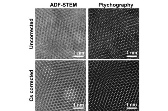
Reimagining electron microscopy: Bringing high-end resolution to lower-cost microscopes
Researchers at the University of Illinois at Urbana-Champaign have shown for the first time that expensive aberration-corrected microscopes are no longer required to achieve record-breaking microscopic resolution.
The field of microscopy is in the middle of a great revolution. Since the 1800s and the invention of the compound light microscope, there have only been a few major jumps in resolution to see different length scales: from bacteria and cells, to viruses and proteins, and even down to single atoms.
Generally, as resolution has been making these incredible jumps, so has the price of the microscopes used to achieve that resolution. Such hefty price tags severely limit the accessibility of these instruments. The current jump in resolution comes from a new technique called electron ptychography—a method that uses computation to boost the resolution of electron microscopes—which has taken the field by storm in the last 5-6 years.
Read more.
20 notes
·
View notes
Photo

High-resolution images of terminals in water chains.
Shiotari, A., Sugimoto, Y. Ultrahigh-resolution imaging of water networks by atomic force microscopy. Nat Commun 8, 14313 (2017). https://doi.org/10.1038/ncomms14313
153 notes
·
View notes
Text

Produce bricks anywhere and anytime
SnPC Machines: Factory of brick on wheel
Fully automatic mobile brick making machine by SnPC Machines, First of its kind of machine in the world, our brick-making machine moves on wheels like a vehicle and produces bricks while the vehicle is on move. This allows kiln owners to produce bricks anywhere and anytime, as per their requirements. Fully automatic Mobile brick-making machine can produce up to 12000 bricks/hour with a reduction of up to 45% in production cost in comparison with manual and other machinery as well as 4-times (as per testing agencies report) more in compressive strength with standard shape, sizes and another extraordinary provision exist i.e. (that is) machine produced several brick sizes and it can be changed as per customer requirements from time to time. SnPC machines India is selling 04 models of fully automatic brick making machines: BMM160 brick making machine,BMM310, BMM400, and BMM410, (semi-automatic and fully automatic ) to the worldwide brick industry which produce bricks according to their capacities and fuel requirements. Raw material required for these machines is mainly clay, mud, soil or mixture of both. These moving automatic trucks are durable and easy to handle while operating. These machines are eco-friendly and budget-friendly as only one-third of water as compared to other methods is required and minimum labour is enough for these machines. We are offering direct customers access to multiple sites in both domestic and international stages, so they can see the demo and then will order us after satisfaction.
#snpcmachines#factory of brick on wheel#brick manufacturer#brick making machine India#brick making machine Haryana#brick making machine Delhi#mobile brick production#fast brick production#SnPC Machines#SnPC Factory#Team SnPC<
9 notes
·
View notes
Text
An interdisciplinary research team at Institute for Molecular Science, has successfully observed infrared vibrational spectra of single proteins using infrared near-field optical microscopy. This method utilizes light confined at the nanometer scale, allowing for the detailed analysis of extremely small samples, which was challenging with conventional infrared spectroscopy.
6 notes
·
View notes
Note
feel free to ignore if you don’t want to answe, but I was curious—what is your thesis about?? Or what are you getting a PhD in?
structural biology! i've switched labs so the particular target of study has changed, but basically i'm always thinking "how can i figure out the actual shape of this group of molecules and how they fit together to do their job" and i (mostly) use cryo-electron microscopy to do it. that means taking my complex of interest, making as much of it as i can, freezing it on teeny tiny little 3mm wide grids, and then using an electron microscope to get millions upon millions of images of the complex at every possible angle. then i use software to combine all those images into a nice high-resolution 3D structure! then i do some mutational studies by mutating some of the spots on my proteins that look like they'd be important based on my structure, and i see how that alters the function of the complex in actual cells. then i write a paper about it and my university says "good job on the science! here's a phd" and i live happily ever after
as for the actual subject of my structural biology work, my lab focuses on epstein-barr virus, and i'm investigating how the virus hijacks the host's proteins to replicate its own genome :)
#anon#sam answers stuff#literally ALWAYS happy to talk about my science now that i actually like my lab
3 notes
·
View notes
Text
Powerhouse of Precision: Exploring the Key Features of Piezo Stacks
Piezo stacks are innovative devices that utilize the piezoelectric effect to achieve precise and controlled movements. These stacks consist of multiple layers of piezoelectric ceramic materials stacked together, enabling them to generate high forces and displacements with nanoscale precision. In this infographic, we explore the key features, working principle, and applications of piezo stacks.
For More Information Please visit, pzt stacks
Key Features of Piezo Stacks:
Nanoscale Precision: Piezo stacks offer exceptional precision, allowing for nanometer-level movements and control. This precision is crucial in applications such as microscopy, semiconductor manufacturing, and optical alignment.

High Force Output: Despite their compact size, piezo stacks can generate significant amounts of force. The stacked design allows for the amplification of force, enabling precise control even in applications that require high force requirements.
Rapid Response: Piezo stacks can respond to electrical signals in microseconds, enabling fast and dynamic adjustments. Their rapid response is vital in applications that require quick positioning or movement changes.
Direct Drive and Non-Magnetic Operation: Piezo stacks operate without the need for mechanical gears or magnetic components. This direct drive mechanism eliminates backlash and allows for precise control, even in non-magnetic environments.
Working Principle of Piezo Stacks:
Piezoelectric Effect: Piezo stacks utilize the piezoelectric effect, which refers to the ability of certain materials to generate an electric charge when subjected to mechanical stress. This effect allows the stack to convert electrical signals into precise mechanical movements.
Layered Structure: Piezo stacks consist of multiple thin layers of piezoelectric ceramics. Each layer is carefully engineered to maximize the piezoelectric effect.
Voltage Application: When a voltage is applied to the stack, an electric field is created within the piezoelectric layers. This field causes the layers to expand or contract, resulting in precise mechanical displacements.
Applications of Piezo Stacks:
Precision Positioning: Piezo stacks are extensively used in positioning systems for microscopy, atomic force microscopy (AFM), lithography, and optical alignment. Their precise control allows for accurate positioning and scanning.
Micro-dispensing and Jetting: Piezo stacks find application in micro-dispensing and jetting systems, enabling precise control of fluid flow. They are utilized in industries such as inkjet printing, pharmaceuticals, and biotechnology.

Active Damping and Vibration Control: Piezo stacks are employed in active damping and vibration control systems to minimize unwanted vibrations. They find use in precision manufacturing equipment, semiconductor fabrication, and optical instruments.
Adaptive Optics: In astronomy and laser applications, piezo stacks are used in adaptive optics systems to compensate for atmospheric distortions. They allow for real-time adjustments of deformable mirrors, improving image quality and resolution.
Conclusion:
Piezo stacks provide an effective solution for achieving precision and control in various industries. Their nanoscale precision, high force output, rapid response, and direct drive operation make them invaluable in applications requiring accurate positioning, micro-dispensing, vibration control, and adaptive optics. With ongoing advancements in technology, piezo stacks are poised to continue empowering innovation and driving progress in precision engineering and control systems.
2 notes
·
View notes
Text
Illuminating a critical step in initiating DNA replication in eukaryotes - Technology Org
New Post has been published on https://thedigitalinsider.com/illuminating-a-critical-step-in-initiating-dna-replication-in-eukaryotes-technology-org/
Illuminating a critical step in initiating DNA replication in eukaryotes - Technology Org
Brandt Eichman and Walter Chazin, professors of biochemistry, have worked together to better understand how DNA replication is initiated in eukaryotes. Using Vanderbilt’s state-of-the-art instrumentation in the Center for Structural Biology’s Cryo-Electron Microscopy Facility, Eichman, Chazin, and their colleagues provided detailed visualizations of a multi-functional protein in action, which sheds light on how DNA replication is initiated in humans.
Cryo-EM structures of polα–primase reveal a remarkable range of motion between two sub-complexes.
Eichman and Chazin shared reflections on this research, newly published in Nature Structural & Molecular Biology:
What issue does your research address?
We are interested in the molecular details of human DNA replication, one of the most fundamental processes of life; it is repeated millions of times each day as we make new cells. The new copies of DNA are synthesized by polymerases, which read the sequence of an existing DNA strand one nucleotide at a time and add the complementary nucleotide to the nascent DNA strand. Specific polymerases perform the bulk of DNA synthesis but cannot function without first having a short “primer” segment of the new strand.
This work addresses the molecular mechanisms of DNA polymerase α–primase (polα–primase), the enzyme responsible for synthesizing the primers. Polα–primase is an essential enzyme as it is the only polymerase that can initiate DNA synthesis by generating the primers that the other polymerases need to duplicate the genome.
Despite polα–primase being the first human polymerase discovered, the way it synthesizes very specific lengths of RNA and DNA in a single strand remained unclear for more than fifty years. How does it know that it has synthesized a specific number ofnucleotides of RNA before transitioning to DNA synthesis? How does it transition between the two modes? How does it know that it has synthesized a certain number of nucleotides of DNA before stopping?
Understanding the mechanisms behind polα–primase’s ability to “count” the length of the RNA and DNA segments of the primer is important because primers must be kept to a very short length, as they contain RNA in the new DNA strand and the DNA synthesized by polα is littered with mutations. Thus, the primers would be highly detrimental to the cell if they became a substantial part of the new DNA strand that persisted in the genome after replication.
To answer these outstanding questions, we used cryo-electron microscopy to capture snapshots of this multi-functional protein at various stages as it generates a primer. The high-resolution structures we determined illuminated the mechanisms of RNA and DNA counting by polα–primase. They also provide a starting point for design of novel small molecule modulators of polα–primase function that would provide new ways to investigate DNA replication in cells.
What was unique about your approach to the research?
The Eichman and Chazin labs have collaborated for many years to understand how polα–primase works. We visualized some of the first structures of polα–primase bound to nucleic acid substrates. It was the highly strategic design of primer/template substrates that allowed our team to “trap” the enzyme at several specific points along the pathway to synthesizing the primer. Importantly, this research was made possible by access to the state-of-the-art instrumentation in the CSB Cryo-Electron Microscopy Facility.
What were your findings?
Our data directly show that polα–primase holds on to one end of the primer throughout all stages of synthesis. This observation is critical to understanding how the initial RNA-primed template is handed off from the primase active site in one subunit (where RNA synthesis occurs) to the DNA polymerase active site in another subunit (where DNA synthesis occurs). The sustained attachment also serves to increase polα–primase’s ability to remain bound to the template and to regulate both RNA and DNA composition. Importantly, the detailed analysis of the structures revealed how flexibility within this four-subunit complex is critical to being able to synthesize the primer strand across two active sites.
In addition, our research suggests that termination of DNA synthesis is facilitated by reduction polα and primase affinities for the template as more DNA is synthesized.
What do you hope will be achieved with the research results?
We hope our research findings will illuminate to the field a more complete understanding of replication initiation and contribute to the growing understanding that complex molecular machinery requires flexibility and dynamics to function. The inherent flexibility within this complex, multi-subunit polymerase is essential to primer synthesis and to its ability to dynamically interact with multiple other enzymes present in the replisome (for the handoff of the primer to the replicative polymerase for bulk DNA synthesis, for example).
We also hope that this work will lead to a better understanding of how current polα–primase inhibitors work and more broadly pave the way for future designs of small molecule modulators to serve as tools for studying DNA replication in cells. Tool compounds of this type can also be used to evaluate the therapeutic potential of targeting specific replication proteins with roles in diseases of genome instability.
Source: Vanderbilt University
You can offer your link to a page which is relevant to the topic of this post.
#amp#Analysis#approach#Art#biochemistry#Biology#Biotechnology news#Capture#cell#Cells#Composition#cryo-electron microscopy#data#Design#details#Diseases#DNA#DNA replication#dynamics#electron#enzyme#enzymes#eukaryotic cells#Fundamental#Future#Genetic engineering news#genome#High-Resolution#how#human
0 notes
Photo

Ubiquitous, increasing ferric iron on lunar surface The Moon has been considered extremely reductive since the Apollo era, as estimated by the low ferric iron content in lunar samples returned in the 1970s. In addition, it has long been a mystery whether a large amount of ferric iron exists on the Moon and how it is formed. Recently, however, a research team led by Profs. XU Yigang and HE Hongping from the Guangzhou Institute of Geochemistry of the Chinese Academy of Sciences has discovered that high ferric iron content is present in agglutinate glass from lunar soil returned by China's Chang'e-5 mission. The ferric iron was formed from a charge disproportionation reaction of ferrous iron in micrometeoroid impact processes. This revelation has challenged previous knowledge about the form, content, and evolution of lunar ferric iron. The study was published in Nature Astronomy on Jan. 9. To estimate the ferric concentration in tiny agglutinate particles (~100 μm) formed during micrometeoroid impacts, the researchers observed the surface morphology of particles using scanning electron microscopy. They sampled a micron-scale foil using a focused ion beam and then examined it using high-resolution transmission electron microscopy (HRTEM) and electron energy loss spectroscopy (EELS). Results showed that a large amount of nanophase metallic iron (npFe0) spheres were randomly dispersed in the high-ferric-iron-content agglutinate glass. The scientists estimated the three-dimensional distribution of iron species with various valences using EELS-based electron tomography technology. "Based on the quantitative results, we proposed that the ferric iron and ~63% of the total npFe0 were formed from charge disproportionation reaction during micrometeoroid impacts, while the rest of the npFe0 was formed by solar wind irradiation," said Prof. HE. The researchers inferred that the newly discovered charge disproportionation reaction is ongoing on the Moon as micrometeorites are bombarding the surface of the Moon. "The repetitive micrometeoroid impacts on the lunar surface suggest that the impact-induced ferric iron content is progressively increasing," said Prof. XU. "The high content of ferric iron discovered in the agglutinate glass, however, does not represent an oxidized Moon, as the charge disproportionation reaction does not introduce extra oxygen to or remove any electrons from the Moon. Nevertheless, the impact-induced evolution of the iron valence could shed light on the environmental evolution of the surface of airless bodies," added Prof. XU.
3 notes
·
View notes
Text
The landed interior design Singapore service provided by Surebuild provides custom solutions made to improve the appeal and usefulness of landed properties. Experience ingenuity and sophistication in every aspect, from conception to completion.
0 notes
Text
[PDF] High-Resolution Electron Microscopy John C. H. Spence
The discovery of the Nanotube in 1991 by...
https://pdfelite.com/product/pdf-high-resolution-electron-microscopy-john-c-h-spence/?feed_id=6306&_unique_id=662ebd13d4df5
0 notes
Text
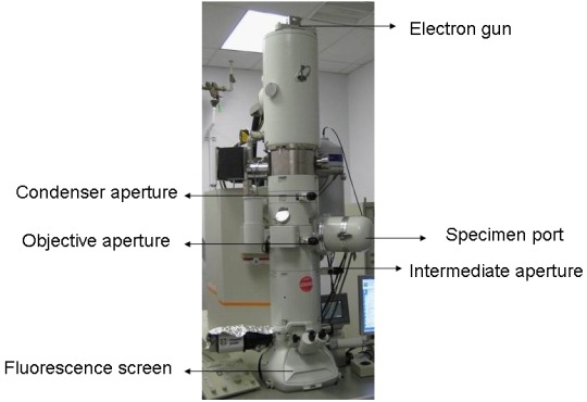


Materials Characterization: Transmission electron microscopy
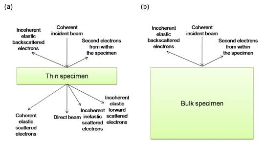
As with other forms of electron microscopy, transmission electron microscopy (TEM) uses a beam of electrons to 'illuminate' the specimen of interest and provide information and images. As the name would suggest, the electrons used in this technique are transmitted through the sample, requiring ultrathin specimens (typically less than 100nm in thickness). The thinner the specimen, the clearer the resulting images. The resolution of TEM can be less than a nanometer, though the specific machine resolution can vary based on a number of factors.
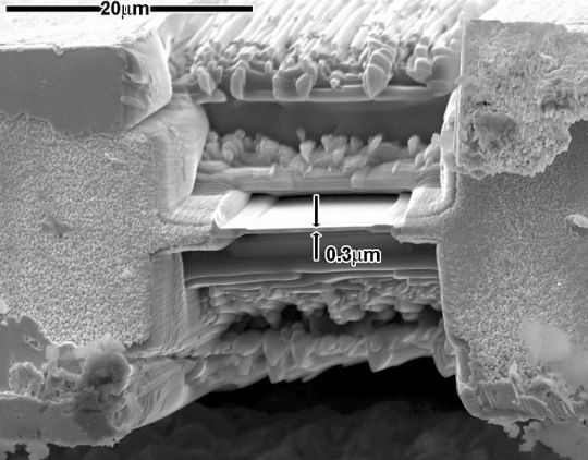
There are several techniques to produce TEM samples. For solid, bulk inorganic specimens, historically, mechanical polishing, chemical etching, and electropolishing have been used, though these techniques are time consuming and cannot easily be used to extract TEM foils from specific locations. These days, focused ion beam milling is often used to extract foils from targeted areas, as shown in image 2 above.
In addition to images such as the one shown in image 4 above, TEM can also produce diffraction patterns (as in image 3) which can be used to determine the site-specific crystal structure, as well as its orientation. TEM can also provide chemical information, including, if the resolution is high enough, on an atom-by-atom basis.
Many variations of the technique exist, including scanning transmission electron microscopy (STEM), cryo-TEM, aberration corrected TEM (more common in modern instruments), and the ability to conduct various in-situ/environmental experiments during imaging. While TEM can be used to collect extremely detailed small scale information about a sample, it should be noted that the size of the specimens mean that, particularly for bulk specimens, results may not be representative of the sample overall.
Sources/Further reading: ( 1 - images 1, 4, 5 ) ( 2 - images 2 and 3 ) ( 3 ) ( 4 ) ( 5 )
#Materials Science#Science#Transmission electron microscopy#Electron microscopy#Materials Characterization#MyMSEPost
34 notes
·
View notes
Last Seen Blogs

dewoowoo
Untitled

asgards-princess-of-mischief
Evelyn Kingsley

daisies-in-thedark
Daisies in the Dark

jihucy
ִֶָ

onlysexygirls13
Daddy's Sexy Girls