#Blasts
Text
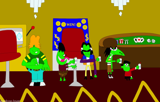
The Gangreen Gang all enjoying Sonic ice cream blasts at a casino here!
Made by me! (x)
#gangreen gang#casino#sonic#sonic drive-in#ace#snake#big billy#grubber#little arturo#ice cream#blasts#sonic blasts#ice cream blasts#slots#gambling#roulette#poker#jackpot#chandeliers#acedcop-lover
3 notes
·
View notes
Text

#gun shot#blasts#murder#death#kill#killer#gun#guns#hand gun#blow your brains out#gif#gifs#dead girl#girl#girls#skull girls#tumblr girls#kill a motherfucker#murderer#killa#dead by daylight#she's dead#blast to the dome#straight up murder#killing#killed#alternate realities#alt girl#alternate universe#alternative
4 notes
·
View notes
Text

Got to move in fast.
18 notes
·
View notes
Text
byler: *exists*
conan gray’s songs: *exist*
me: *making them exist together*


#byler#blasts#blasts the story because who needs therapy when self destruction is an option#mike wheeler#will byers#cleradin#your therapist doesn’t want you to know this but suffering is always free!
24 notes
·
View notes
Text


Mysterio effect’s
#mysterio#MCU#powers#spiderman#far from home#jake gylenhall#Quinton beck#magic#cgi#special effects#levitation#smog#lightning#blasts
30 notes
·
View notes
Text
I’ve been saying “AUTISM BLAST!!!!“ like it’s a special attack I have to shoot a beam using my autism and honestly I think I might keep saying it
Warrior’s Most Powerful Ability
Autism Blast
9 notes
·
View notes
Text
Sky News Breakfast: Kyiv hit by series of explosions
Sky News Breakfast: Kyiv hit by series of explosions
On Sky News Breakfast with Kay Burley:
– Ukrainian capital Kyiv targeted by missile strikes for the first time in months
– Could the warning on winter blackouts lead to our coal power stations being fired up again?
– And as Taiwan’s President says the country will bolster its defences amid tensions with China, we speak to the island’s Deputy Foreign Minister.
Plus all the day’s headlines and…
View On WordPress
#Beijing#BLACKOUTS#BLASTS#Breakfast#Breakfast in full#bridge#china#COAL#Conservative#COST OF LIVING#cost of living uk#CRIMEA#ELECTRICITY#ENERGY#energy bills#explosions#gas#Katie Piper#Kay Burley#Kyiv#kyiv explosions today#Labour#LATEST NEWS#mental health#Petrol station#PRESIDENT PUTIN#putin#russia#Service station#Sky
11 notes
·
View notes
Text
team rocket names?
13 notes
·
View notes
Photo


Meagu Blasts 🍂
Higher quality on Imgur -> LINK
BDO Official Forum Gif Topic - BDO various gif sets
[EU] Unikornu
#BlackDesert#black desert online#black desert#bdo#black desert gif#gif#gif set#character#meagu#new class#twin class#fox#charm fox#charms#explosive charms#blasts#meagu skills#slow mo skills#she made the gifs to big i could barely optimize them under 10MB#rip#family unikornu#eu
5 notes
·
View notes
Text
Blasts shouldn't be seen in peripheral blood.
Rule out leukemia, myelodysplastic syndrome, chronic myeloid leukemia.
Blasts may be other immature hematopoeitic cells. High monocyte count might actually be blasts, not monocytes.
If auto differential is abnormal, get a manual one.
Blasts newly elevated is more suggestive of acute leukemia. Pancytopenia suggests bone marrow failure. Febrile neutropenia is also concerning.
Make sure pt is stable with vitals, oxygen if needed, fluids.
Rule out emergencies and then contact heme/once.
APL is a subtype of AML. AML has 7 morphological categories using a system called fab. APL is the third subtype, M3. It has unique history and treatment. It presents with DIC and has early mortality. People hemorrhage and die. You can prevent it by giving all trans retinoic acid (ATRA). It helps APL cells differentiate into normal cells. APL is curable. Give all trans retinoic acid (ATRA). Diagnose by looking at source of the tumor cells. Look at peripheral blood smear. It separates ALL from AML. Auer rods are seen in AML. APL cells have multiple auer rods and folded nuclei that are very granulated.
CML has maturation of all the white cells, mature granulocytes, basophilia.
MDS (myelodysplastic syndrome) = elderly pts; hypogranular neutrophils, bilobed neutrophils, red cell shape/size changes (anisopoikilocytosis)*.
Do the peripheral blood smear first. Next do a peripheral blood flow cytometry or cell markers. It classifies cells based on the markers they express. Blasts express CD34. If CD34 is elevated, then the pt has increased blasts.
The definitive test to diagnose acute leukemia is a bone marrow biopsy. AML = more than 20% myeloblasts on bone marrow biopsy. ALL = more than 20% lymphoblasts on bone marrow biopsy.
APL has a characteristic genetic translocation--chromosome 15 and 17.
Before talking to heme/onc, also get bloodwork to rule out DIC (fibrinogen level, INR, PTT, d-dimer). If pt is febrile, work up includes blood culture, chest X-ray, urine culture for febrile neutropenia.
Also get CMP, phosphorus, magnesium, calcium, uric acid level to look for tumor lysis syndrome.
Transfuse platelets if platelets less than 10,000 and bleeding. If symptomatic anemia, transfuse RBCs.
Leukostasis = elevated WBCs such that blood flow to organs is impeded. Immature cells clog up the vasculature. Causes hypoxia, pulm infiltrates, SOB, AMS, HA, dizziness. WBC can be 50 to 100 or even less than that in leukostasis.
Other hematologic emergencies: cauda equina (spinal cord compression) is seen in lymphoproliferative disorders (lymphoma-> mass effect on spinal cord); myeloma (alters bone integrity-> compression fractures). Sometimes myeloma deposits can compress the spinal cord. So assess lower extremity reflexes, tone, Babinski sign, saddle anesthesia, rectal tone, bowel or bladder incontinence.
Top 5 clinical pearls
1) blasts are worrisome in peripheral blood, should not be in peripheral blood. Get heme/onc on board fast.
2) get a manual differential, review the film manually
3) acute leukemia can present with pancytopenia and no circulating blasts, OR with just circulating blasts. So either of those should be explored.
4) if you think the pt has acute leukemia, suspect APL, which is a medical emergency that can be treated with all trans retinoic acid immediately.
5) hematologic emergencies: severe cytopenia, febrile neutropenia, DIC, tumor lysis syndrome, leukostasis, cord compression. So screen for these.
*Anisopoikilocytosis is when you have red blood cells that are of different sizes and shapes.
The term anisopoikilocytosis is actually made up of two different terms: anisocytosis and poikilocytosis. Anisocytosis means that there are red blood cells of varying sizes on your blood smear. Poikilocytosis means that there are red blood cells of varying shapes on your blood smear.
So basically: get a peripheral blood smear, which let's you look at the cells. Flow cytometry can further differentiate the specific cell type that is elevated. Bone marrow biopsy is the definitive way to diagnose.
#heme#AML#APL#CML#febrile neutropenia#blasts#leukostasis#monocytosis#M3#leukemia#all trans retinoic acid#ATRA#Auer rods#Auer rod#hemeonc
3 notes
·
View notes
Text
Something about the colour 'violet' makes me think of Nuclear Blasts and Lilac Fields at the same time.
Maybe it's because both of them never fail to get my attention, do they?

#light academia#chaotic academia#dark academia#academia#desi dark academia#dark acadamia aesthetic#desi academia#desi aesthetic#aesthetic#desiblr#seventh#colour#seventh colour#rainbow#pride#violet#nuclear#blasts#lilac aesthetic#lilacs#fields#attention#symbolism#violet moodboard#violet aesthetic#compare#contrast#opposites
3 notes
·
View notes
Text

The Gangreen Gang all on an airplane while enjoying Sonic ice cream blasts there!
Made by me! (x)
#gangreen gang#airplane#sonic#sonic drive in#ice cream#ace#snake#big billy#grubber#little arturo#ice cream blasts#travelinh#cheesecake#reece's#m&ms#chocolate chip cookie dough#oreo cookies#blasts#acedcop-lover
3 notes
·
View notes
Photo

BON-BON THUNDER BALLS!
OVER 400 BLASTS!
#stalone#capgun#blasts#rocky#rambo#bootleg#blacktubbootlegs#nonflammable#bon-bon#retro#junk#knock off toys#Bootleg Toys#motuko#motu
5 notes
·
View notes
Text
I imagine this is how Joe Keery actualy sings.
4 notes
·
View notes
Text
the funniest meltdown ive ever had was in college when i got so overstimulated that i could Not speak, including over text. one of my friends was trying to talk me through it but i was solely using emojis because they were easier than trying to come up with words so he started using primarily emojis as well just to make things feel balanced. this was not the Most effective strategy... until. he tried to ask me "you okay?" but the way he chose to do that was by sending "👉🏼👌🏼❓" and i was so shocked by suddenly being asked if i was dtf that i was like WHAT???? WHAT DID YOU JUST SAY TO ME?????????? and thus was verbal again
#yeehaw#1k#5k#10k#posts that got cursed. blasted. im making these tag updates after... 19 hours?#also i have been told it should say speech loss bc nonverbal specifically refers to the permanent state. did not know that!#unfortunately i fear it is so far past containment that even if i edited it now it would do very little. but noted for future reference#edit 2: nvm enough ppl have come to rb it from me directly that i changed the wording a bit. hopefully this makes sense#also. in case anyone is curious. though i doubt anyone who is commenting these things will check the original tags#1) my friend did not do this on purpose in any way. it was not intended to distract me or to hit on me. im a lesbian hes a gay man. cmon now#he felt very bad about it afterwards. i thought it was hilarious but it was very embarrassed and apologetic#2) “why didn't he use 🫵🏼?” didn't exist yet. “why didn't he use 🆗?” dunno! we'd been using a lot of hand emojis. 👌🏼 is an ok sign#like it makes sense. it was just a silly mixup. also No i did not invent 👉🏼👌🏼 as a gesture meaning sex. do you live under a rock#3) nonspeaking episodes are a recurring thing in my life and have been since i was born. this is not a quirky one-time thing#it is a pervasive issue that is very frustrating to both myself and the people i am trying to communicate with. in which trying to speak is#extremely distressing and causes very genuine anguish. this post is not me making light of it it's just a funny thing that happened once#it's no different than if i post about a funny thing that happened in conjunction w a physical disability. it's just me talking abt my life#i don't mind character tags tho. those can be entertaining. i don't know what any of you are talking about#Except the ppl who have said this is pego/ryu or wang/xian. those people i understand and respect#if you use it as a writing prompt that's fine but send it to me. i want to see it#aaaand i think that's it. everyday im tempted to turn off rbs on it. it hasn't even been a week
144K notes
·
View notes
Text
Tumblr: No NSFW! You know how it is we banned it because of the bots in 2018!
Also tumblr:

#Edited because someone said they're not bots#i think they are because i did click on their streams and it was loop r extremely obscene#and i didn't LOOK for them#it's being blasted to ALL users#i don't think this is safe for them like this
104K notes
·
View notes
Last Seen Blogs


brothertedd
#brothertedd

sweer-tomato
The Puffy Zone

myradicora
"Ah, the sweet taste of power."

itsnanaunderfire-blog
fmaiqm